
Lymphoma & related disorders
General
Flow cytometry
Authors: Abdallah Flaifel, M.D., Reid Wilkins, M.D., Nicholas Ward, M.D., Tamar C. Brandler, M.D., M.S.
Editorial Board Members: Alexa J. Siddon, M.D., Julie Feldstein, M.D.


Last author update: 11 July 2023
Last staff update: 14 November 2023
Copyright: 2021-2025, PathologyOutlines.com, Inc.
PubMed Search: Flow cytometry

Table of Contents
Definition / general | Essential features | Terminology | CPT coding | Sites | Diagrams / tables | Overview | Advantages of flow cytometry | Disadvantages of flow cytometry | Effectiveness | Clinical applications | Cytology images | Flow cytometry images | Practice question #1 | Practice answer #1 | Practice question #2 | Practice answer #2Cite this page: Flaifel A, Wilkins R, Ward N, Brandler TC. Flow cytometry. PathologyOutlines.com website. https://www.pathologyoutlines.com/topic/cytopathologyflowcytometry.html. Accessed August 14th, 2025.
Definition / general
- Flow cytometry is a technique that measures various parameters (optical characteristics and emitted fluorescence) of cells in a flowing fluid suspension
Essential features
- Sensitive method for simultaneously obtaining information on various parameters, including optical characteristics and fluorescence of cells in suspension
- Components include fluidics, optical systems and electronic systems
- Applications are limited to cells in suspension and provide no information about cell to cell interactions
- Provides valuable clinical application in hematologic diseases and body fluids diagnostics
Terminology
- Cell sorting: separating cells, identified by specified characteristics
- Compensation: mathematical algorithm for removing bleeding / spillover of 1 fluorophore into multiple detectors
- Forward scatter: light scattered in the forward direction after interacting with a particle
- Gating: specifying populations of cells with common characteristics to investigate further
- Side scatter: light scattered at 90 degrees after interacting with a particle
CPT coding
- Fresh tissue or body fluids
Sites
- Peripheral blood
- Bone marrow
- Body cavity fluids (pleural, peritoneal, pericardial, joints)
- Cerebrospinal fluids
- Tissue (e.g., lymph nodes)
Diagrams / tables
Contributed by Abdallah Flaifel, M.D.
Cell block TdT
Images hosted on other servers:

Schematic of a flow cytometer
Overview
- Basic principles of flow cytometry (Crit Rev Biotechnol 2017;37:163)
- Single cells are dissociated in a liquid medium
- Cells are then stained with 1 or more fluorochrome tagged markers
- Laser beams with specific wavelengths strike the cells
- Cells tagged with fluorochrome dye absorb photons and emit fluorescence
- Light scatter and fluorescence from individual cells are detected by photomultiplier tubes
- Electronic impulse is converted to digital data (analog to digital) and is recorded by the computer
- Data acquisition software stores digital data as flow cytometry standard file
- Software packages display data as uni / bivariate histograms, scatter / dot plots, contour plots or density plots
- Components (Curr Protoc Immunol 2018;120:5.1.1, Crit Rev Biotechnol 2017;37:163)
- Fluidics
- Composed of a coaxial system: inner (sample) and outer (sheath)
- The aim of the system is to maintain a stable flow of cells
- Sheath fluid protects the sample from turbulence caused by resistance to the flow by the tube walls
- Air pressure of the sample controls the rate of flow of cells
- Increasing sample fluid pressure increases the rate of flow
- Lowering the sample fluid pressure decreases rate of flow
- Composed of a coaxial system: inner (sample) and outer (sheath)
- Optical system
- Light source
- Laser (light amplification by stimulated emission of radiation)
- Emits light with a specific wavelength
- Primary laser (488 nm wavelength) or secondary red diode laser (635 nm wavelength) may be utilized
- Excitation of the fluorochrome from laser beam results in fluorescence; the selection of fluorochrome should be appropriately matched with the wavelength of the laser used
- Laser (light amplification by stimulated emission of radiation)
- Light scattering
- Forward scatter (FSC): directly proportional to the size and surface area of the cell; it is collected by the FSC detector located on the same axis as the laser beam
- Side scatter (SSC): directly proportional to the granularity and internal complexity of the cell
- Fluorescence emission
- Fluorochrome dye can be used to characterize various properties in cells
- Fluorochrome is commonly tagged with an antibody
- Fluorochrome will absorb and emit light at a specific wavelength
- Light collection: via a set of special filters and optical mirrors
- Light source
- Electronic system (Methods Mol Biol 2011;699:1)
- Photons of light, generated by light scatter and fluorophore emission, hit the photodetectors
- Photon signal is converted into an electrical current called a photocurrent
- Photocurrent is then converted into a voltage pulse
- Voltage pulse is directly proportional to forward / side scatter and the number / brightness of fluorophores
- Voltage pulse may then be amplified (linear or logarithmic) or digitized by an analog digital converter (ADC)
- Newer flow cytometers (termed digital systems) digitize photocurrent early without prior amplification or processing
- Digitization involves organization of the continuous analog data into digital channels through process known as binning
- Analog digital converter outputs a data file in a standard FSC format
- Fluidics
Advantages of flow cytometry
- Sensitive method for simultaneously obtaining information on various parameters and processes, including expression of surface markers or the presence of intracellular cytokines and proteins
- High throughput system that can excel in characterizing heterogeneous cell populations
- Capable of sorting cells based on specific features
- Indispensable tool for the classification and immunophenotyping hematologic neoplasms
- Reference: J Invest Dermatol 2012;132:1
Disadvantages of flow cytometry
- Limited to cells in suspension, so information on tissue architecture and cell - cell interactions is not available
- Analyses requiring more fluorophores are subject to signal spillover
- Analysis is complicated by the amount of data generated
- Results are limited to the specific immunofluorescence panels that are being tested
- Less accurate in diagnosing certain lymphomas (i.e., Hodgkin lymphoma)
- Limited utility in evaluating infectious etiologies (e.g., EBV, HHV8)
- Reference: J Invest Dermatol 2012;132:1
Effectiveness
- Efficient flow cytometry satisfies the following
- High sensitivity
- Threshold: the capability to distinguish dim cells from the particle free background
- Resolution: the ability to separate dim cells from unstained ones
- Relative measured values of fluorescence (which depends on the instrument’s linearity accuracy)
- Reproducibility of the results
- High sensitivity
- Quality control
- Periodically: laser time delay, laser alignment
- Every day: photomultiplier voltage settings, compensation, gating control for multicolor flow
- External quality assessment
- Reference: Clin Lab Med 2007;27:671
Clinical applications
- Hematologic diseases
- Diagnosis and sub classifying non-Hodgkin lymphoma (NHL)
- WHO approaches to classify the lymphomas based on the lineage of the cells: B cell and T / NK cell; precursor versus mature
- Useful in immunophenotyping different lymphomas
- B cell markers: CD19, CD20, CD79a, PAX5, CD10, CD23
- T cell markers: CD2, CD3, CD4, CD5, CD7, CD8
- NK cell markers: CD2, CD3, CD56
- Plasma cell markers: CD38, CD138, CD56
- Fine needle aspiration of lymph node with flow cytometry
- Rapid onsite evaluation
- Material processed for
- Routine smears
- Cell block
- Flow cytometry
- Cytogenetics
- False negative in flow cytometry after fine needle aspiration (FNA) can be due to
- Predominantly necrotic tissue or fibrosed lymph node
- Material diluted with blood
- Few and scattered atypical cells as in Hodgkin lymphoma
- Assessment of minimal residual disease (Blood Cancer J 2020;10:108)
- Ultrasensitive method of determining disease progression, relapse and response to therapy
- May be used with or without next generation sequencing to detect specific populations with a sensitivity between 10-5 and 10-6
- Usually performed on bone marrow aspirate fluid (multiple myeloma and acute leukemias) but may also use peripheral blood in certain instances (e.g., CLL or ALL)
- Limitations include hemodilution and sampling error
- One major application of flow cytometry is demonstrating the clonality of lymphoma
- Light chain restriction: monoclonal B cell proliferation has altered kappa to lambda chain ratio
- Diagnosis and sub classifying non-Hodgkin lymphoma (NHL)
- Flow cytometry of cytology samples
- Effusion
- Detection of carcinoma in fluid
- Higher synthetic phase (S phase) cells may be used as indirect evidence of malignancy
- Epithelial cell adhesion molecule (EpCAM [CD326]) or its antibody (BerEP4) can be used to identify epithelial cells (Appl Immunohistochem Mol Morphol 2009;17:202)
- Possible pitfalls
- Poorly differentiated carcinoma may not express any markers
- Benign cells may express epithelial markers, especially in peritoneal washings and instrumentalizations
- Effusion due to involvement by a lymphoreticular neoplasm
- Cytology along with flow cytometry have 100% sensitivity and 94% specificity in diagnosing lymphoma (Diagn Cytopathol 2006;34:335)
- Some panels suggested include
- Acute leukemia: MPO, Tdt, CD45 / CD34 / CD7 / CD13 and CD33 / CD56 / CD19
- Lymphoma: CD45 / CD3 / CD4 / CD8 and CD19 / CD20 / CD10
- For suspected T cell lineage, must include: CD3 / CD2 / CD5 / CD7 / CD8, CD26
- For suspected B cell lineage, must include CD5 / CD23 / CD10 / CD38 / CD138 / FMC7 / surface kappa and lambda light chain
- For suspected myeloma: CD20 / CD19 / CD38 / CD138 / CD27 / CD45 / cytoplasmic kappa / lambda
- Primary effusion lymphoma
- Cytomorphology shows large cells with nuclear pleomorphism and nucleoli
- Flow cytometry
- Positive: CD45, CD30, CD38, CD138, CD43
- Negative for B cell markers: CD19, CD20 and no light chain restriction
- Absent or aberrant expression of T cell markers: CD2, CD3, CD5, CD7
- Detection of carcinoma in fluid
- Bronchoalveolar lavage (BAL) (Am J Respir Crit Care Med 2012;185:1004)
- Cellular analysis may provide useful adjunct information for patients who lack radiographic evidence of usual interstitial pnuemonia
- Predominant inflammatory cellular pattern could help clinicians determine etiology of interstitial lung disease
- Effusion
- Identification of therapeutic targets
- Selection of antibody based therapies
- Rituximab: anti-CD20
- Epratuzumab: anti-CD22
- Gemtuzumab: anti-CD33
- Blinatumomab: directed against CD19 and CD3
- Selection of antibody based therapies
- Nonhematologic disease
- Assessment of nonhematologic neoplasms (Am J Clin Pathol 2020;153:99)
- May utilize antibodies directed against epithelial markers such as MOC31, Ber-EP4, CK5 / 8 and MUC1
- Targeted panel of CD45, CD33 and EpCAM (CD326) has been used to distinguish monocytes / macrophages, mesothelial cells and epithelial cells
- Assessment of nonhematologic neoplasms (Am J Clin Pathol 2020;153:99)
- DNA content analysis
- Use of propidium iodide that interposes into the DNA helical structure
- Fluorescence is directly proportional to the amount of DNA
- DNA content aneuploidy can be determined in a tumor cell
- Use of propidium iodide that interposes into the DNA helical structure
Cytology images
Contributed by Abdallah Flaifel, M.D.
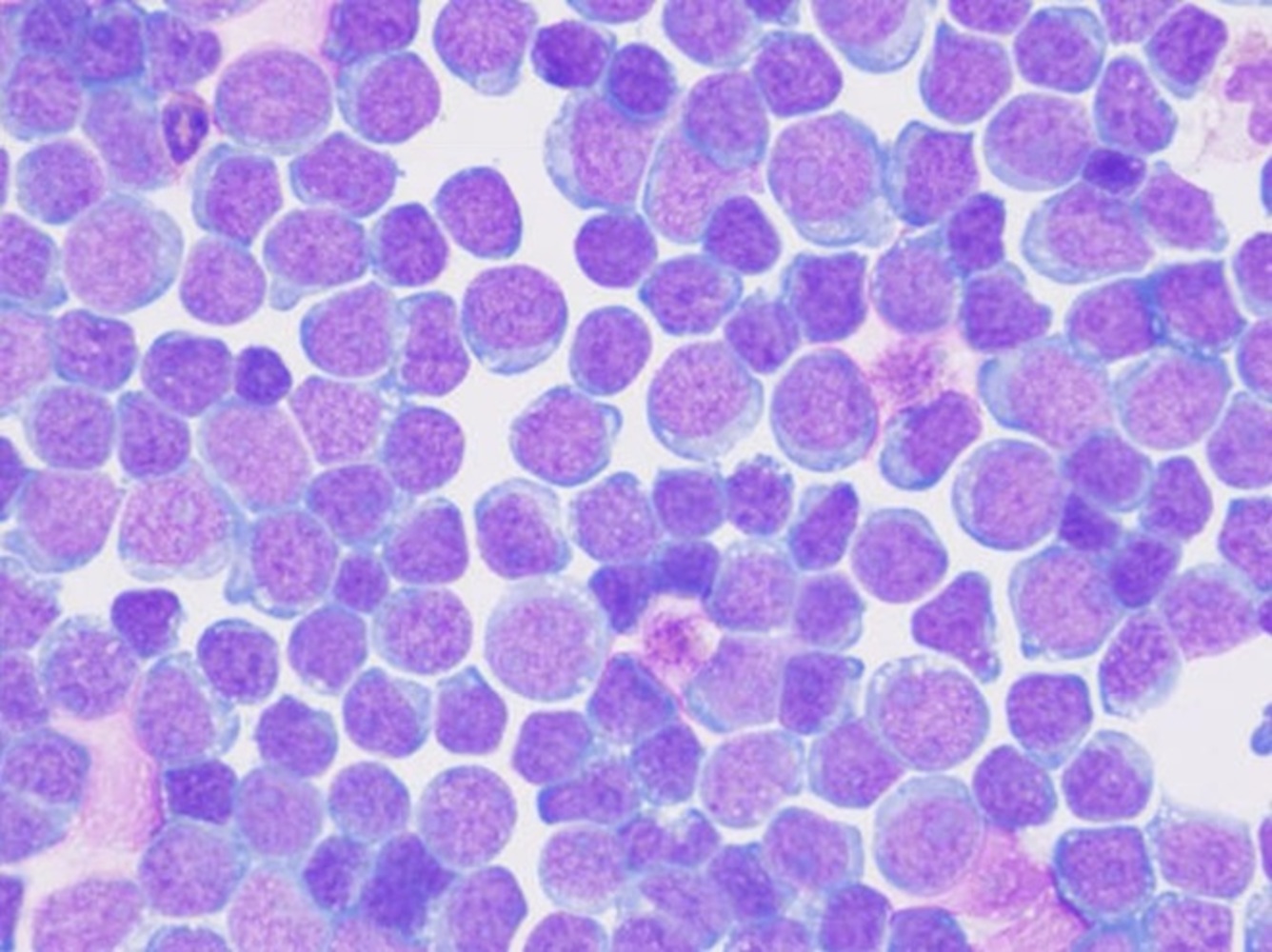
Pericardial fluid cytospin
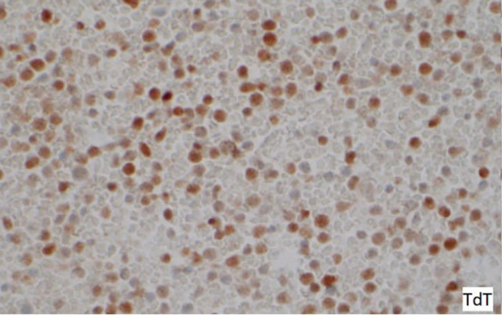
Cell block TdT
Flow cytometry images
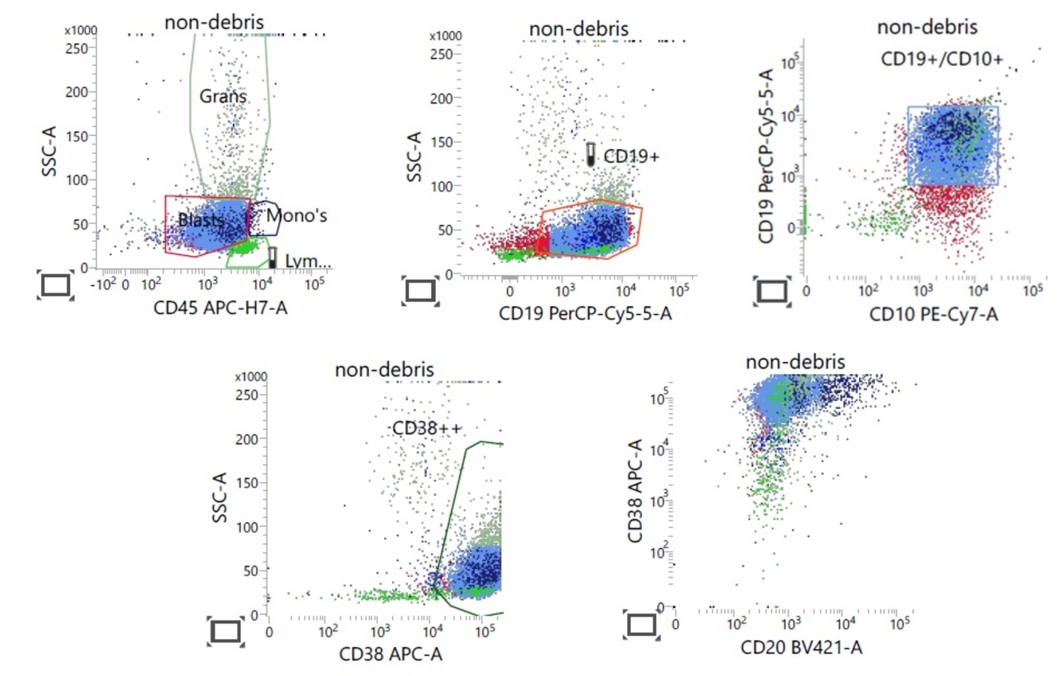
Contributed by Nicholas Ward, M.D.
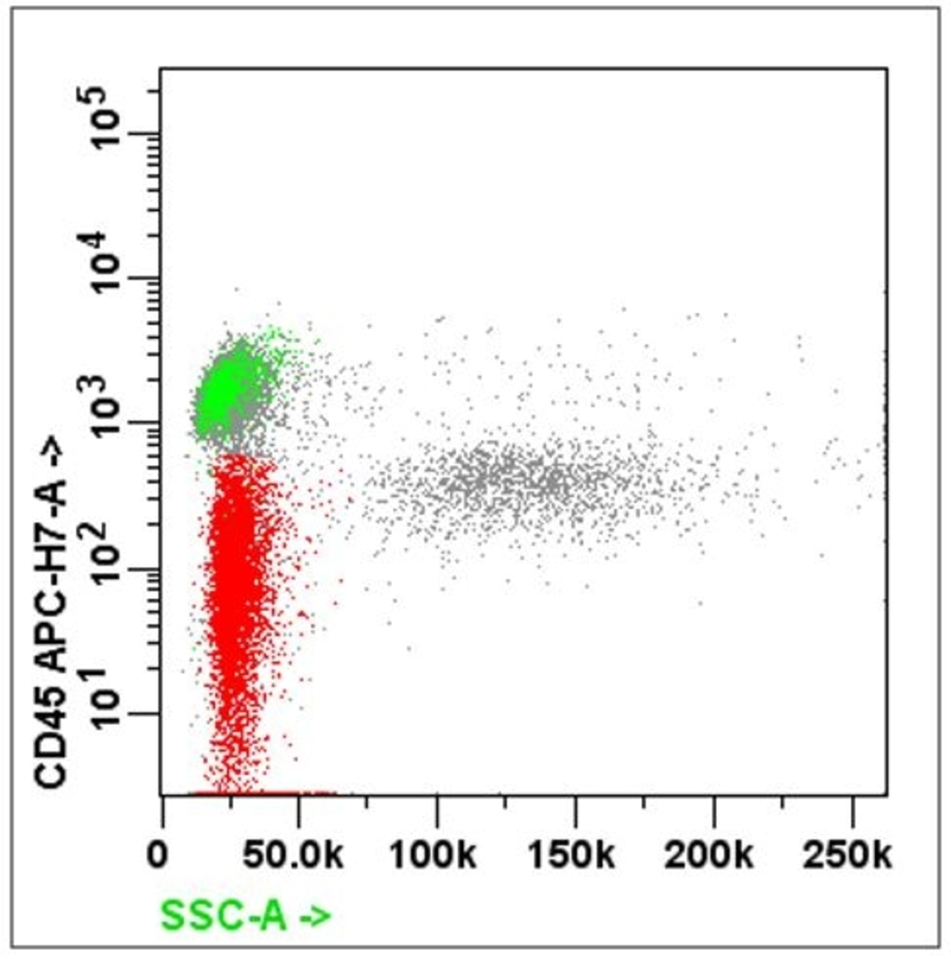
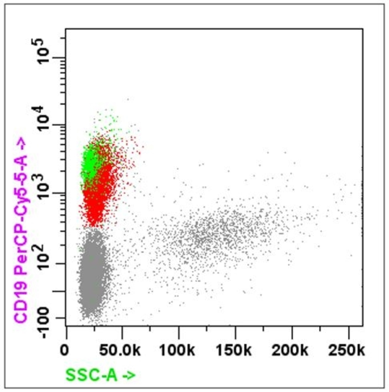
B ALL flow cytometry
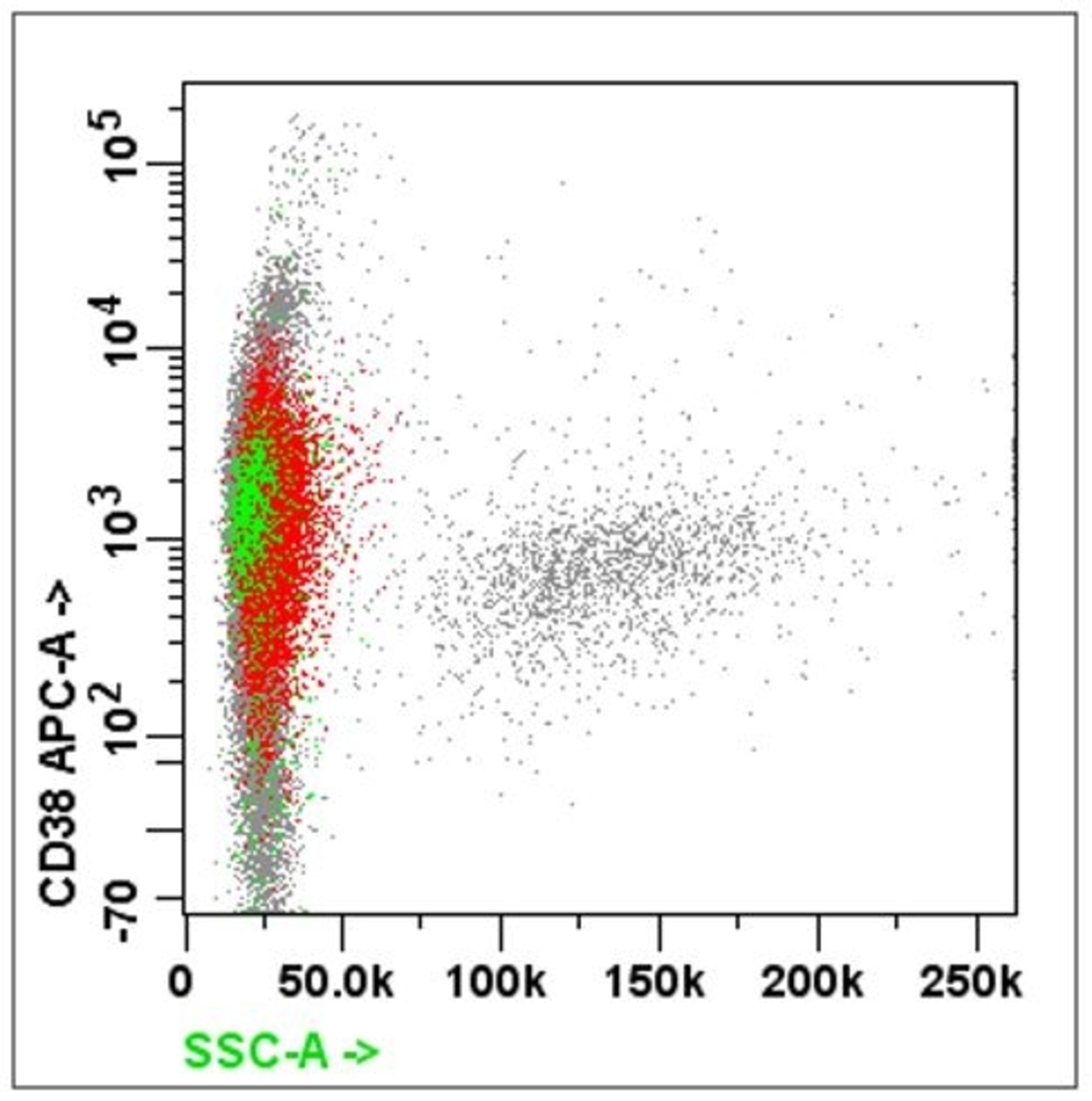
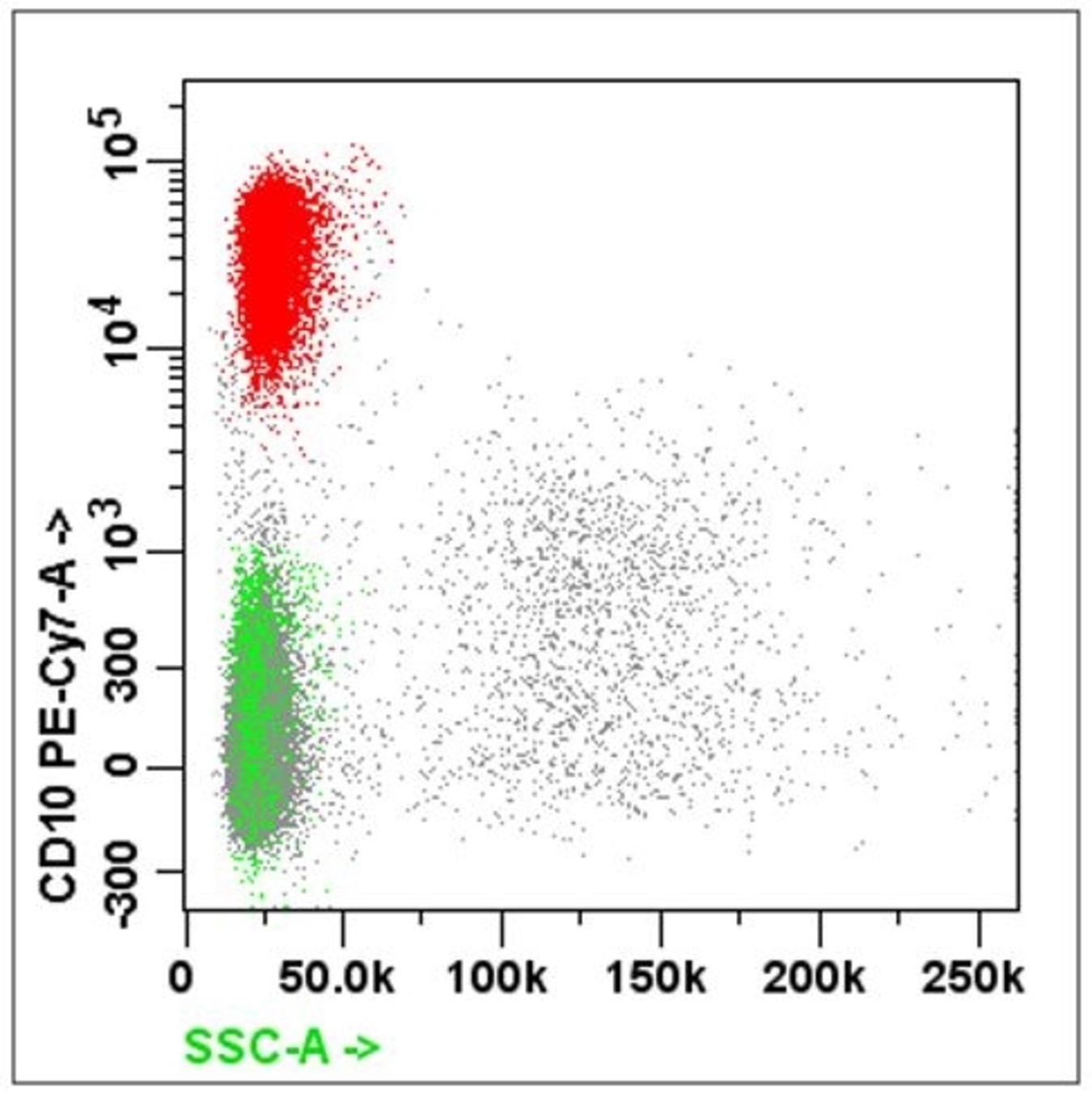
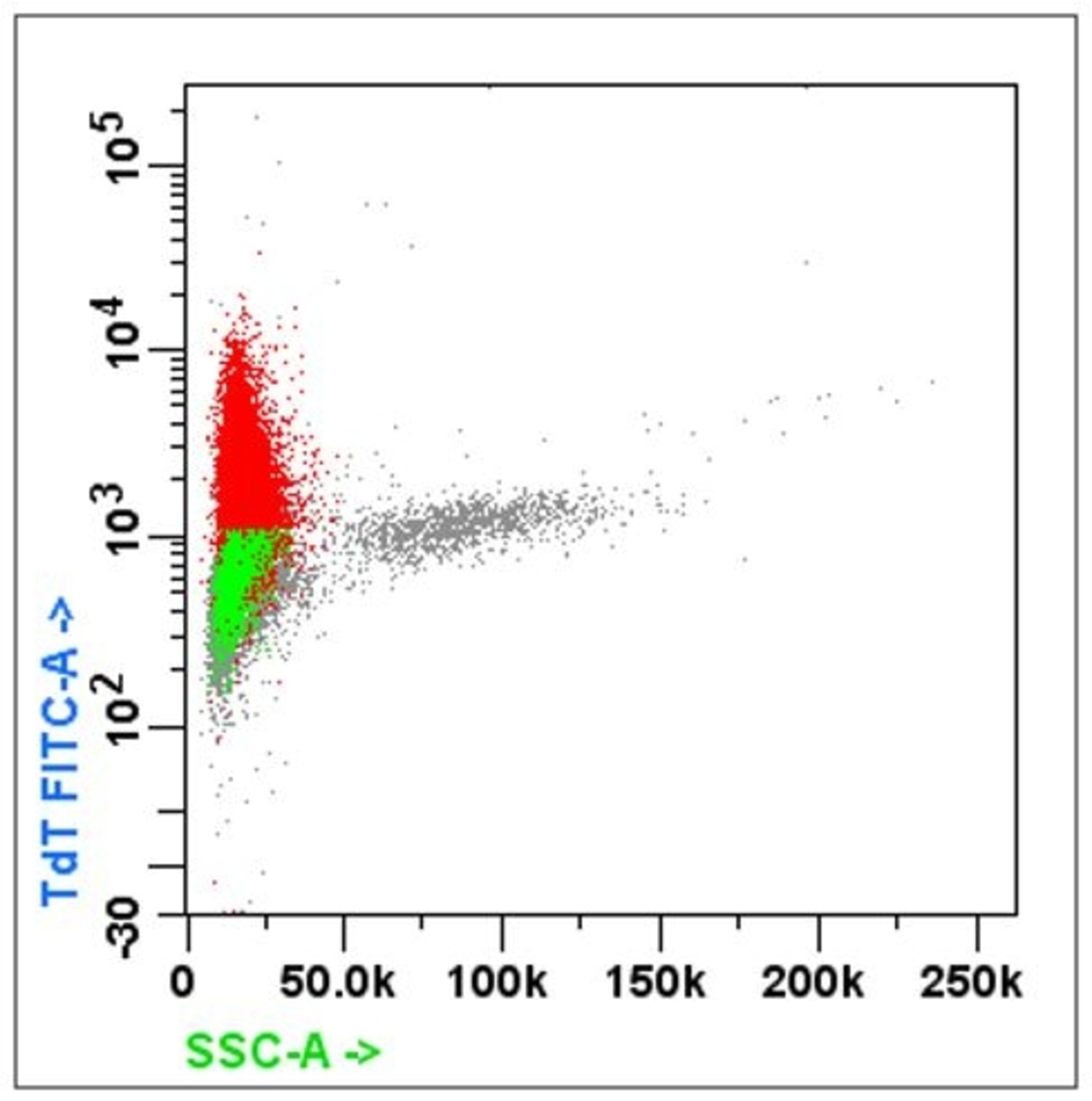
B ALL flow cytometry
Practice question #1
In a typical flow cytometry, the side scatter (SSC) and forward scatter (FSC) provide information about which of the following?
- Architecture and granularity
- Cytoplasmic complexity and size
- Intracellular signaling and viability
- Size and surface markers
Practice answer #1
Practice question #2
Which of the following is a clinical application of flow cytometry?
- Demonstrating clonality of lymphoma
- Easily identifying Hodgkin lymphoma
- Identifying benign cells in instrumented effusion fluid
- Identifying relevant information about architecture of carcinoma
Practice answer #2
A. Demonstrating clonality of lymphoma. Answer B is incorrect because Reed-Sternberg cells are admixed in a rich inflammatory background which consists mainly of T cells, B cells, eosinophils, histiocytes and plasma cells. Answer C is incorrect because benign cells are scant in instrumented fluid. Answer D is incorrect because flow cytometers analyze suspensions of single cells.
Comment Here
Reference: Flow cytometry
Comment Here
Reference: Flow cytometry
