
Eye
Orbit & optic nerve
Glioma-optic nerve
Author: Nat Pernick, M.D.

Last author update: 1 February 2014
Last staff update: 18 May 2022
Copyright: 2004-2024, PathologyOutlines.com, Inc.
PubMed Search: Glioma optic nerve
Table of Contents
Definition / general | Radiology description | Radiology images | Treatment | Gross description | Microscopic (histologic) description | Microscopic (histologic) imagesCite this page: Pernick N. Glioma-optic nerve. PathologyOutlines.com website. https://www.pathologyoutlines.com/topic/eyeorbitgliomaoptic.html. Accessed April 26th, 2024.
Definition / general
- Relatively rare
- Slow growing tumor within orbital segment of optic nerve
- Usually ages 0 - 9 years with symptoms of minimal exophthalmos, optic nerve atrophy or papilledema
- Associated with neurofibromatosis type 1
Radiology description
- Thickening of nerve on CT scan
- May enlarge optic canal
Radiology images
AFIP images
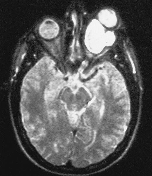
MR of large retrobulbar optic nerve tumor
Treatment
- Resection for tumors limited to optic nerve
- Also radiation therapy for more extensive lesions
Gross description
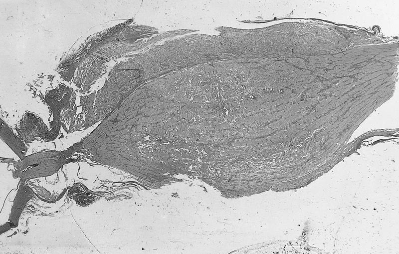
- Small tumors are limited to optic nerve
- Larger tumors form bulbous enlargement of nerve, often infiltrate pia causing arachnoid thickening
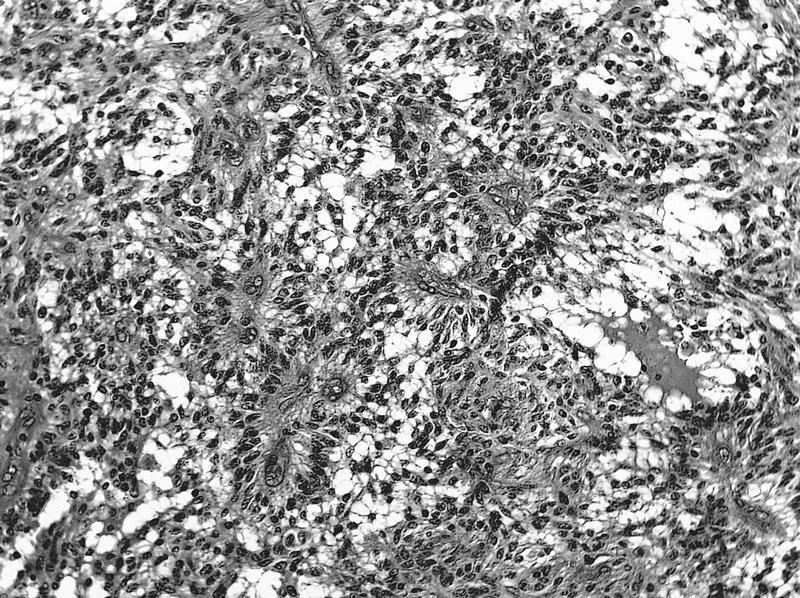
Microscopic (histologic) description
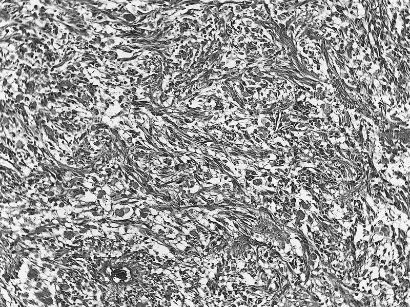
- Variable cytology and cellularity, even within same tumor, but usually are low grade pilocytic astrocytomas similar to cerebellar and third ventricle tumors with round to spindled nuclei and dendrite-like cytoplasmic processes
- Often Rosenthal fibers (fusiform, cigar shaped eosinophilic structures within astrocyte cytoplasmic processes, are a nonspecific degenerative change)
- Rarely are hypercellular with brisk mitotic activity, marked pleomorphism, necrosis and vascular proliferation
- Difficult to differentiate reactive vs. neoplastic resection margins
- Typically has intense mucinous degeneration with tumor cells in pools of mucin
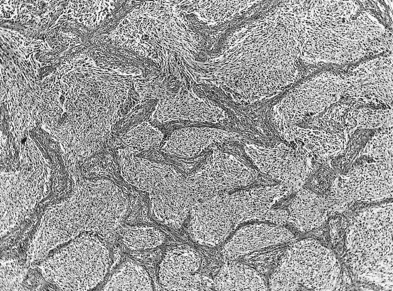
- Infiltrating tumor may cause reactive proliferation of arachnoid cells resembling meningioma
Microscopic (histologic) images
AFIP images
Pilocytic astrocytoma
Intraparenchymal tumor

Numerous Rosenthal fibers
Astrocytes infiltrate meninges
High grade astrocytoma
Images hosted on other servers:

Elongated or hair-like appearance

Rosenthal fibres
