
Eye
General
Anatomy & histology-retina
Author: J. Stephen Nix, M.D.

Last author update: 22 September 2022
Last staff update: 22 September 2022 (update in progress)
Reviewed by: Abberly Lott Limbach, M.D.
Copyright: 2004-2025, PathologyOutlines.com, Inc.
PubMed Search: Retina anatomy histology

Table of Contents
Definition / general | Essential features | Terminology | Anatomy and physiology | Diagrams / tables | Clinical features | Microscopic (histologic) description | Microscopic (histologic) images | Additional references | Practice question #1 | Practice answer #1 | Practice question #2 | Practice answer #2Cite this page: Nix JS. Anatomy & histology-retina. PathologyOutlines.com website. https://www.pathologyoutlines.com/topic/eyeretinageneral.html. Accessed August 24th, 2025.
Definition / general
- Specialized nervous tissue of the posterior eye that is responsible for the detection of light
Essential features
- Embryologic derivative of the optic vesicle, which arises from the diencephalon and enables detection of light (Dev Biol 2007;305:1)
- Composed of photoreceptors, first order neurons, second order neurons and glia (including Müller cells), the latter of which account for gliotic reaction
- Lines the innermost layer (or coat) of the eye, extending posteriorly from the ora serrata and situated between the vitreous body and choroid
Terminology
- Macula: highest density of photoreceptors and ganglion cells, responsible for high acuity vision and identified histologically by an increased number of ganglion cells and increased layer thickness
- Fovea centralis: small depression located in the center of the macula that is responsible for highest acuity vision, lacks rods and blood vessels, and relies on choroidal circulation (StatPearls: Anatomy, Head and Neck, Eye Fovea [Accessed 6 April 2022])
- Ora serrata: point of transition between anterior ciliary body and posterior retina
- Bruch membrane (not part of retina): separates choroid from overlying retinal pigment epithelium and features a 5 layer structure (basement membrane of retinal pigment epithelium, inner collagenous layer, elastin layer, outer collagenous layer and basement membrane of choriocapillaris); thickens with age and acquires focal extracellular deposits known as drusen (Prog Retin Eye Res 2010;29:1)
Anatomy and physiology
- Light is detected by the rods and cones of the photoreceptor layer and signals are transmitted through the layered retina to the optic nerve
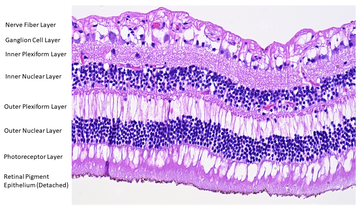
- Anatomical structure of retina and surrounding structures (outer to inner):
- Orientation
- Outer: nearest the limits of the globe (sclera)
- Inner: nearest the center of the globe (vitreous body)
- Retinal pigment epithelium: single layer of microvillous, cuboidal cells containing melanosomes that aid in light absorption by melanin, nutrient and fluid transport and phagocytosis that assists in photoreceptor turnover; forms the outer limit of the retina adjacent to Bruch membrane (Curr Mol Med 2010;10:802)
- Photoreceptor (rods and cones) layer: composed of the outer segments of the rods and cones (the photoreceptors that convert light into signal impulses) (J Cell Sci 2015;128:4039)
- External (outer) limiting membrane: permeable adherent junctions between photoreceptors and Müller cells (J Comp Neurol 1990;295:155)
- Outer nuclear layer: contains the photoreceptor nuclei
- Outer plexiform layer: located between the outer nuclear and inner nuclear layers and contains axons and dendrites of photoreceptors and bipolar cells (first order neurons), including Henle fibers (photoreceptor axons) that are prominent in the perifoveolar region and features a line of synapses known as the middle limiting membrane (Dev Ophthalmol 2016;55:7)
- Inner nuclear layer: contains bipolar cells, horizontal cells, amacrine cells and Müller cells (Invest Ophthalmol Vis Sci 2021;62:22, Physiol Rev 2019;99:1527)
- Inner plexiform layer: network of neuronal processes between the inner nuclear layer and the ganglion cell layer
- Ganglion cell layer: accommodates the ganglion cells, varying in thickness and density up to 5 - 8 layers in the macula; it is responsible for various functions in the relaying of received visual information out of the retina (Front Neurol 2021;12:661938)
- Nerve fiber layer: comprised of the axons of ganglion cells
- Internal limiting membrane: the only true basement membrane of the retina; it is located adjacent to the vitreous body where it serves as an anchor point for collagen fibers of the vitreous cortex (Exp Eye Res 2021;206:108545)
- Orientation
- Relevant vasculature to the retina:
- Central retinal artery: a branch of the ophthalmic artery that supplies the inner retina, including the nerve fiber layer, ganglion cell layer, inner plexiform layer and a portion of the inner nuclear layer
- Choriocapillaris: supplies the outer retina, including the photoreceptor layer and the innermost portion of the choroid adjacent to Bruch membrane (Prog Retin Eye Res 2022;87:100997)
- Central retinal vein and branches: drain the inner retina
- Retina lacks lymphatics
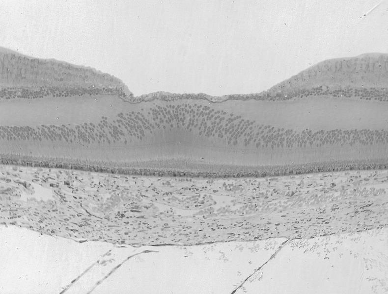
Diagrams / tables
Contributed by J. Stephen Nix, M.D.
Retina anatomy
Clinical features
- Central retinal artery occlusion: often presents with sudden, painless vision loss and damage to the inner aspect of the retina (Eye (Lond) 2013;27:688)
- Central retinal vein or branch occlusion: features cystoid macular edema as well as superficial and deep retinal hemorrhages, and in chronic cases, gliosis and inner ischemic retinal atrophy (Eagle: Eye Pathology - An Atlas and Text, 2nd Edition, 2011, Trans Am Ophthalmol Soc 1981;79:371)
- Hemorrhages of the nerve fiber layer appear streaked or flame shaped, while hemorrhages of deeper layers appear dot-like or blot-like on ophthalmoscopic examination (Pediatr Emerg Care 2018;34:665)
Microscopic (histologic) description
- Gliotic reaction due to the presence of glia (including Müller cells) are seen as a hypercellularity of variably spindled cells with oblong nuclei
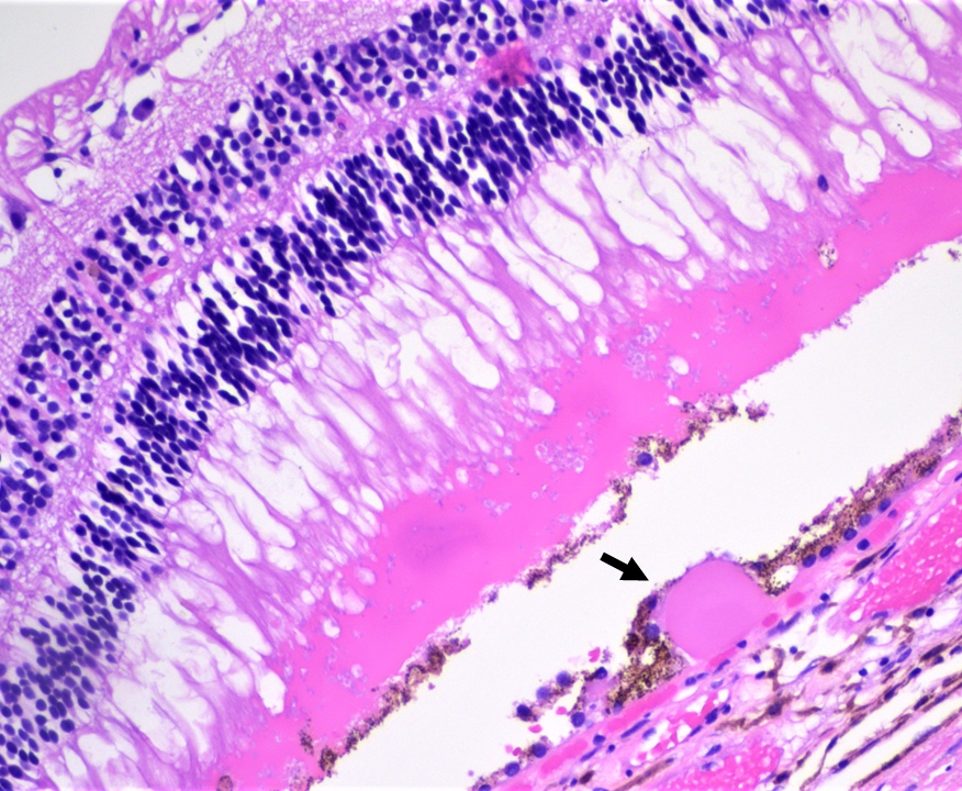
- Retinal detachment: separation of neurosensory retina (rods and cones) from retinal pigment epithelium
- Artifactual retinal detachment: common artifact of processing and lacks the subretinal proteinaceous fluid or hemorrhage found in true retinal detachments (Eagle: Eye Pathology - An Atlas and Text, 2nd Edition, 2011)
- Peripheral microcystoid degeneration: commonly encountered cystoid spaces of the outer plexiform layer near the ora serrata in adults > 20 years of age
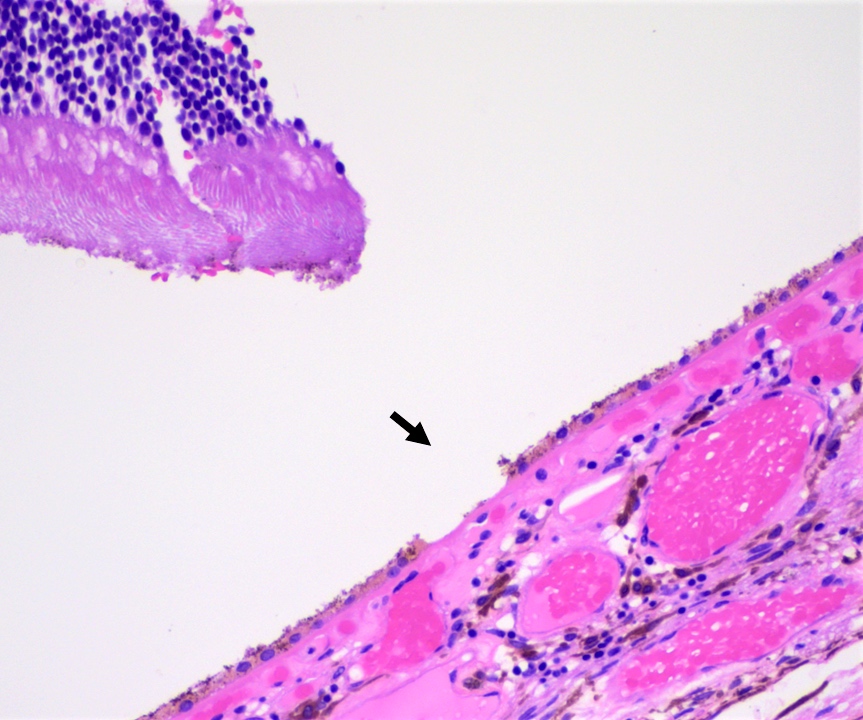
- Hard drusen are eosinophilic nodules that are observed between the retinal pigment epithelium and Bruch membrane and are encountered in aging
Microscopic (histologic) images
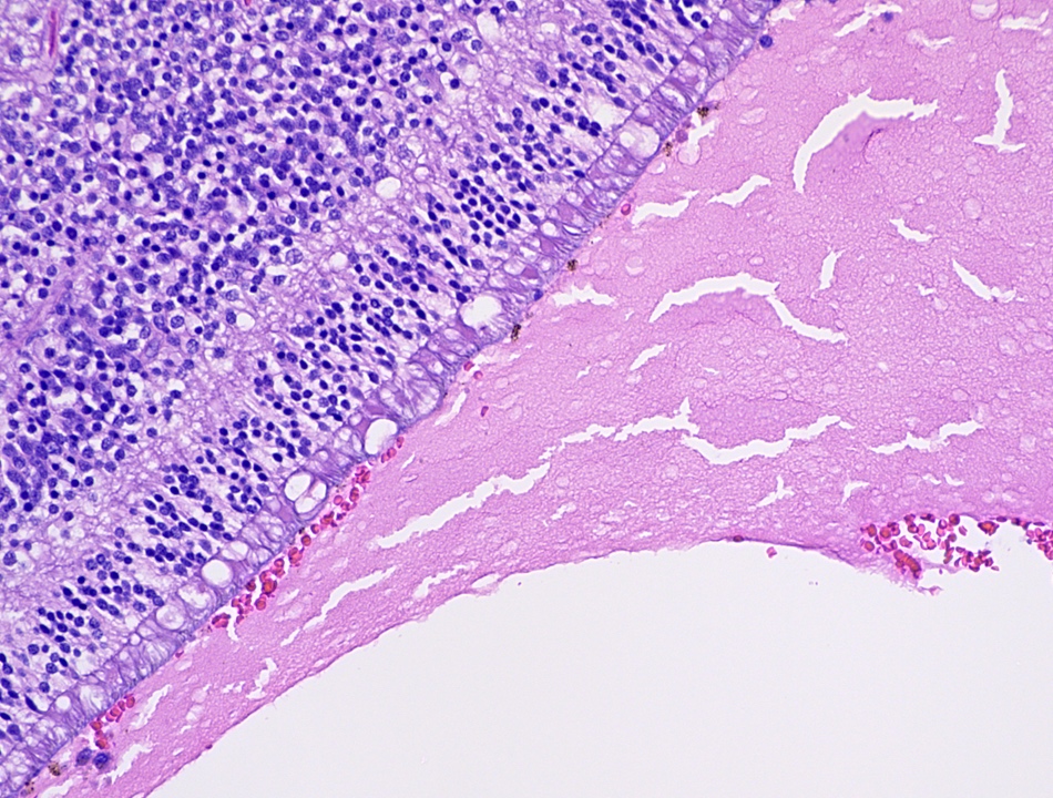
Contributed by J. Stephen Nix, M.D. and AFIP images

Macula
Choroid
Hard drusen
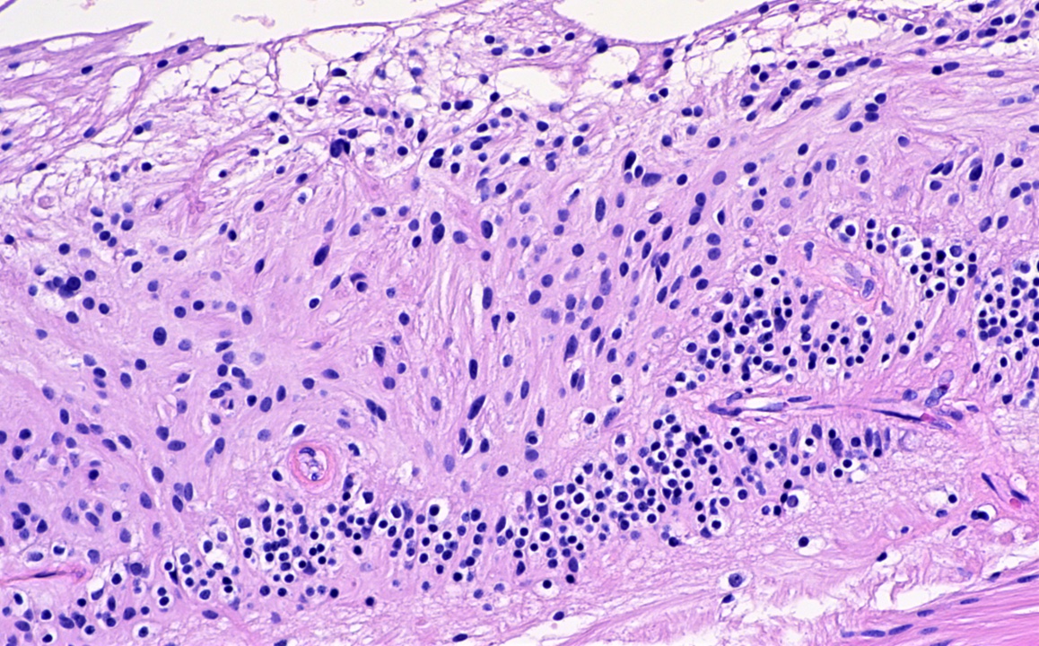
Retinal gliosis
Serosanguinous retinal detachment
Fovea centralis
Additional references
Practice question #1
Which cell type is responsible for the pathologic change seen in this image?
- Cones
- Ganglion cells
- Müller cells
- Rods
Practice answer #1
C. Müller cells. Glial cells, including Müller cells, cause retinal gliosis in response to injury.
Comment Here
Reference: Anatomy & histology-retina
Comment Here
Reference: Anatomy & histology-retina
Practice question #2
A patient with sudden, painless vision loss is diagnosed with central retinal artery occlusion. What layer of the retina would be expected to be affected?
- Ganglion cell layer
- Outer nuclear layer
- Outer plexiform layer
- Photoreceptor layer
Practice answer #2
A. Ganglion cell layer. The central retinal artery supplies the inner layers of the retina, including much of the inner nuclear layer, the inner plexiform layer, the ganglion cell layer and the nerve fiber layer.
Comment Here
Reference: Anatomy & histology-retina
Comment Here
Reference: Anatomy & histology-retina
