Lung
Infectious
Viral
CMV
Author: Elliot Weisenberg, M.D.

Last author update: 1 September 2011
Last staff update: 20 July 2020
Copyright: 2003-2025, PathologyOutlines.com, Inc.
PubMed search: CMV pneumonia / pneumonitis
Table of Contents
Definition / general | Epidemiology | Sites | Etiology | Clinical features | Treatment | Gross description | Microscopic (histologic) description | Microscopic (histologic) images | Cytology description | Positive stains | Electron microscopy description | Differential diagnosis | Additional referencesCite this page: Weisenberg E. CMV. PathologyOutlines.com website. https://www.pathologyoutlines.com/topic/lungnontumorcmv.html. Accessed September 9th, 2025.
Definition / general
- Lung injury caused by infection by CMV, a β herpes virus
- Usually immunocompromised patients
- Often associated with pneumocystis and other infections
- Xray: 2 - 4 cm peripheral nodules, miliary pattern, diffuse interstitial process
Epidemiology
- 70% of Americans in large cities have latent infection
- In immunocompetent individuals, primary infection is generally asymptomatic, but mononucleosis-like illness may occur; rarely, self limiting CMV pneumonia may occur during mononucleosis-like illness (eMedicine)
- In organ transplant recipients, infection may occur when the donor organ is latently infected with CMV
- When infection occurs in immunosuppressed patients, the risk of organ damage is higher the greater the immunosuppression
- In AIDS patients, CMV disease generally occurs when the CD4+ cell count < 50/mm3
- CMV is the most common life threatening infection in hematopoietic stem cell transplant recipients and is the most common opportunistic viral infection in AIDS
- Prophylactic therapy and preemptive therapy, where CMV DNA levels are monitored by PCR and antiviral therapy against CMV is given prior to end organ damage, has greatly reduced the incidence of CMV induced disease in vulnerable patients
- Highly active antiretrovirus therapy has reduced the incidence of end organ damage by 80% in HIV positive patients
Sites
- In addition to lung, CMV causes severe disease in retina, brain, kidneys, GI
Etiology
- Disease may occur with primary infection or secondarily when latent disease is reactivated
- Virus can be spread transplacentally; neonatal disease can occur through exposure to cervical or vaginal secretions or from breast milk when active infection is present
- In children and adults, infection is spread through saliva, respiratory droplets or sexual activity
- Fecal oral transmission has also been documented
- CMV causes down regulation of expression of MHC class I and II molecules, which helps it elude host immune responses
Clinical features
- In immunocompetent patients, cough with interstitial infiltrates on chest xray
- Immunosuppressed patients present with cough, fever, fatigue, shortness of breath, malaise, night sweats, muscle and joint pain
- Note: positive culture, serology or PCR may not represent evidence of illness
- Diagnosis is based on identification of viral inclusions in biopsy material
- Acute CMV infection leads to transient but severe immunosupression
- Prevention is key as CMV pneumonia has high mortality rate even with aggressive antiviral therapy
Treatment
- Ganciclovir, valganciclovir, foscarnet, cidofovir, maribavir
Gross description
- Lung is firm (similar to interstitial lung disease) with hemorrhagic nodules that may become confluent
Microscopic (histologic) description
- Mononuclear infiltrates, usually mild edema and pneumocyte hyperplasia
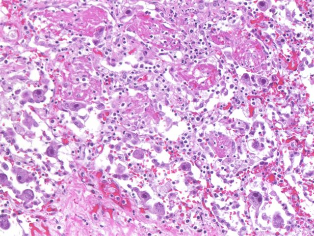
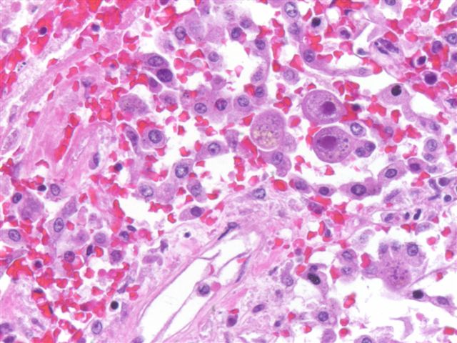
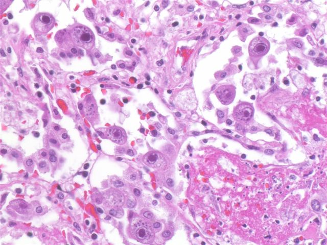
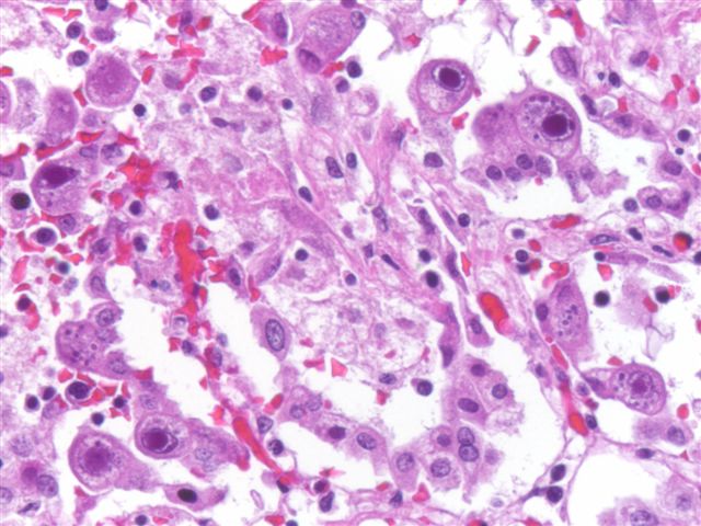
- Infected cells are generally large with a prominent basophilic nuclear inclusion, often with a clear halo, and smaller basophilic cytoplasmic inclusions are also present
- Hemorrhagic necrosis may be present
- In lung, usually infects endothelial and epithelial cells and alveolar histiocytes
Microscopic (histologic) images
Contributed by Dr. Michael P. Orejudos and Dr. Rosemarie Rodriguez

Cytology description
- Diagnostic inclusions may be seen in brochiolalveolar lavage fluid, but diagnostic yield is very low
Positive stains
Electron microscopy description
- Particles are 150 - 200 nm with round core and double membrane
Differential diagnosis
- Herpes simplex virus and herpes varicella zoster produce similar lung disease, but characteristic CMV inclusions are not present
- Drug induced injury, particularly in bone marrow transplant recipients, may cause changes similar to CMV; in problematic cases, immunohistochemical studies are useful
