Oral cavity & oropharynx
Other nonneoplastic
Amalgam tattoo
Author: Sarah H. Glass, D.D.S.
Editorial Board Member: Molly Housley Smith, D.M.D.
Deputy Editor-in-Chief: Kelly Magliocca, D.D.S., M.P.H.

Last author update: 16 May 2022
Last staff update: 16 August 2024
Copyright: 2002-2025, PathologyOutlines.com, Inc.
PubMed Search: Amalgam tattoo
Table of Contents
Definition / general | Essential features | Terminology | ICD coding | Epidemiology | Sites | Pathophysiology | Etiology | Clinical features | Diagnosis | Radiology description | Radiology images | Prognostic factors | Case reports | Treatment | Clinical images | Gross description | Gross images | Microscopic (histologic) description | Microscopic (histologic) images | Sample pathology report | Differential diagnosis | Additional references | Practice question #1 | Practice answer #1 | Practice question #2 | Practice answer #2Cite this page: Glass SH. Amalgam tattoo. PathologyOutlines.com website. https://www.pathologyoutlines.com/topic/oralcavityamalgamtattoo.html. Accessed August 20th, 2025.
Definition / general
- Iatrogenic implantation of exogenous foreign material, specifically dental amalgam, into the tissues of the oral cavity
Essential features
- Deposition often occurs during oral procedures involving amalgam
- Gray, blue or black macule on clinical exam in the oral cavity
- Large black deposits or fine black granules in the connective tissue with affinity for reticulin fibers
Terminology
- Foreign body tattoo
ICD coding
- ICD-10: M79.5 - residual foreign body in soft tissue
Epidemiology
- Person with history of amalgam tooth restorations (dental fillings)
- Affects 3.3% of the U.S. adult population (Head Neck Pathol 2019;13:47)
Sites
- Any location in the oral cavity, with gingiva and alveolar mucosa being the most common location due to proximity to the teeth
Pathophysiology
- Implantation of dental amalgam into oral mucosa
Etiology
- Implantation of dental amalgam can occur several ways, including contaminated mucosal abrasion during dental procedure, broken amalgam pieces in an extraction site and endodontic retrofill procedure (Neville: Oral and Maxillofacial Pathology, 4th Edition, 2015)
Clinical features
- Gray, blue or black macule that is rarely raised
- Multiple macules may occur
- Borders may be well defined, irregular or diffuse
- Enlargement can occur (Head Neck Pathol 2019;13:47)
- May be useful as a means for person identifications (J Forensic Odontostomatol 1991;9:17)
Diagnosis
- In some cases, the diagnosis may be presumed on clinical exam with corresponding historical information and radiographic features
- If the diagnosis cannot be made confidently on clinical exam, a biopsy is recommended for definitive diagnosis (Head Neck Pathol 2019;13:47)
Radiology description
- Radiopaque fragments may be noted on radiographic exam if amalgam particles are large; however, this is not common
Radiology images
Contributed by Sarah H. Glass, D.D.S. and Duane Schafer, D.D.S., M.S.
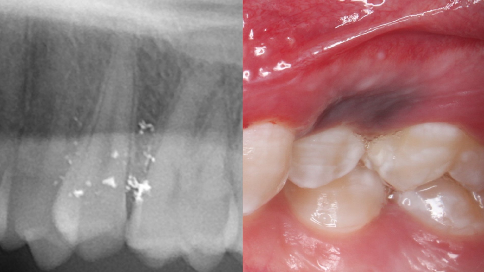
Radiopaque amalgam fragments
Prognostic factors
- Excellent prognosis
Case reports
- 38 year old woman with amalgam in mucoperiosteal flap from endodontic surgery (J Conserv Dent 2016;19:280)
- 49 year old woman with a brown-gray macule on the ventral tongue noted after dental restoration (N Engl J Med 2016;374:e21)
- 53 year old man with black macule on alveolar ridge and remote history of tooth extraction (Indian J Med Res 2018;148:240)
Treatment
- No treatment is needed once diagnosis is established
- Conservative surgical excision can be recommended for esthetic concerns, especially for the anterior maxilla (J Esthet Restor Dent 2020;32:770)
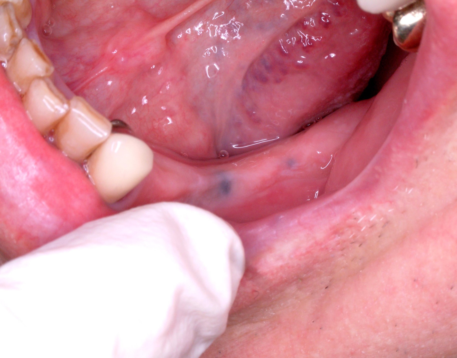
Clinical images
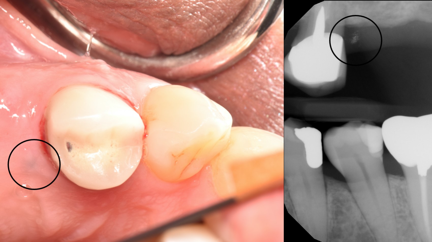
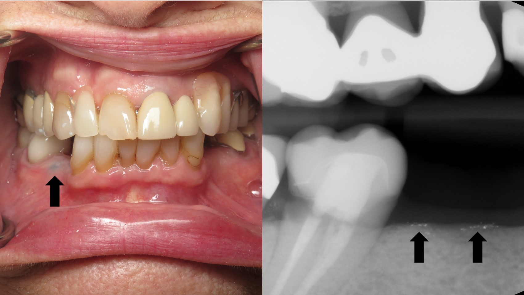
Contributed by Sarah H. Glass, D.D.S. and Duane Schafer, D.D.S., M.S.
Buccal mucosa amalgam tattoo

Large maxillary amalgam tattoo
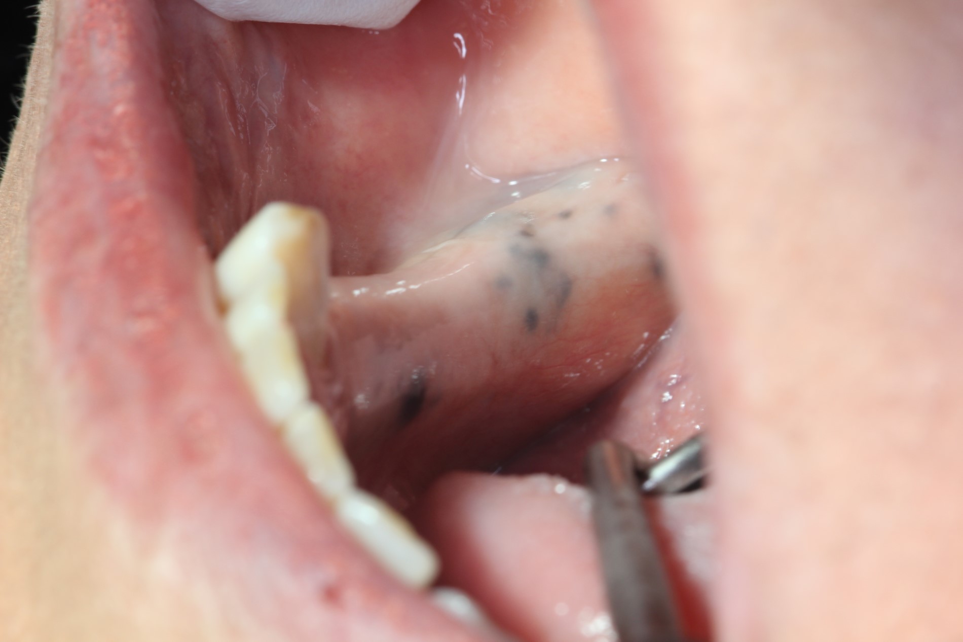
Alveolar ridge amalgam tattoos
Gross description
- Well defined or diffuse black, gray or blue pigmentation in connective tissue on cross section
Gross images
Contributed by Sarah H. Glass, D.D.S.
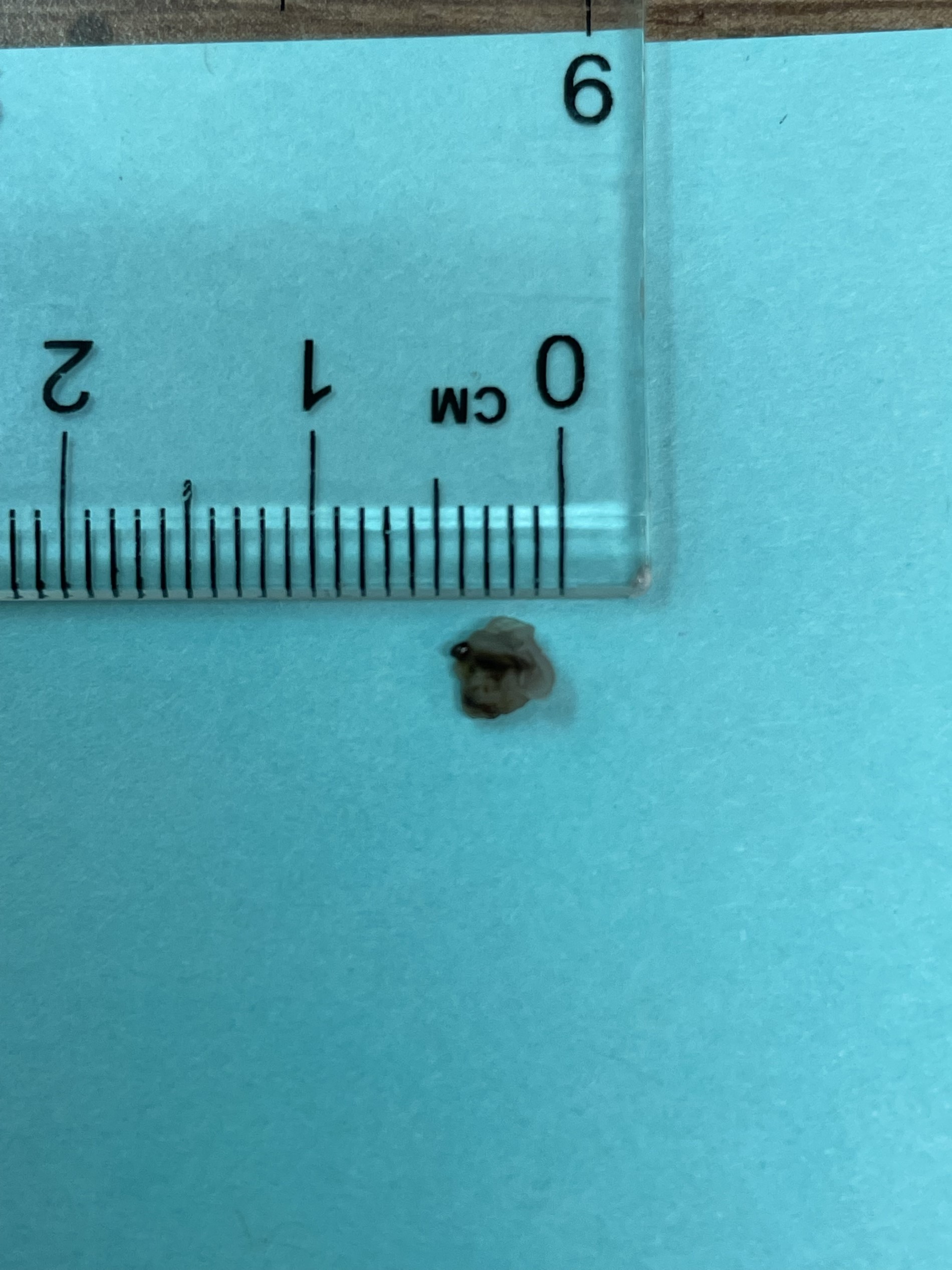
Excisional biopsy specimen
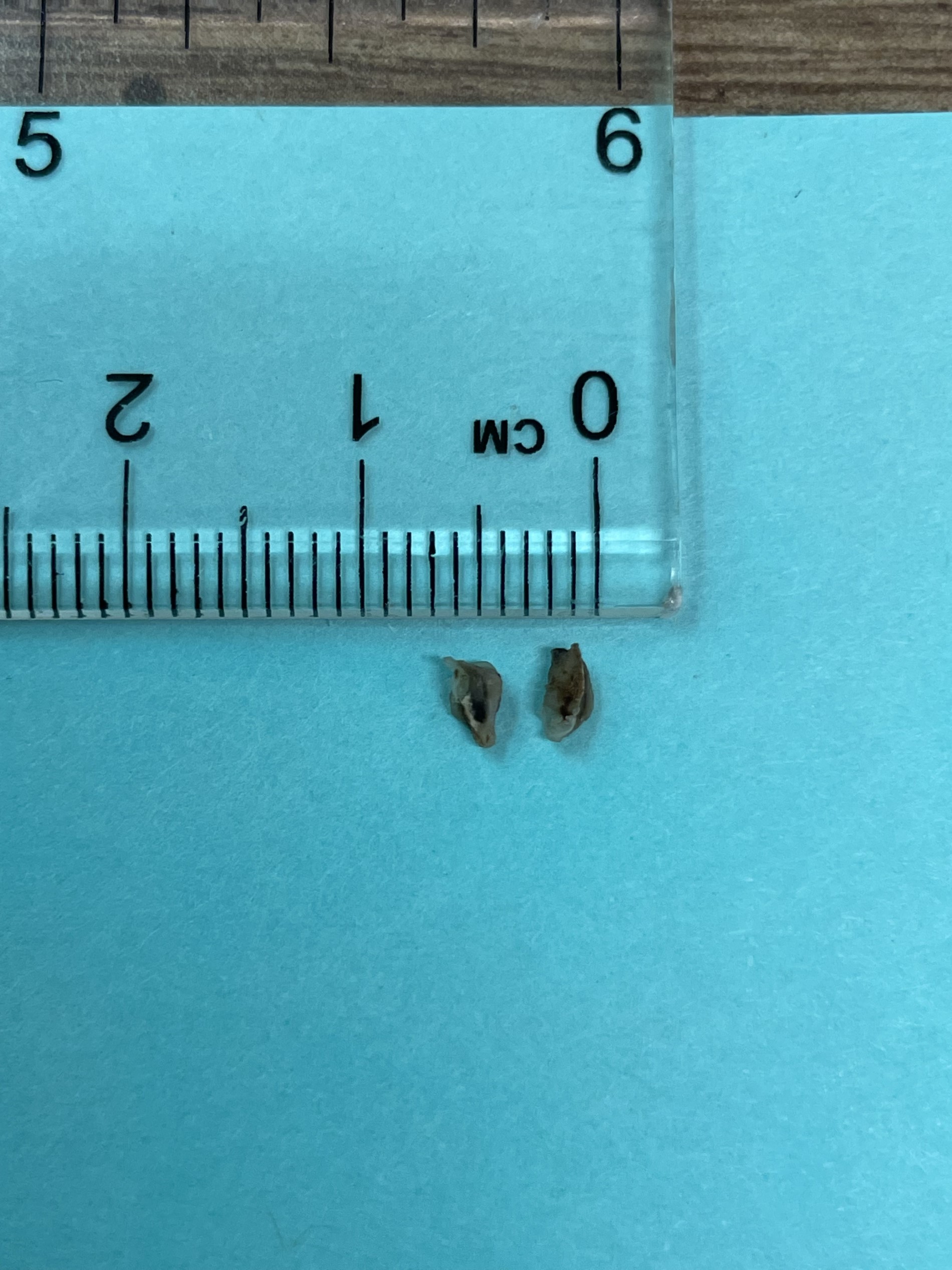
Cross section
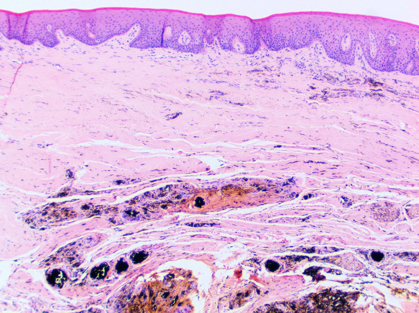
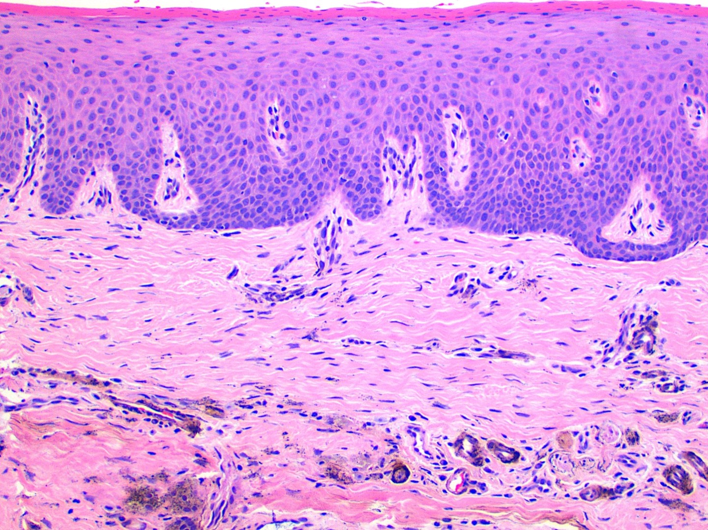
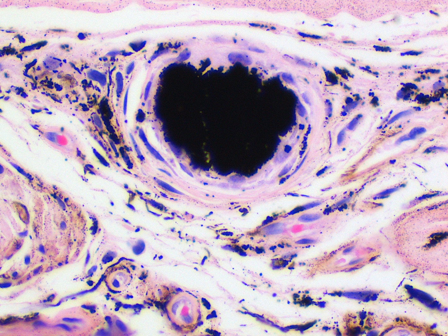
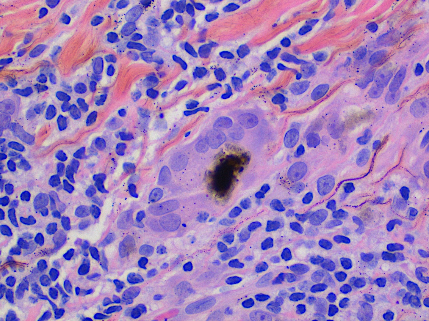
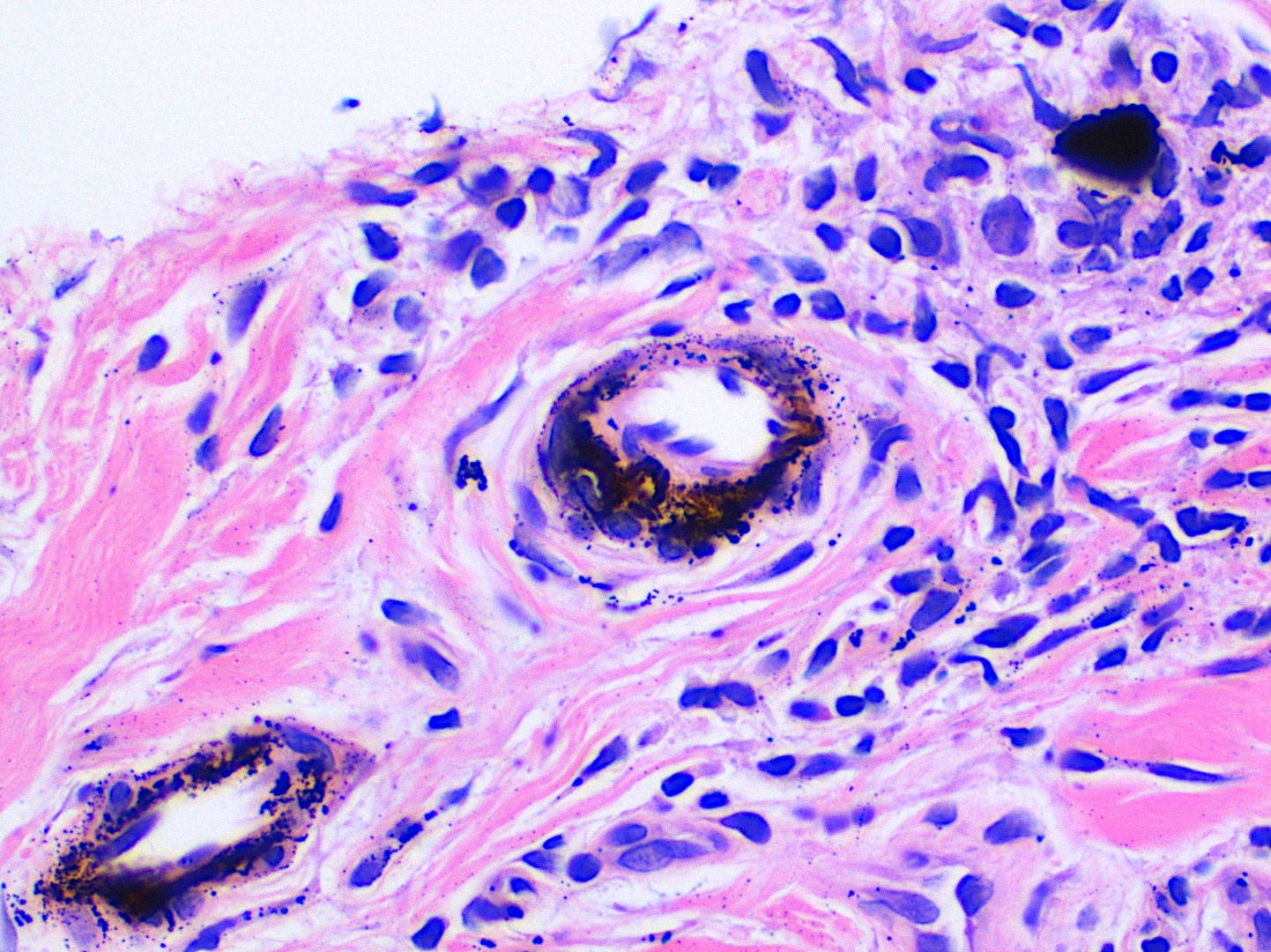
Microscopic (histologic) description
- Large black deposits of foreign material with or without a chronic inflammatory response in the connective tissue
- Fine black granules in the connective tissue; may demonstrate affinity for reticulin fibers (Head Neck Pathol 2019;13:47)
Microscopic (histologic) images
Sample pathology report
- Oral cavity, excisional biopsy:
- Foreign body tattoo (see comment)
- Comment: The histopathology and clinical information are consistent with an amalgam tattoo.
Differential diagnosis
- Oral melanotic macule:
- Increased melanin in basal cell layer of the epithelium with melanin incontinence in the superficial connective tissue
- Multifocal presentation may be seen in a systemic disease, such as Addison disease or Peutz-Jegher syndrome (Head Neck Pathol 2019;13:47)
- Foreign body tattoo or other exogenous material:
- Intentional tattoo pigment (shown above)
- Graphite from pencil (Dermatol Online J 2015;21:13030)
- Drug related discolorations:
- Depending on the drug, histopathology features mimic those of melanotic macule or brown-yellow granules in connective tissue (Head Neck Pathol 2019;13:47)
- Submucosal hemorrhage:
- Hemosiderin in the connective tissue from trauma
- Positive Prussian blue
Additional references
Practice question #1
A biopsy from the oral cavity shows exogenous black foreign material around blood vessels and along collagen fibers in a patient with a history of amalgam dental fillings. What is the diagnosis?
- Amalgam tattoo
- Oral melanotic macule
- Peutz-Jegher syndrome
- Submucosal hemorrhage
Practice answer #1
A. Amalgam tattoo. Although all answer choices can result in pigmented macules in the oral cavity, amalgam is the only exogenous material. This foreign material has a particular affinity for reticulin fibers and can be seen around blood vessels.
Comment Here
Reference: Amalgam tattoo
Comment Here
Reference: Amalgam tattoo
Practice question #2
What is the etiology of an amalgam tattoo from the oral cavity?
- Drug related discoloration
- Iatrogenic implantation of dental filling material
- Increased melanin production
- Trauma induced hemosiderin deposition
Practice answer #2
B. Iatrogenic implantation of dental filling material. A dental professional may inadvertently implant amalgam, an exogenous foreign material, into the oral mucosa during a dental procedure.
Comment Here
Reference: Amalgam tattoo
Comment Here
Reference: Amalgam tattoo
