Small intestine & ampulla
Benign tumors / tumor-like conditions
Pseudomelanosis duodeni
Author: Hanni Gulwani, M.B.B.S.

Last author update: 1 August 2012
Last staff update: 25 August 2025 (update in progress)
Copyright: 2003-2025, PathologyOutlines.com, Inc.
PubMed Search: Pseudomelanosis duodeni small bowel
Table of Contents
Definition / general | Case reports | Microscopic (histologic) images | Practice question #1 | Practice answer #1Cite this page: Gulwani H. Pseudomelanosis duodeni. PathologyOutlines.com website. https://www.pathologyoutlines.com/topic/smallbowelpseudomelanosis.html. Accessed September 3rd, 2025.
Definition / general
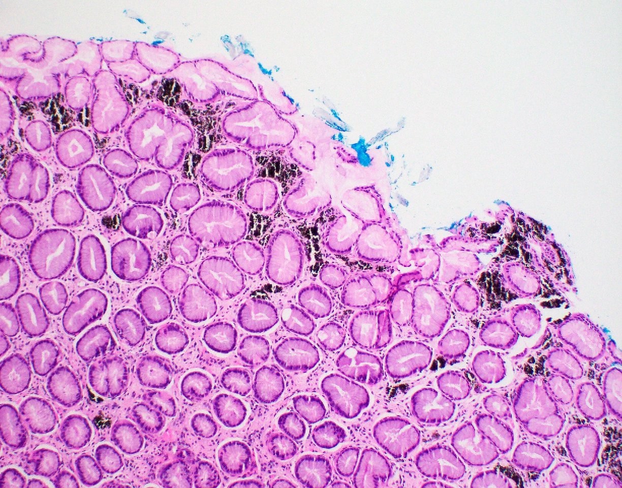
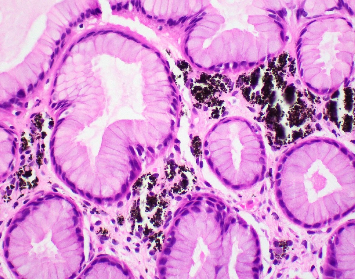
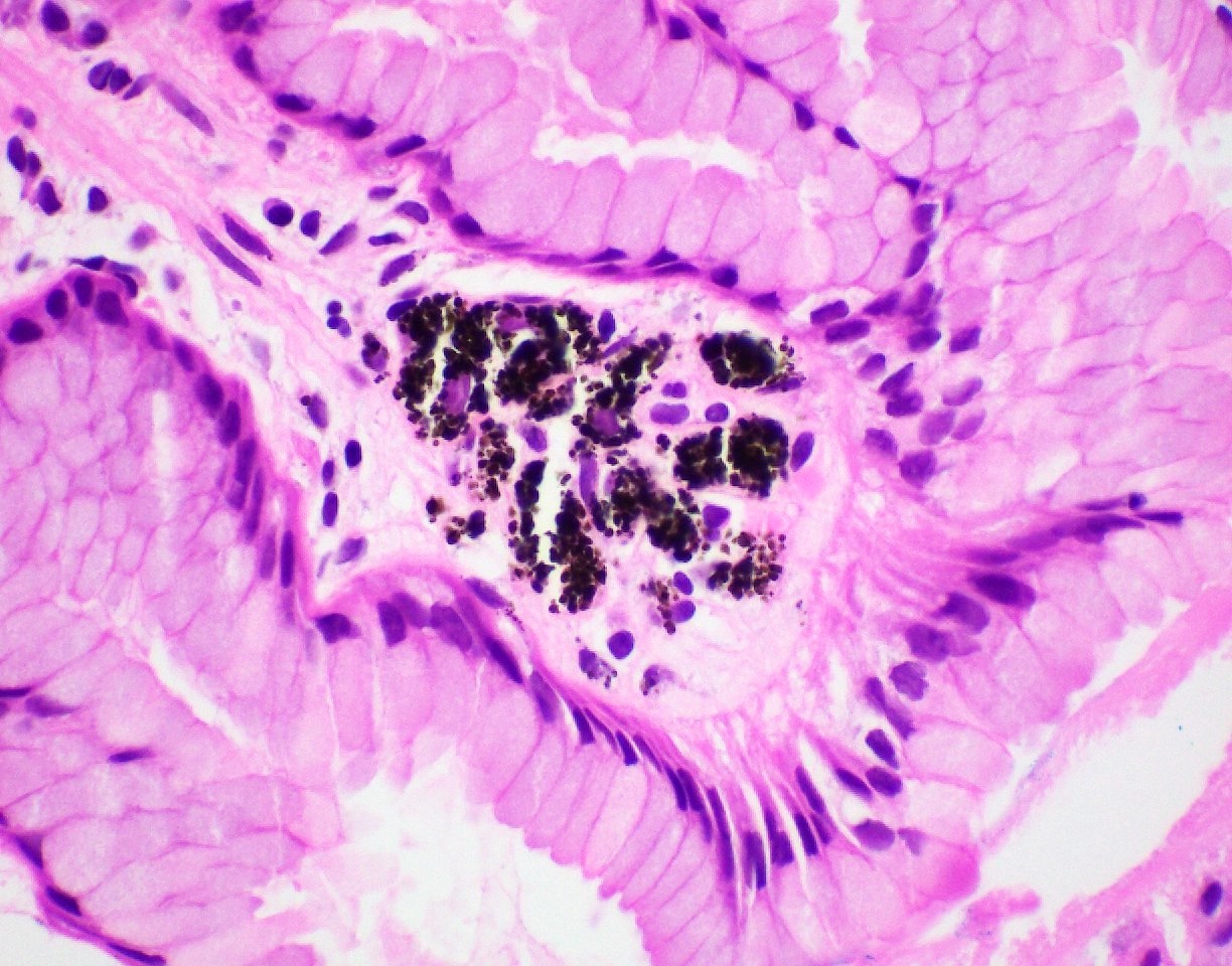
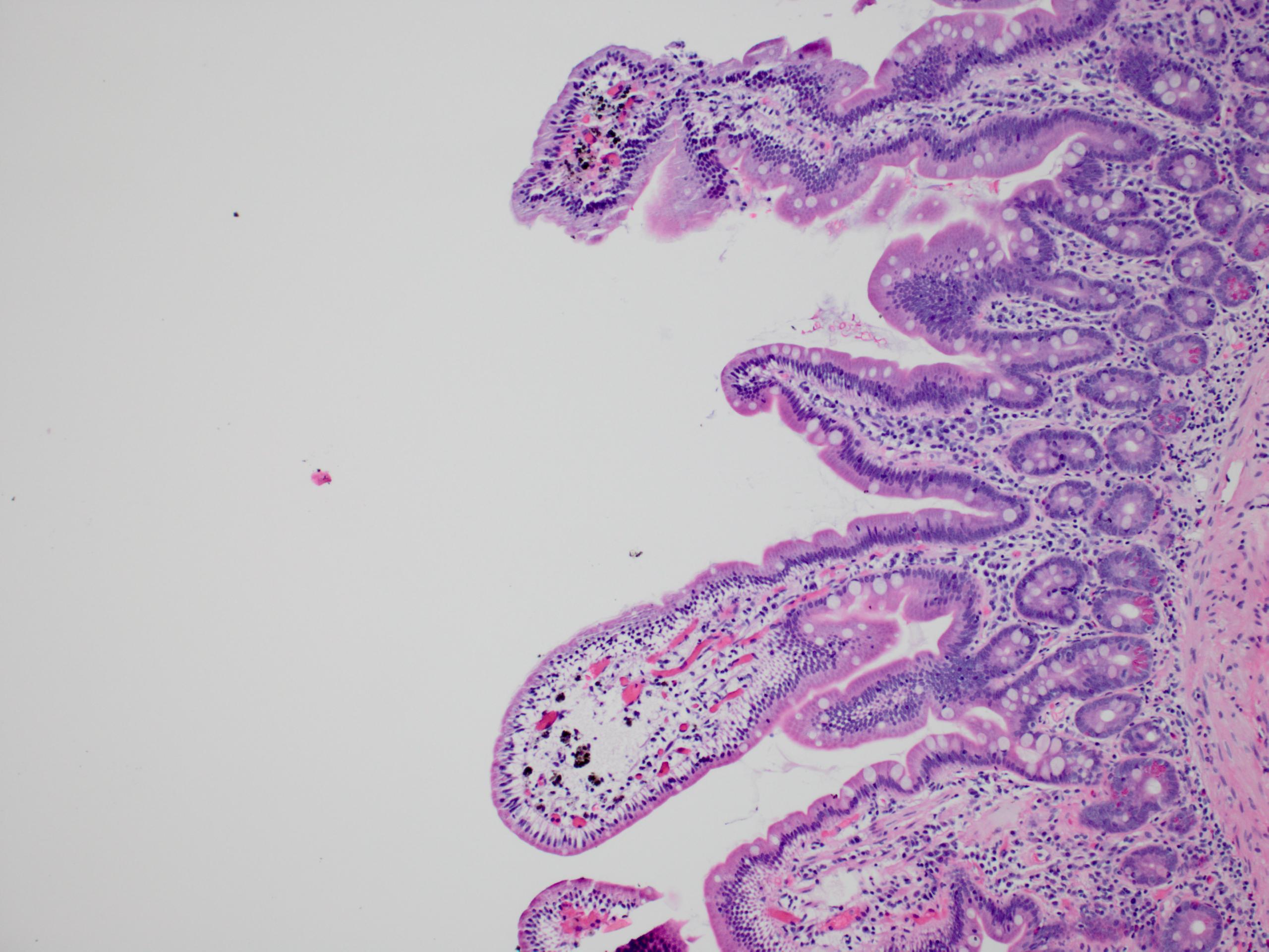
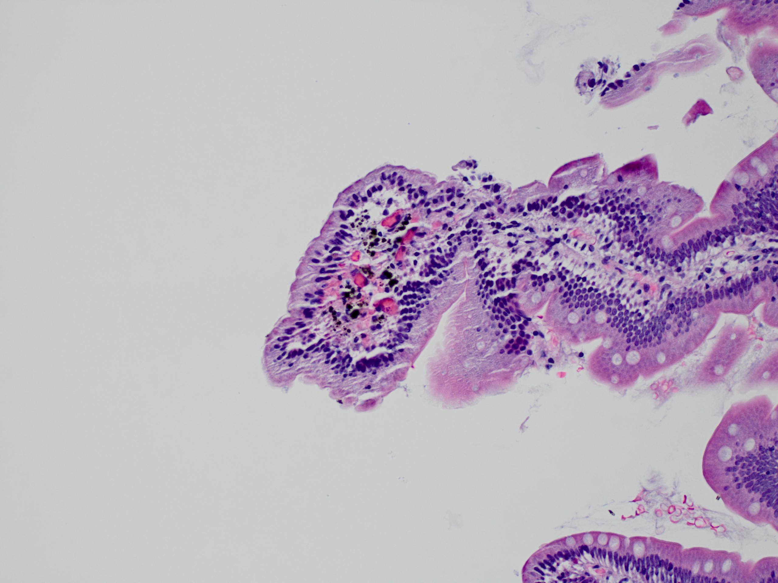
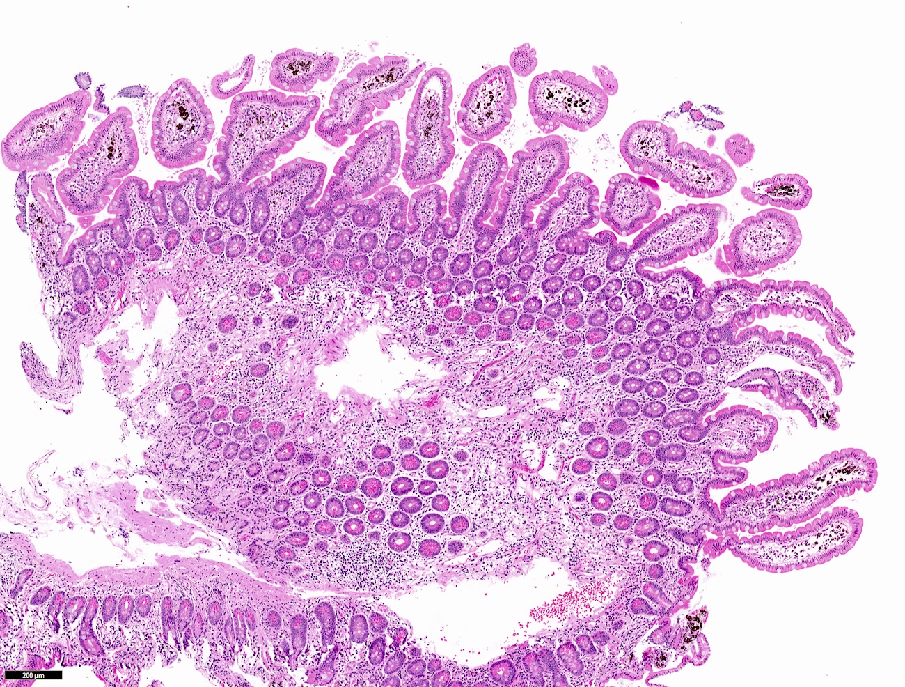
- Brownish black pigment in lamina propria macrophages of proximal duodenum
- Also known as melanosis duodeni
- Due to iron and sulfur (J Formos Med Assoc 1995;94:632) and may be associated with oral iron intake, hypertension, diabetes or end stage renal disease (Endoscopy 2008;40:165, World J Gastroenterol 2012;18:1414)
- Not associated with laxative abuse (J Clin Gastroenterol 1988;10:127)
- Not associated with lipofuscin pigment, which is identified in melanosis coli
- Often identified at endoscopy
- Pigment may originate in atmosphere or diet
- No known clinical significance
Case reports
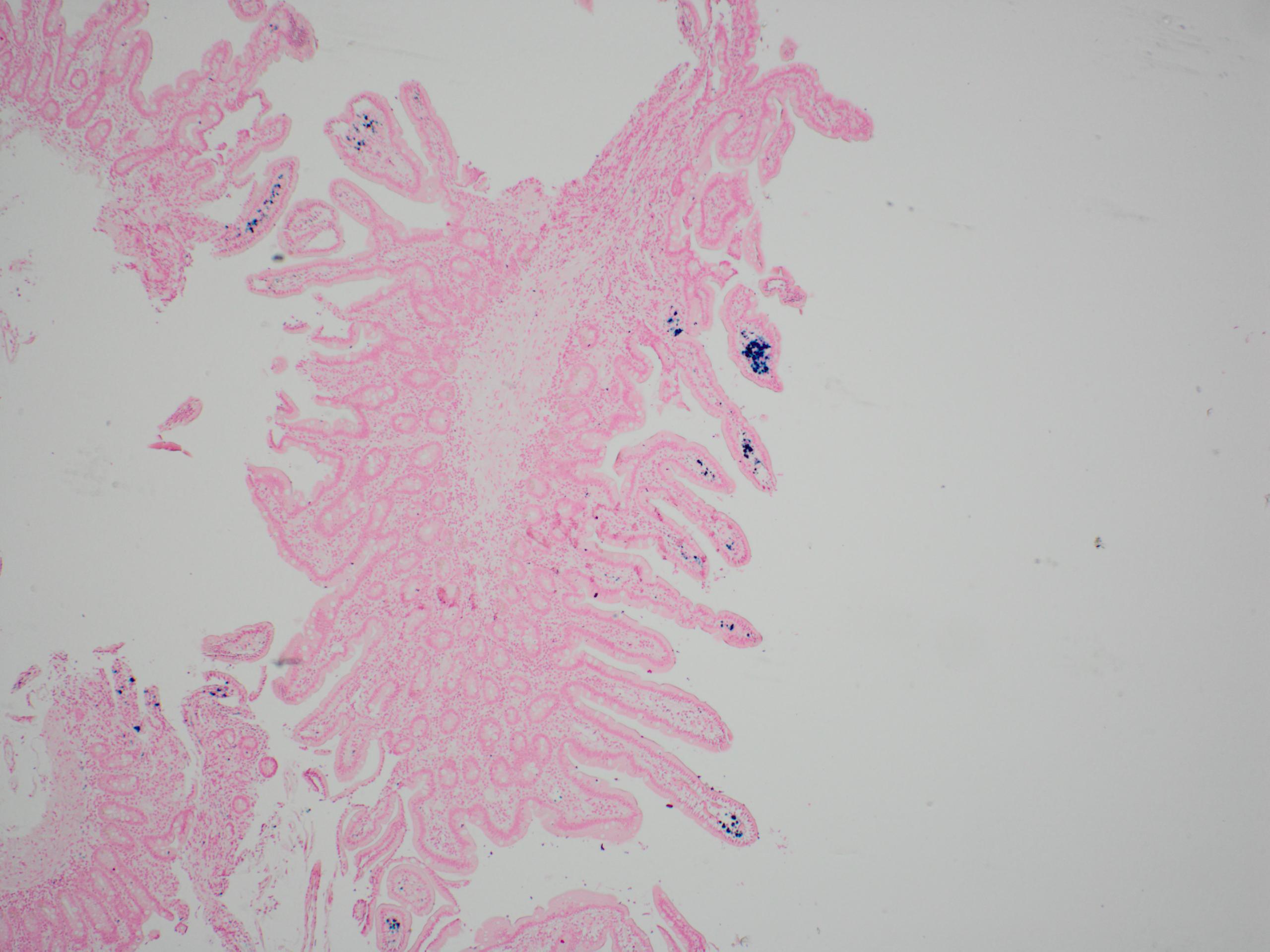
Microscopic (histologic) images


Practice question #1
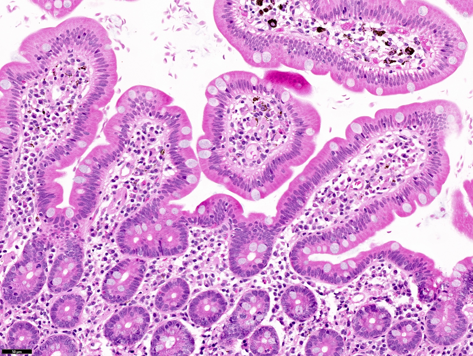
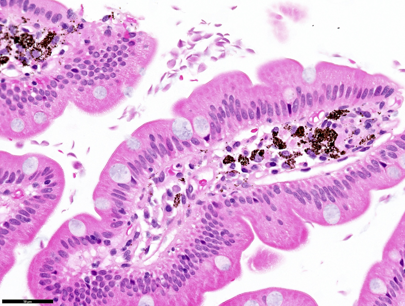
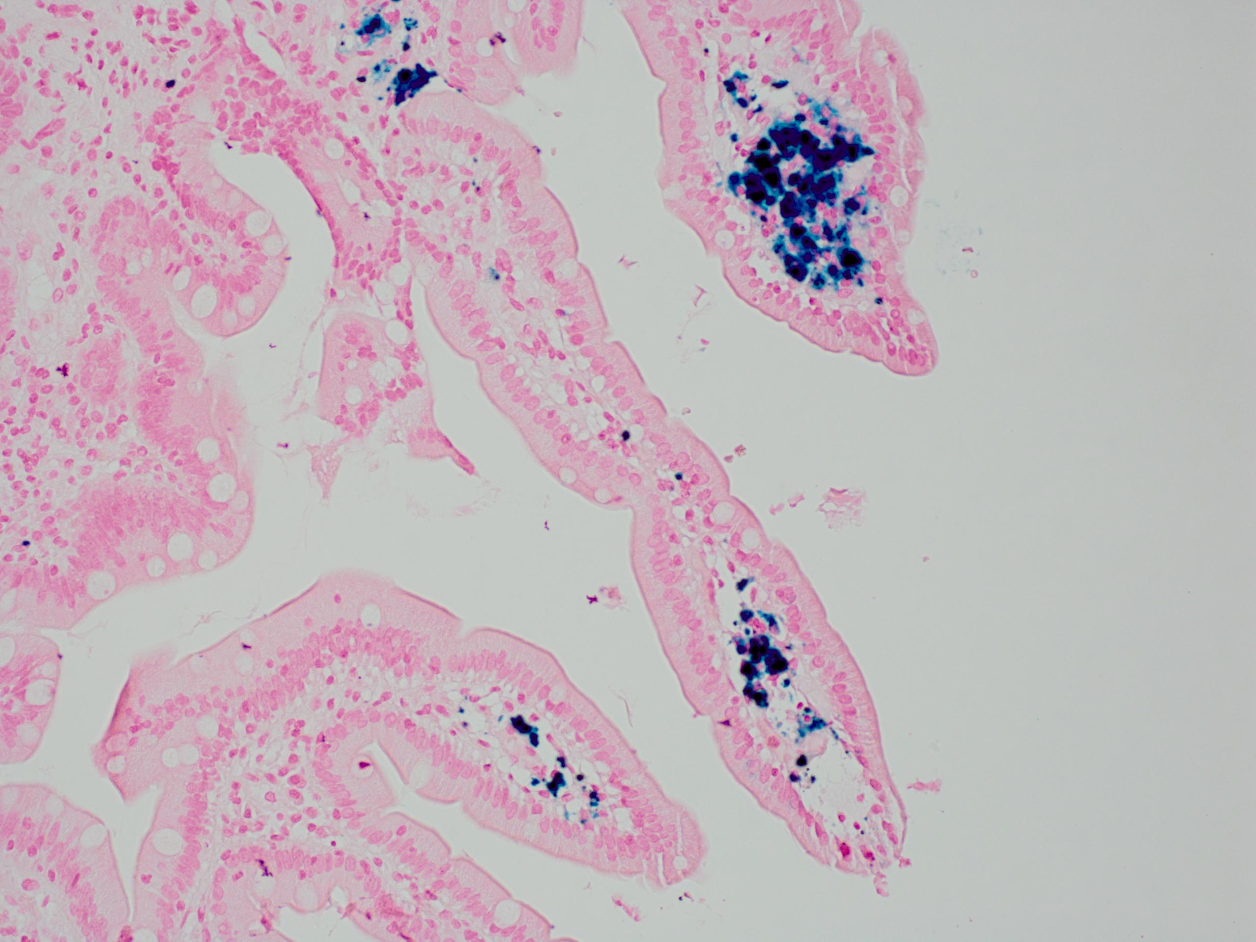
The brown, black pigment shown in these samples would be positive for which of the following stains?
- GMS
- PAS
- Prussian blue
- Von Kossa
Practice answer #1
C. Prussian blue. The brown, black pigment in pseudomelanosis duodeni is thought to contain ferrous sulfide and has been shown to be positive on Prussian blue iron stain and Masson Fontana melanin stain. Answer A is incorrect because GMS stain is used for fungal organisms. Answer B is incorrect because PAS is used to detect glycogen deposits and lipofuscin in melanosis coli. Answer D is incorrect because Von Kossa stains calcium.
Comment Here
Reference: Pseudomelanosis duodeni
Comment Here
Reference: Pseudomelanosis duodeni
