Table of Contents
Definition / general | Essential features | Terminology | Pathophysiology | Interpretation | Uses by pathologists | Prognostic factors | Microscopic (histologic) description | Microscopic (histologic) images | Positive staining - normal | Positive staining - disease | Negative staining | Sample pathology report | Practice question #1 | Practice answer #1Cite this page: Lezcano C, Jungbluth AA, Busam KJ. PRAME. PathologyOutlines.com website. https://www.pathologyoutlines.com/topic/stainsprame.html. Accessed September 30th, 2025.
Definition / general
- PRAME: preferentially expressed antigen in melanoma (Immunity 1997;6:199)
- Cancer testis antigen expressed by melanoma and other malignant neoplasms with expression in normal tissue largely restricted to testis (Immunity 1997;6:199)
Essential features
- Diffusely positive in many melanomas
- Usually negative or nondiffuse / focal immunoreactivity in nevi, other benign neoplasms and most normal adult tissue (except for testis and a few other tissues that express PRAME)
- Other malignant tumors, including certain sarcomas, carcinomas and hematolymphoid neoplasms, can show variable extent of PRAME staining in subsets of cases
Terminology
- Less frequently known as MAPE (melanoma antigen preferentially expressed in tumors) and OIP4 (Opa interacting protein 4) (Mol Cancer 2010;9:226)
Pathophysiology
- PRAME is a cancer testis antigen that was first identified by autologous T cell epitope cloning in a patient with metastatic cutaneous melanoma (Immunity 1997;6:199)
- Belongs to family of non-X cancer testis antigens, mapping to autosome (chromosome 22) versus classical cancer testis antigens, such as MAGE-A, NY-ESO-1, mapping to chromosome X (Nat Rev Cancer 2005;5:615)
- Expressed in most cutaneous and ocular melanomas but also in nonmelanocytic malignant tumors, including some carcinomas, leukemias, lymphomas and sarcomas (Immunity 1997;6:199, Int J Surg Pathol 2021;29:826, Am J Surg Pathol 2022;46:1467)
- Most benign adult tissue shows low or absent PRAME mRNA expression, except for testis (high level expression), ovary, placenta, adrenals and endometrium (Immunity 1997;6:199)
- PRAME acts as a repressor of the retinoic acid receptor (RAR) signaling pathway (Cell 2005;122:835)
Interpretation
- Nuclear immunostain (Am J Surg Pathol 2018;42:1456)
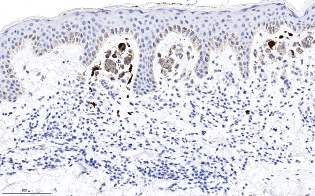
- PRAME's frequently diffuse (positive in > 75% of tumor cells) pattern of immunoreactivity within in situ, invasive and metastatic melanoma contrasts with the infrequent and typically focal staining seen in nevi and normal background melanocytes (Am J Surg Pathol 2018;42:1456, Am J Surg Pathol 2020;44:503, J Cutan Pathol 2021;48:856)
- With anti-PRAME antibody clone EPR20330, consistent cytoplasmic labeling of unclear significance is observed in sebocytes (Am J Surg Pathol 2018;42:1456)
Uses by pathologists
- Supports a suspected diagnosis of melanoma (usually diffusely positive for PRAME) or nevus (usually negative or only focally positive for PRAME) in conjunction with morphologic evaluation on H&E (Am J Surg Pathol 2018;42:1456, J Cutan Pathol 2021;48:1115, Am J Dermatopathol 2023;45:733)
- Assists in the determination of primary tumor staging and Breslow tumor thickness in cases with dermal melanocytes with nevoid features that are difficult to interpret (e.g., possible invasion versus associated nevus, precise extent of invasive melanoma versus a nevus component) (Am J Surg Pathol 2018;42:1456, J Cutan Pathol 2021;48:856)
- Margin assessment, especially for melanoma in situ with broad lentiginous growth and diffuse immunoreactivity for PRAME, as background melanocytes are mostly PRAME negative (Am J Surg Pathol 2018;42:1456, Histopathology 2021;78:1000)
- Assessment of melanocytic deposits in sentinel lymph nodes for melanoma, diffuse PRAME staining is evidence in support of metastatic melanoma; nodal nevi are typically negative for PRAME (Am J Surg Pathol 2020;44:503, Pathol Res Pract 2020;216:153105)
Prognostic factors
- PRAME mRNA expression level has been identified as a biomarker for metastatic risk stratification of uveal melanomas and is part of a 12 gene expression prognostic assay (Clin Cancer Res 2016;22:1234, Oncotarget 2016;7:59209)
- Possible prognostic implications of PRAME expression in other tumor types remain under study
Microscopic (histologic) description
- PRAME expression is characterized as diffuse when positive nuclear staining is present in over 75% of tumor cells (Am J Surg Pathol 2018;42:1456)
- Immunohistochemistry for PRAME can be combined with MelanA in a dual PRAME / MelanA immunostain (e.g., for easier assessment of small melanocytic deposits in lymph nodes) (Am J Surg Pathol 2020;44:503)
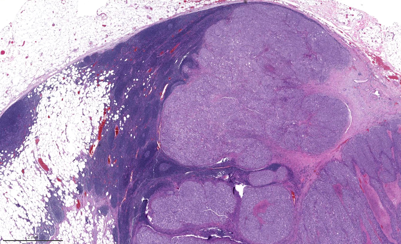
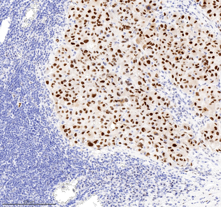
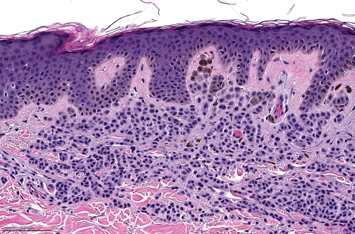
Microscopic (histologic) images
Contributed by Cecilia Lezcano, M.D.

PRAME in normal testis
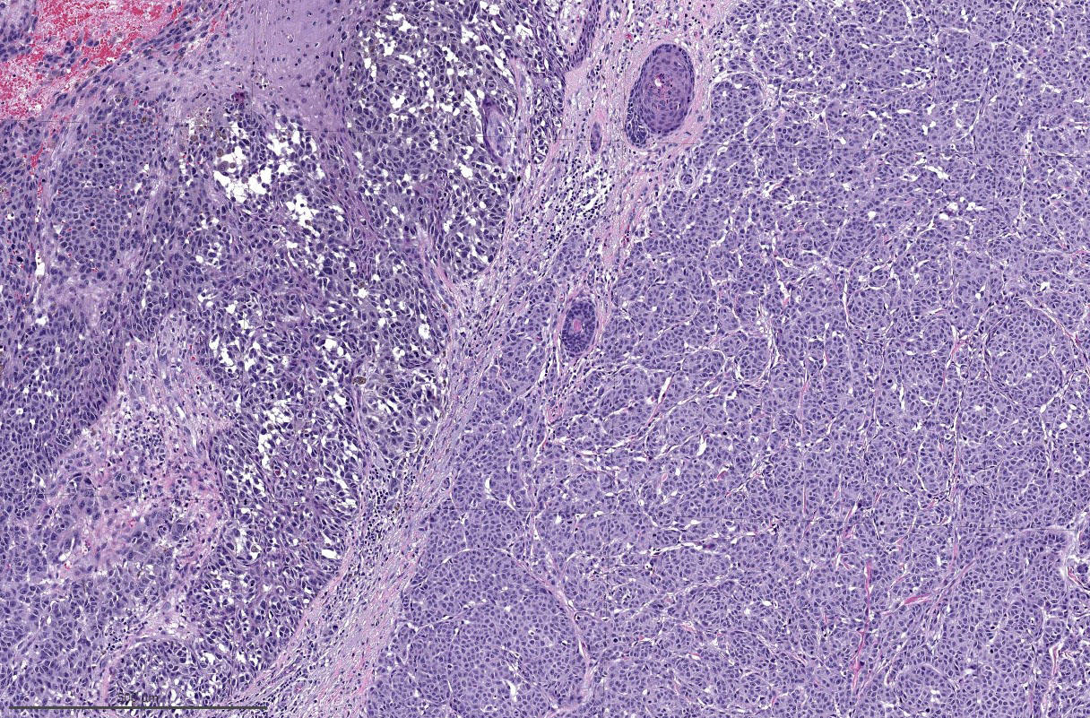
Primary cutaneous melanoma
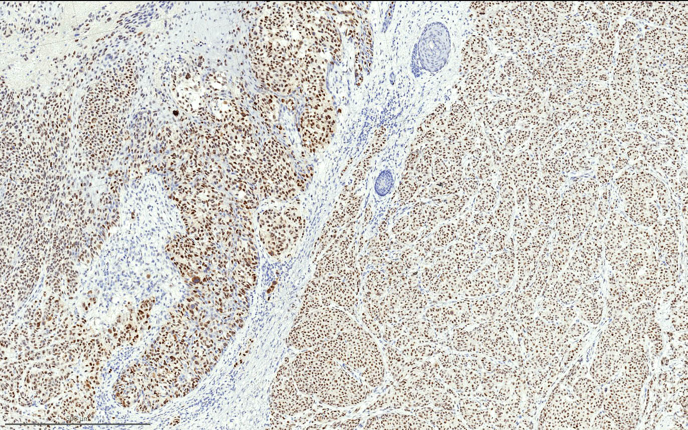
PRAME in primary cutaneous melanoma
Melanoma metastasis

PRAME positive melanoma metastasis
Nevus
PRAME immunostain in nevus
Positive staining - normal
- In testis, cells of early spermatogenesis located close to the basal membrane of the seminiferous tubules show nuclear positive staining (Int J Surg Pathol 2021;29:826)
- Usually expressed in normal sebaceous glands; predominantly in cytoplasm of mature germinative sebocytes (J Cutan Pathol 2021;48:1252, Ocul Oncol Pathol 2024;10:1)
Positive staining - disease
- Metastatic melanoma (~90%) (Am J Surg Pathol 2018;42:1456, Pathol Res Pract 2020;216:153105, J Cutan Pathol 2021;48:479, Am J Surg Pathol 2022;46:1467)
- Primary cutaneous melanomas (diffuse PRAME expression in 70 - 95% of non-spindle cell melanomas, lower rate of ~35% of diffuse PRAME staining in desmoplastic / spindle cell melanomas) (Am J Surg Pathol 2018;42:1456, J Cutan Pathol 2020;47:1123, J Cutan Pathol 2021;48:856, Am J Surg Pathol 2020;44:893, Am J Dermatopathol 2020;42:625, Surg Pathol Clin 2021;14:165, Am J Dermatopathol 2023;45:733)
- Mucosal melanomas (Mod Pathol 2019;32:1727, Am J Surg Pathol 2022;46:1467)
- Most synovial sarcomas and myxoid liposarcomas are diffusely positive for PRAME (Int J Surg Pathol 2021;29:826)
- Most (~65%) of malignant peripheral nerve sheath tumors (MPNST) show at least focal PRAME staining; ~33% of MPNSTs show over 50% of tumor cells positive for PRAME (J Neuropathol Exp Neurol 2021;80:384)
- Subsets of mesenchymal and neuroectodermal tumors (e.g., Wilms tumor, neuroblastoma) can show variable (focal to diffuse) expression of PRAME (Am J Surg Pathol 2022;46:1467)
- Subsets of carcinomas of pulmonary, cutaneous, salivary, ovarian, endometrial and mammary origin as well as germ cell tumors can show variable extent of PRAME staining (Int J Surg Pathol 2021;29:826, J Cutan Pathol 2021;48:1150, Histopathology 2019;75:672, Am J Surg Pathol 2022;46:1467)
Negative staining
- Most nevi are negative for PRAME; however, a subset (~13 - 19%) show nondiffuse (focal or patchy) staining for PRAME
- Diffuse PRAME immunoreactivity in nevi is very rare (Am J Surg Pathol 2018;42:1456, J Cutan Pathol 2021;48:856, J Cutan Pathol 2020;47:1123, Surg Pathol Clin 2021;14:165, J Cutan Pathol 2021;48:1115, Am J Dermatopathol 2023;45:733)
- Benign peripheral nerve sheath tumors (96.4% completely negative; 3.6% show rare weak positive cells) (J Neuropathol Exp Neurol 2021;80:384)
- Clear cell sarcomas (70% negative, focal staining in 30%) (J Cutan Pathol 2020;47:1226, Int J Surg Pathol 2021;29:826, Am J Surg Pathol 2022;46:1467)
Sample pathology report
- Skin, temple, shave biopsy:
- Melanoma in situ (see comment)
- Comment: Sections show a lentiginous intraepidermal proliferation of atypical melanocytes with occasional nests, multifocal pagetoid spread and diffuse immunoreactivity for PRAME, which is consistent with this diagnosis.
Practice question #1
Which of the following statements regarding PRAME immunohistochemistry is correct?
- All melanomas are positive for PRAME
- Benign nevi never show immunoreactivity for PRAME
- Diffuse nuclear immunoreactivity for PRAME is seen in a majority of melanomas
- Melanoma is the only malignant neoplasm that expresses PRAME
Practice answer #1
C. Diffuse nuclear immunoreactivity for PRAME is seen in the majority of melanomas (positive in ~75% or more of primary and metastatic tumors). Answer A is incorrect because some primary and metastatic melanomas can be completely negative for PRAME or show only focal expression. Answer B is incorrect because nevi can occasionally show staining for PRAME, usually in a nondiffuse pattern. Answer D is incorrect because the majority of synovial sarcomas and myxoid liposarcomas diffusely express PRAME. Other tumors, including carcinomas of various origins, can also show variable extent of staining for PRAME.
Comment Here
Reference: PRAME
Comment Here
Reference: PRAME