
Stains & CD markers
Alpha fetoprotein (AFP)


Copyright: 2003-2024, PathologyOutlines.com, Inc.
PubMed Search: Alpha fetoprotein[TI] review[PT] pathology
Alpha fetoprotein (AFP)
Authors: João Lobo, M.D., Rui Henrique, M.D., Ph.D.
Editor-in-Chief: Debra L. Zynger, M.D.
Last author update: 20 April 2020
Last staff update: 18 June 2021
Copyright: 2003-2024, PathologyOutlines.com, Inc.
PubMed Search: Alpha fetoprotein[TI] review[PT] pathology

Table of Contents
Definition / general | Essential features | Terminology | Pathophysiology | Clinical features | Interpretation | Uses by pathologists | Prognostic factors | Microscopic (histologic) images | Virtual slides | Positive staining - normal | Positive staining - disease | Negative staining | Board review style question #1 | Board review style answer #1Cite this page: Lobo J, Henrique R. Alpha fetoprotein (AFP). PathologyOutlines.com website. https://www.pathologyoutlines.com/topic/stainsafp.html. Accessed April 27th, 2024.
Definition / general
- Alpha fetoprotein is a marker related to embryonic development (yolk sac), mainly used for diagnosing germ cell tumors and liver tumors
- Overall specific but not sensitive marker
Essential features
- Positive immunostaining in a subset of hepatocellular carcinomas and hepatoblastomas but also found to be positive in other liver diseases
- Positive immunostaining can be found in the various patterns of yolk sac tumor but also can be positive in teratoma
- Positive in various tumors with hepatoid differentiation
Terminology
- Alpha fetoprotein (AFP)
- First detected in high concentrations in human embryonal and fetal serum, named alpha-1 globulin (Scand J Clin Lab Invest 1956;8:174)
Pathophysiology
- Part of the albuminoid gene superfamily, gene mapped on chromosome 4 (4q11-q22) (Nucleic Acids Res 1985;13:8007)
- Major protein in serum of early mammalian embryos, synthesized at the site of embryonal hematopoiesis: the yolk sac (J Clin Invest 1967;46:1010)
- Function: transport and binding of ligands, growth regulator during embryonic development, immune regulatory functions (Exp Biol Med (Maywood) 2001;226:377, Annu Rev Med 1977;28:453)
Clinical features
- High serum levels in several conditions:
- Pregnancy
- Hepatic disorders
- Malignancies (hepatocellular carcinomas, germ cell tumors - especially with yolk sac tumor components, neoplasms of endodermal origin - lung, gastric, colon - especially if associated hepatoid foci, liver metastases, pediatric tumors such as pancreatoblastoma)
- History of gastrointestinal / hepatic surgery
- Hereditary ataxia telangiectasia, in cases of liver damage by chemotherapy or anesthetics (Annu Rev Med 1977;28:453, Hum Pathol 2008;39:1115, Gastroenterology 1979;77:787, Nat Rev Urol 2016;13:715, JOP 2007;8:55)
- Specific serum isoforms: L1, in nonneoplastic liver disease; L2, in yolk sac tumor; L3, in liver cancer
- Both L2 and L3 can be elevated in pediatric yolk sac tumor, reflecting both yolk sac and hepatic differentiation (J Pediatr Surg 1989;24:350)
- Elevations in maternal serum and amniotic fluid may indicate fetal abnormalities, including neural tube defects (Am J Obstet Gynecol 2006;195:1623)
- Serum levels drop after birth (still elevated until around 6 months of age, needing care in interpretation); small amounts still produced in adults (Annu Rev Med 1977;28:453)
- Serum AFP increases in 50 - 70% of patients with nonseminoma germ cell tumors; half life is 5 to 7 days; prominent elevations in pure seminomas usually indicate yolk sac differentiation in metastatic locations; patients should be treated as having mixed germ cell tumor (Nat Rev Urol 2020;17:201)
Interpretation
- Membranous or cytoplasmic staining is expected; granular
Uses by pathologists
- Supporting the diagnosis of germ cell tumors (testicular, ovarian or extragonadal), especially those with identifiable yolk sac tumor foci (Mod Pathol 2005;18:S61)
- Supporting the diagnosis of hepatocellular disease (including nonneoplastic diseases such as chronic hepatitis, and neoplastic diseases such as hepatocellular carcinoma and hepatoblastoma) (World J Gastroenterol 2005;11:5015)
- Confirming suspected hepatoid foci within other neoplasms or confirming that tumor cells produce AFP in case of remarkable elevations of this marker in patient serum (Mod Pathol 1997;10:686)
Prognostic factors
- Preoperative levels are prognostic for hepatocellular carcinoma (World J Surg Oncol 2013;11:212)
- Normal AFP hepatocellular carcinoma patients were significantly older, had fewer chronic liver conditions such as viral hepatitis or cirrhosis and had better survival; tumors were smaller, less vascular and well differentiated (J Clin Med 2019;8:E1736)
- Part of staging and prognostic grouping in germ cell tumors, correlation with response to treatment, both in testis and ovary (Biomed Res Int 2019;2019:5030349, Mol Clin Oncol 2015;3:125)
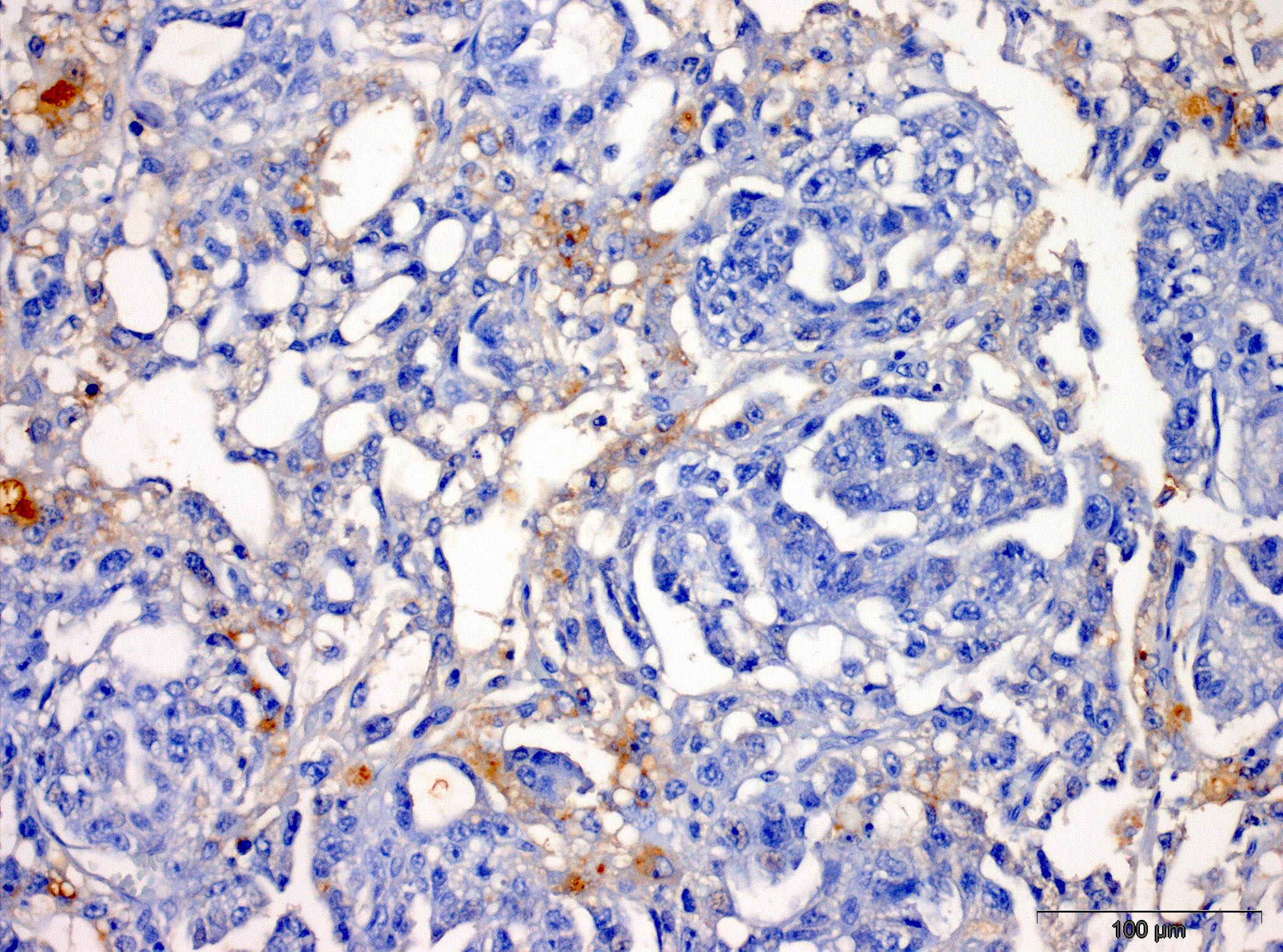
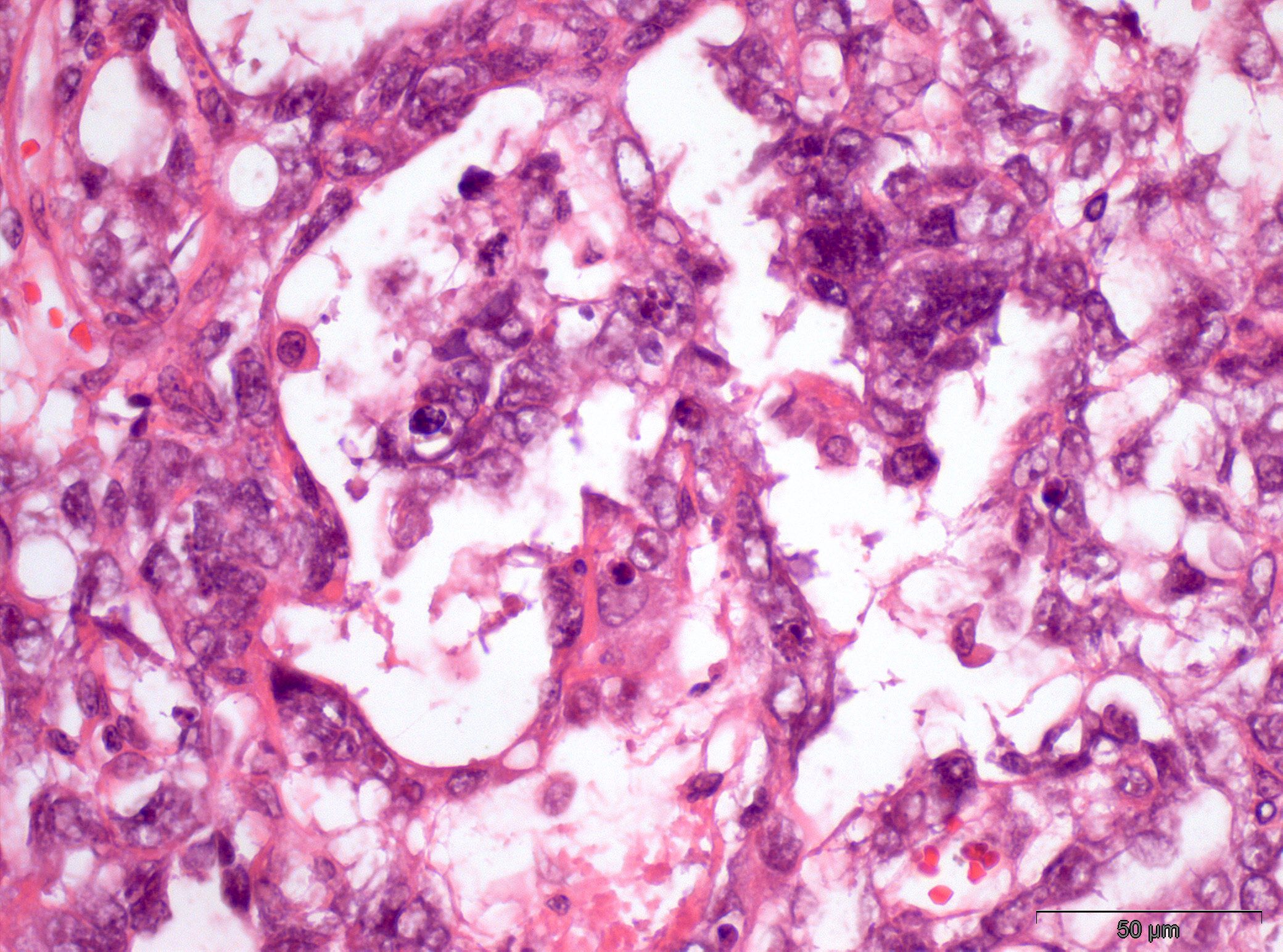
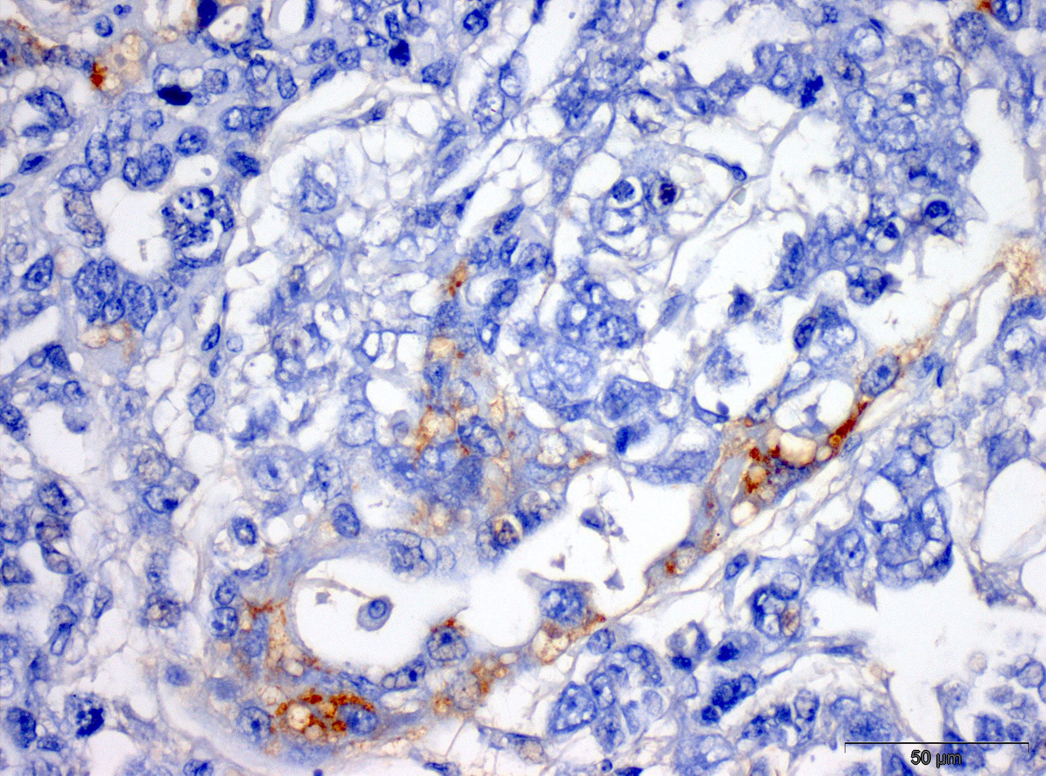
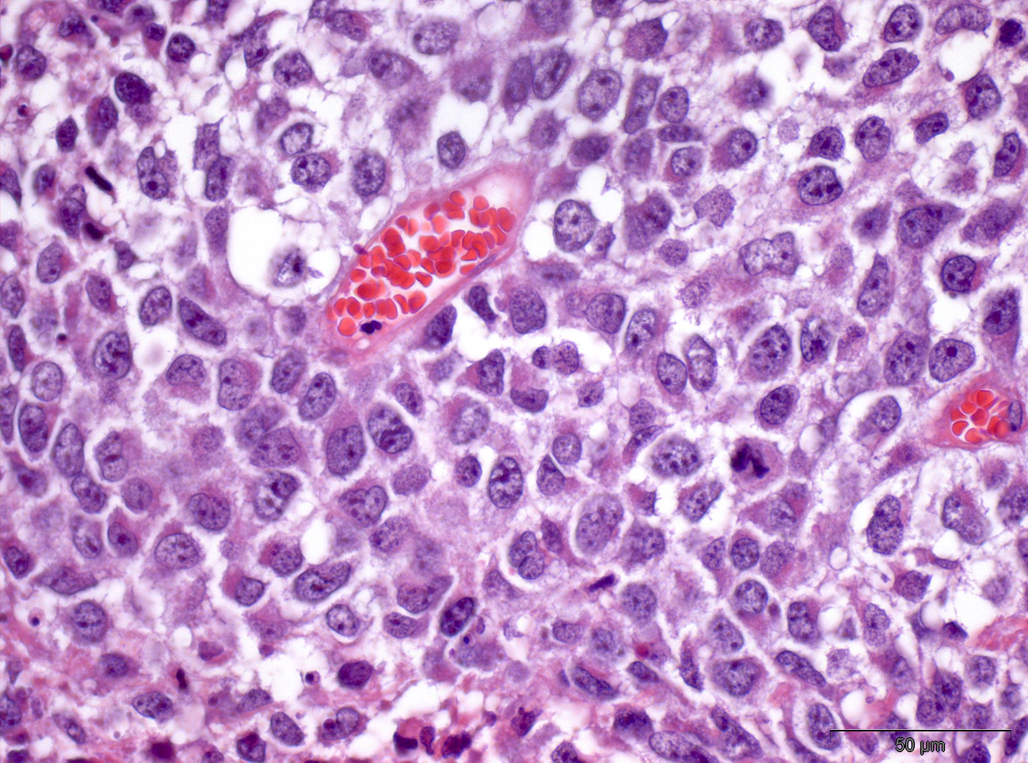
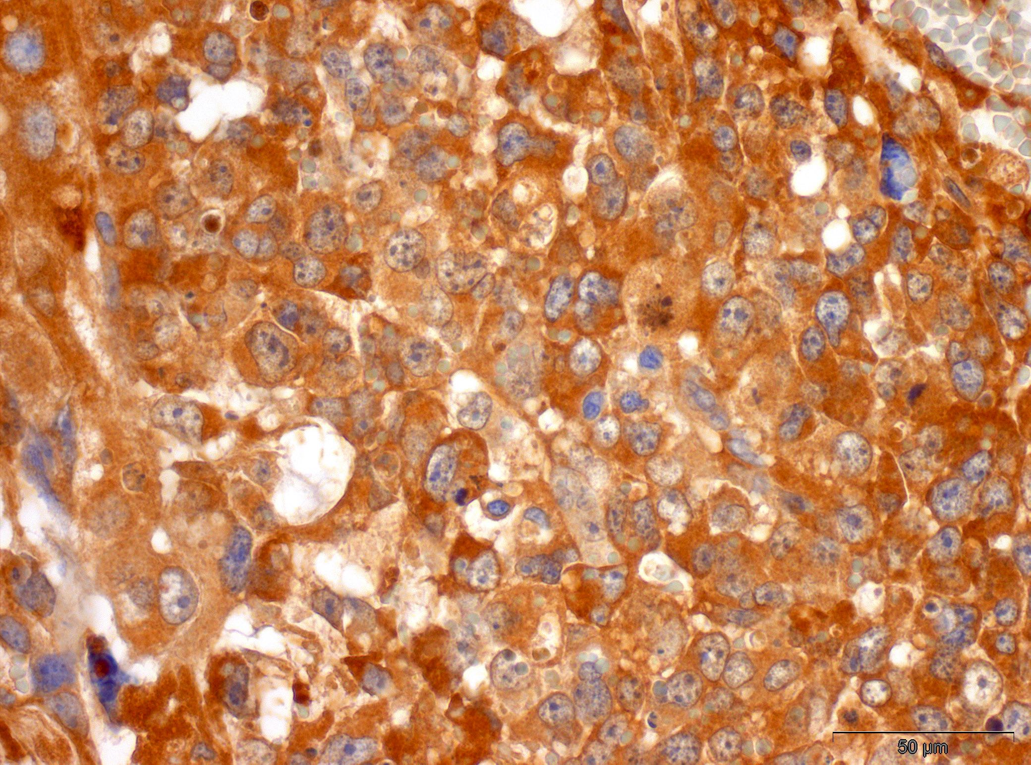
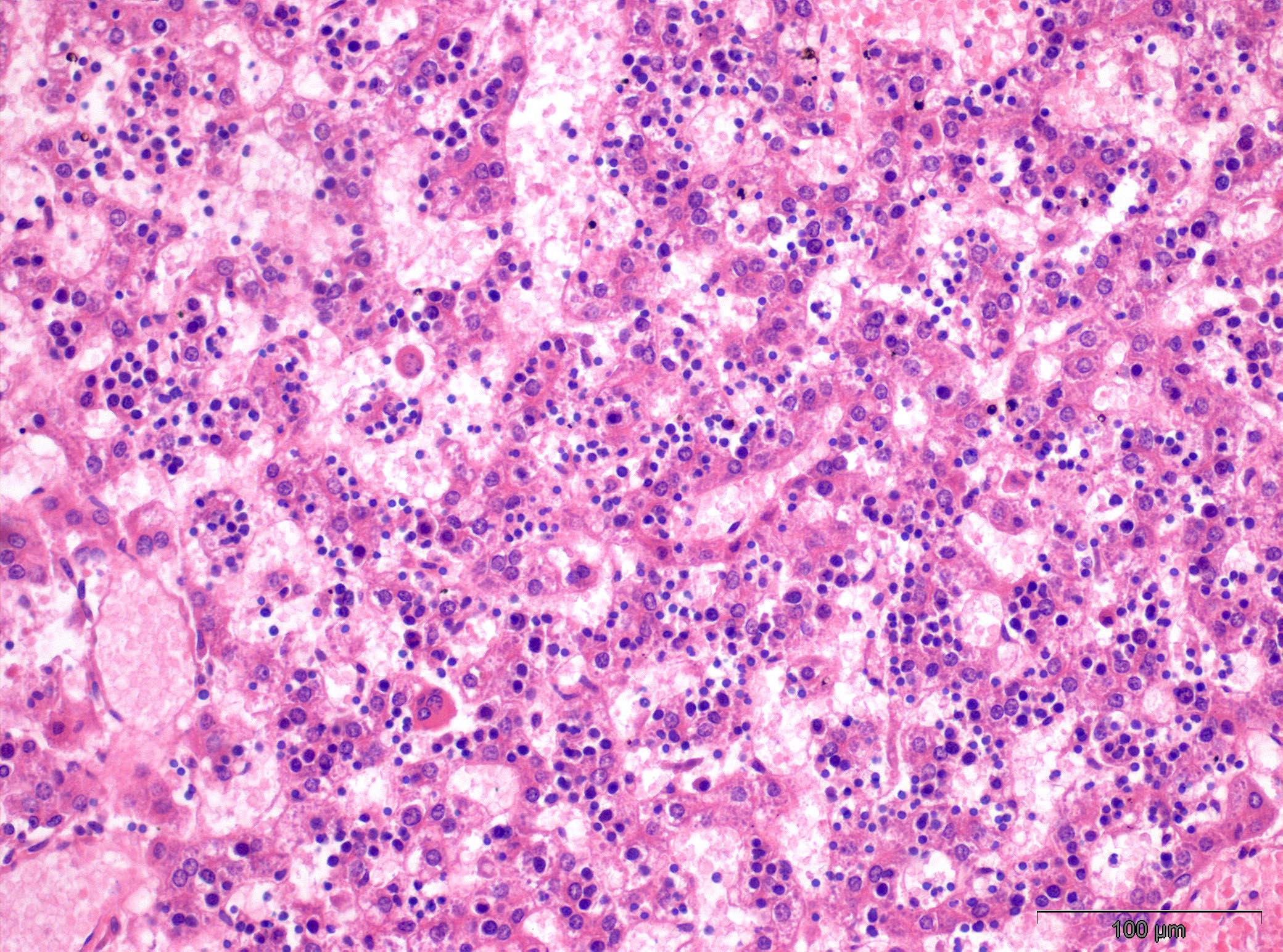
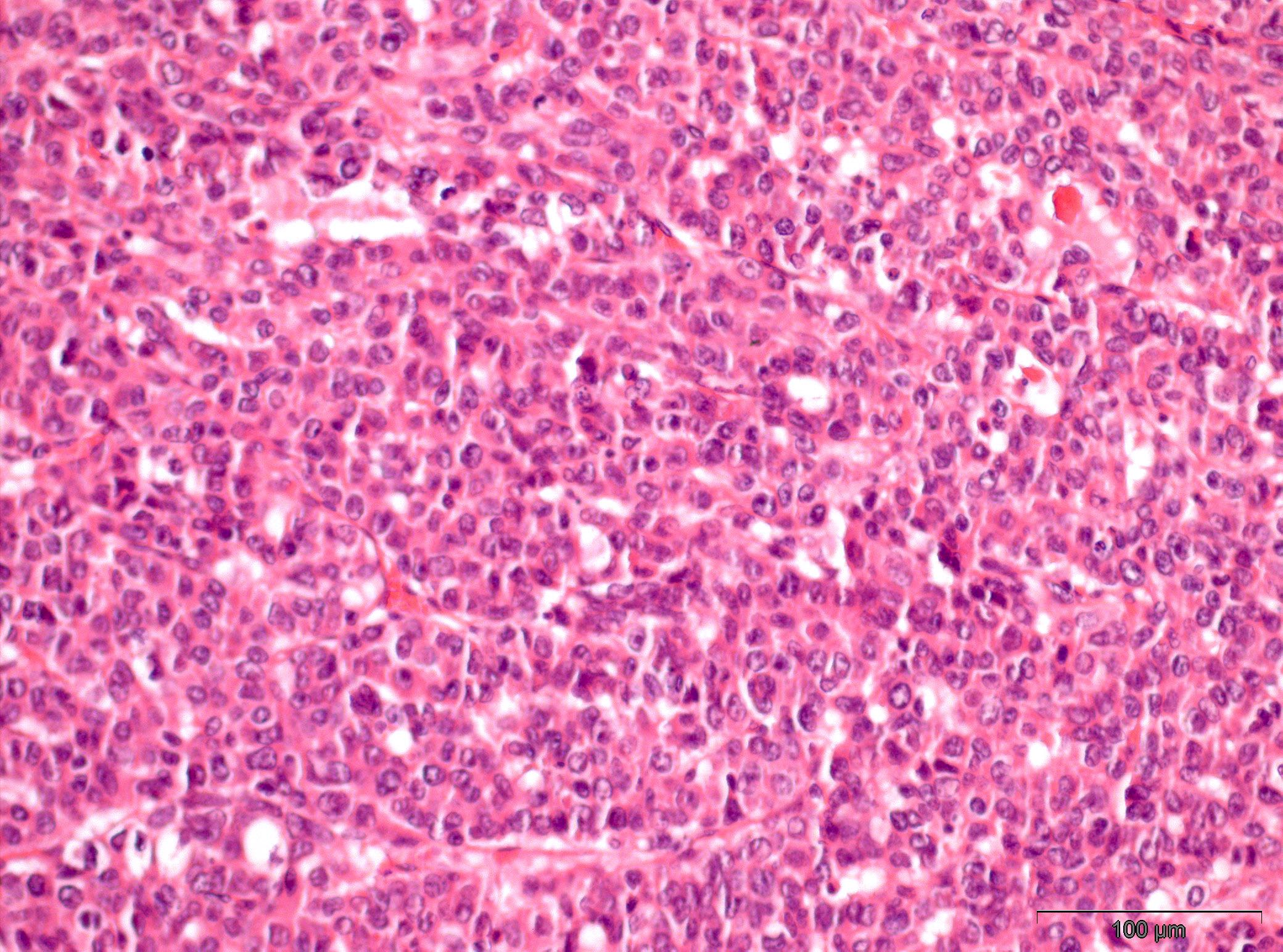
Microscopic (histologic) images
Contributed by João Lobo, M.D., Rui Henrique, M.D., Ph.D.

Prepubertal testicular yolk sac tumor
Prepubertal testicular yolk sac tumor, AFP
Prepubertal testicular yolk sac tumor
Prepubertal testicular yolk sac tumor, AFP
Metastatic yolk sac tumor
Metastatic yolk sac tumor, AFP

Testicular mixed germ cell tumor
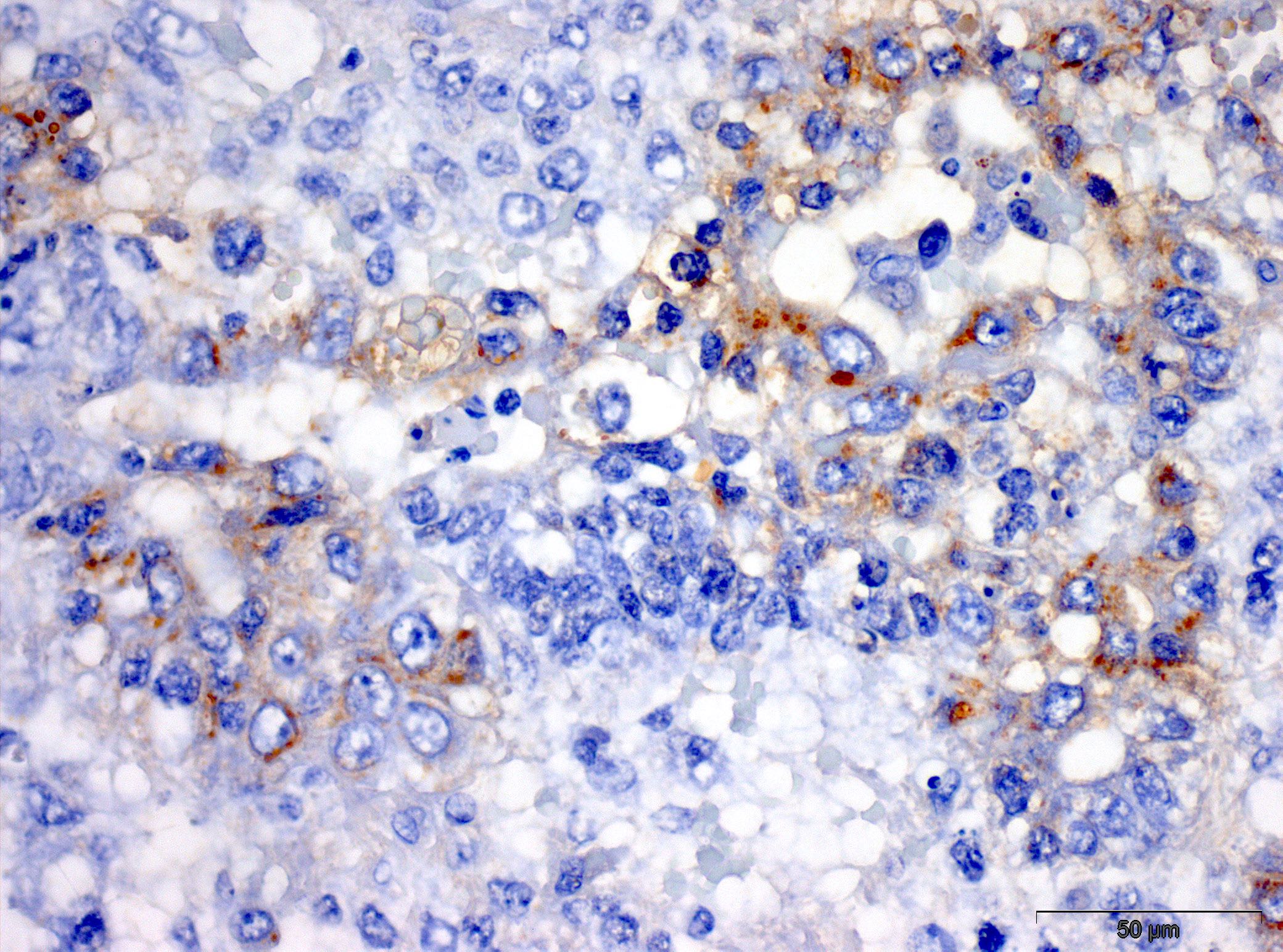
Testicular mixed germ cell tumor, AFP
Fetal liver
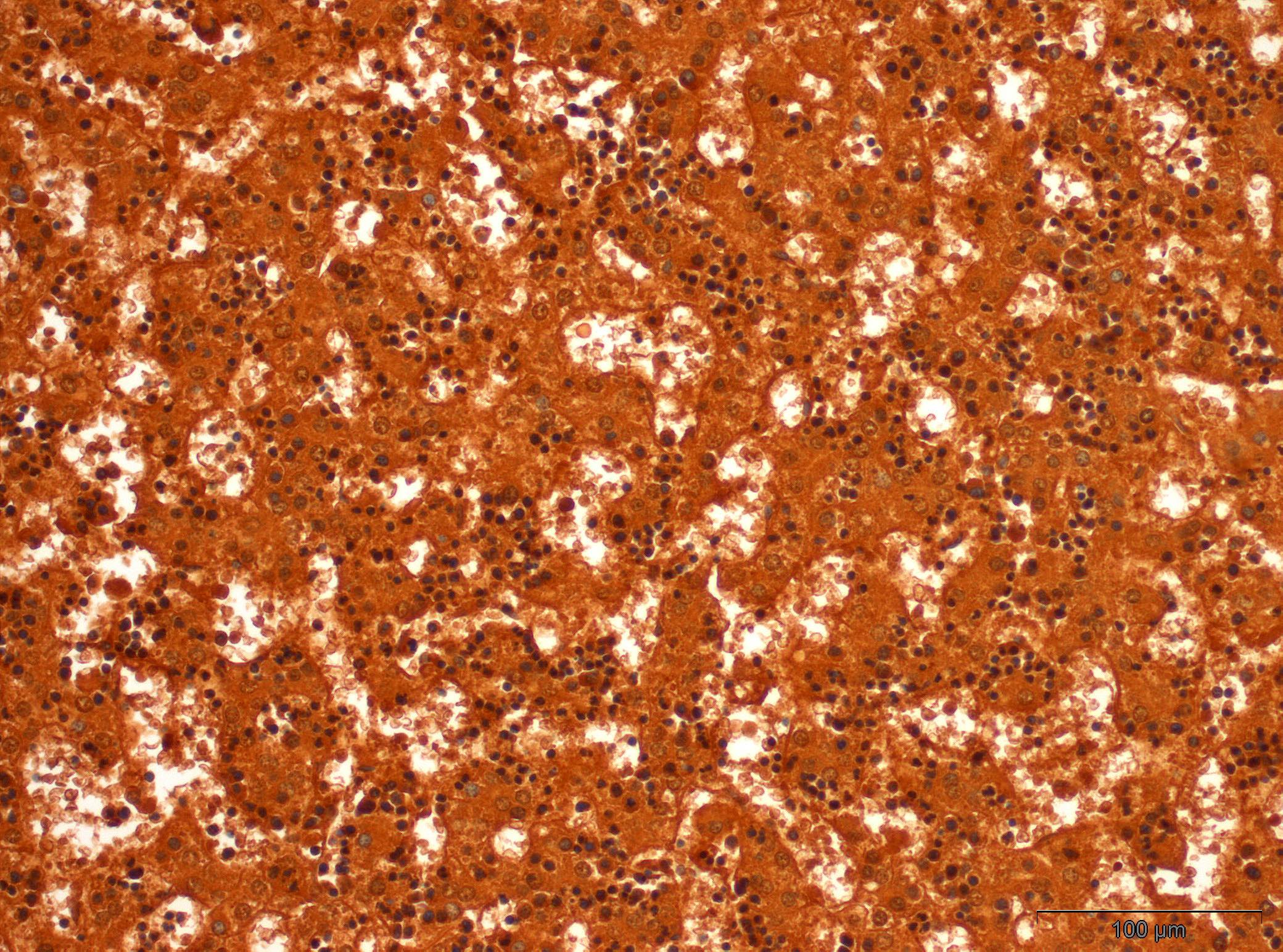
Fetal liver, AFP
Hepatoblastoma

Hepatoblastoma, AFP
Virtual slides
Images hosted on other servers:

Ovarian yolk sac tumor, AFP

Yolk sac tumor in undescended testis, AFP
Positive staining - normal
- Well fixed and nonautolyzed embryonal liver is a reliable positive control
- Early gut, yolk sac; nonspecific background reaction frequent in necrotic and cystic areas, since it is a protein secreted in serum (Int J Dev Biol 2012;56:755)
- Low expression in normal adult liver (EBioMedicine 2018;33:57)
Positive staining - disease
- Yolk sac tumor (62%)
- Specific marker but less sensitive than glypican 3 (glypican 3 stains 97% of solid yolk sac tumors, which can mimic seminoma) (Pathology 2018;50:88)
- In primitive patterns, usually with a strong granular cytoplasmic positivity, especially in intracellular lumina; can be positive in hyaline globules, although this is not frequent (Hum Pathol 1988;19:995)
- In classical patterns, staining may be heterogeneous but is consistent
- In somatic glandular patterns, staining is incomplete, more focal or absent, difficult to identify at low power (Histopathology 2014;65:51)
- Positive foci within embryoid bodies or embryonal carcinoma reflect differentiation into yolk sac tumor (Acta Pathol Microbiol Immunol Scand A 1983;91:165)
- Hepatocellular carcinoma (2 - 62%)
- Specific but not a sensitive marker (Hum Pathol 2002;33:1175)
- Infantile hemangioendothelioma can show expression (Hum Pathol 2010;41:763)
- Cirrhosis, chronic hepatitis B and other liver diseases can show low level expression in liver (EBioMedicine 2018;33:57)
- Reports of positivity in multiple tumors with hepatoid or enteric differentiation or of pediatric origin, such as enteroblastic gastric carcinoma, lung carcinoma, gallbladder carcinoma, colon carcinoma, sex cord stromal tumors like Sertoli-Leydig tumors, among others (Case Rep Pathol 2018;2018:3620293, Respir Med 2016;119:175, JOP 2007;8:55, summarized in World J Gastroenterol 2013;19:321)
Negative staining
- Germ cell neoplasia in situ (GCNIS)
- Seminoma
- Embryonal carcinoma
- Choriocarcinoma
- Gonadoblastoma
- Spermatocytic tumor
- Teratoma: can be positive in some epithelial components (20%) (Acta Pathol Microbiol Immunol Scand A 1983;91:165)
- Hepatoblastoma (15%): not significantly associated with subtype (Eur J Cancer 2012;48:1853)
- Cholangiocarcinoma
- Metastatic adenocarcinomas to the liver (5%)
Board review style question #1
Which of the following is true about the immunohistochemical expression of AFP?
- It can be positive in other liver diseases besides hepatocellular carcinoma and hepatoblastoma
- It is more sensitive than glypican 3 for diagnosing yolk sac tumor
- Nuclear staining is expected
- Seminomas are typically positive
- Teratomas are invariably negative
Board review style answer #1
A. It can be positive in other liver diseases besides hepatocellular carcinoma and hepatoblastoma. It is a specific marker but less sensitive than glypican 3. Seminomas are typically negative. Cytoplasmic or membrane staining is expected. Teratomas may show positivity in certain epithelial elements. Other liver diseases and liver tumors can show positive immunostaining, including metastatic disease to the liver.
Comment Here
Reference: Alpha fetoprotein (AFP)
Comment Here
Reference: Alpha fetoprotein (AFP)

