Actin, alpha smooth muscle type


Copyright: 2003-2025, PathologyOutlines.com, Inc.
PubMed Search: Alpha smooth muscle actin
- Actin is a 43000 kDa ubiquitous protein found in all cells
- Actins are involved in cell motility (alpha, smooth muscle) and the maintenance of the cytoskeleton (beta and gamma, all cells)
- Antibodies to alpha smooth muscle actin do not detect the other actin isoforms
- Involved in cell motility
- Identifies pericytes, myoepithelial cells, smooth muscle cells and myofibroblasts in normal, reactive or neoplastic tissue
- Myofibroblastic staining (tram track) versus smooth muscle staining (block cytoplasmic)
- Also called smooth muscle actin, SMA; clone ASM1 / 1A4 or sm 1
- 3 types: alpha, beta and gamma
- Alpha actins are found in muscle tissues and required for contraction, whereas the beta and gamma actins function as components of the cytoskeleton in many cells
- Expression correlates with the activation of myofibroblasts (Mol Cell Biochem 2008;308:201)
- May play a role in epithelial mesenchymal transition of carcinomas (Rom J Morphol Embryol 2014;55:1383)
- Controversial results on deficiency in intestinal pseudoobstruction (J Clin Pathol 2004;57:1168, Pediatr Surg Int 2008;24:1191, Gut 2004;53:1583)
- Immunoexpression may predict aggressive behavior in cutaneous basal cell carcinoma (Hum Pathol 2010;41:1128)
- Potential prognostic factor in idiopathic pulmonary fibrosis (Clinics (Sao Paulo) 2012;67:1039)
- Membranous or cytoplasmic staining
- Identify smooth muscle cells and myofibroblasts in normal, reactive or neoplastic tissue (Am J Respir Cell Mol Biol 1999;20:582, Am J Dermatopathol 2006;28:105)
- Identify myoepithelial cells in normal, neoplastic or diseased breast, salivary glands or sweat glands
- May be helpful to rule out invasion
- May be particularly important in cytology specimens
- Reference: Anticancer Res 2003;23:4175
- Identify pericytes to correlate with hematogenous metastasis and prognosis (Oncology 2005;69:159)
- Help distinguish pleuropulmonary desmoid tumors (SMA+) from solitary fibrous tumor (SMA-) (Arch Pathol Lab Med 2006;130:1503)
- Note: in breast papillary lesions, p63 is more sensitive and specific because smooth muscle actin also stains myofibroblasts / stromal cells (J Clin Pathol 2007;60:315)
- Myogenic differentiation (either only SMA or SMA+ desmin) in dedifferentiated liposarcoma significantly decreases 5 year disease free survival (Am J Surg Pathol 2020;44:799)
- Myofibroblastic staining (tram track) versus smooth muscle staining (block cytoplasmic)
Contributed by Kemal Kösemehmetoğlu, M.D.
Leiomyoma
SMA expression in leiomyoma
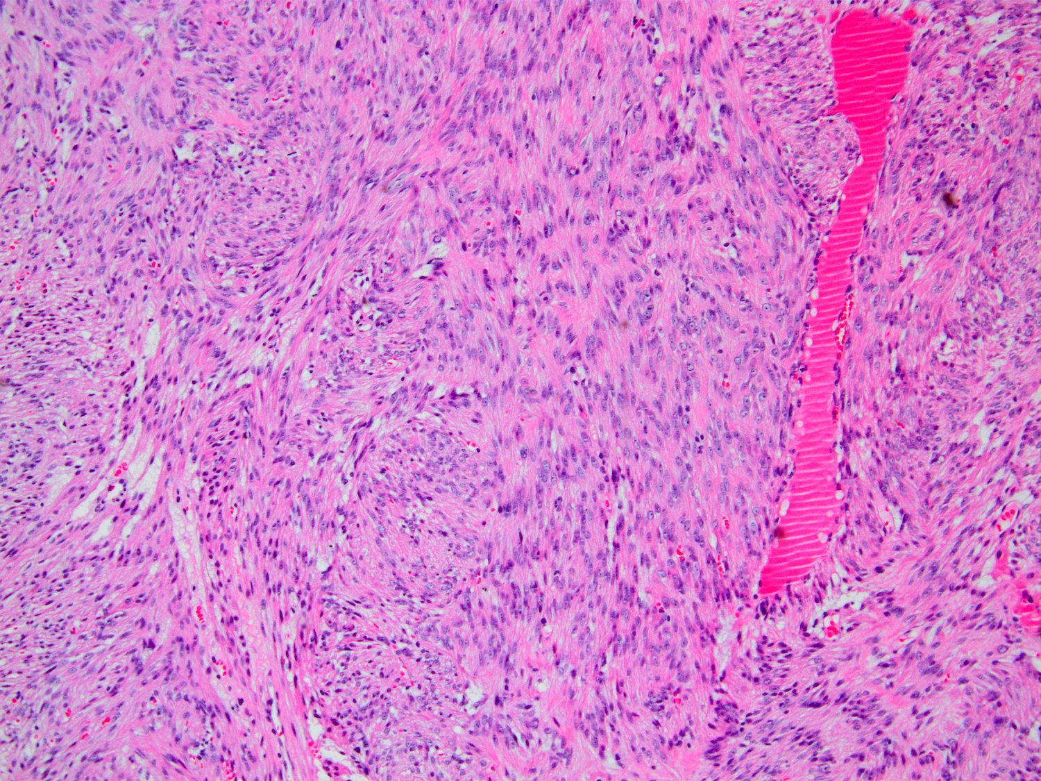
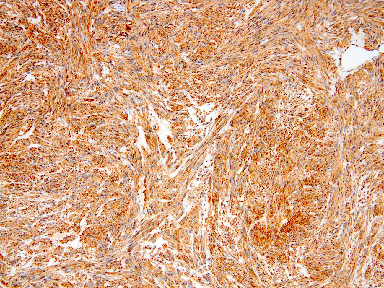
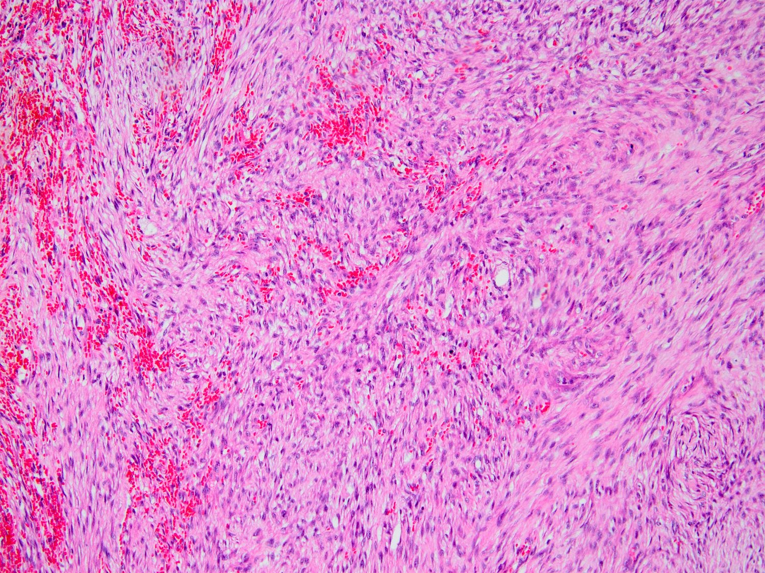
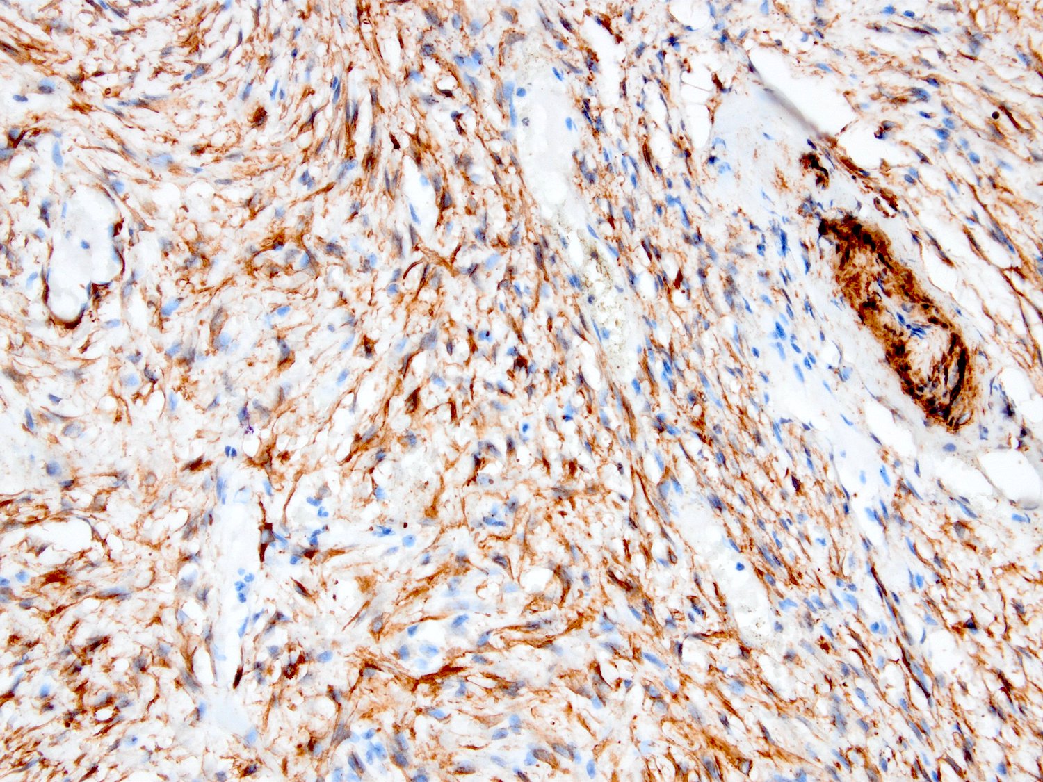
Leiomyosarcoma
Diffuse cytoplasmic block SMA expression
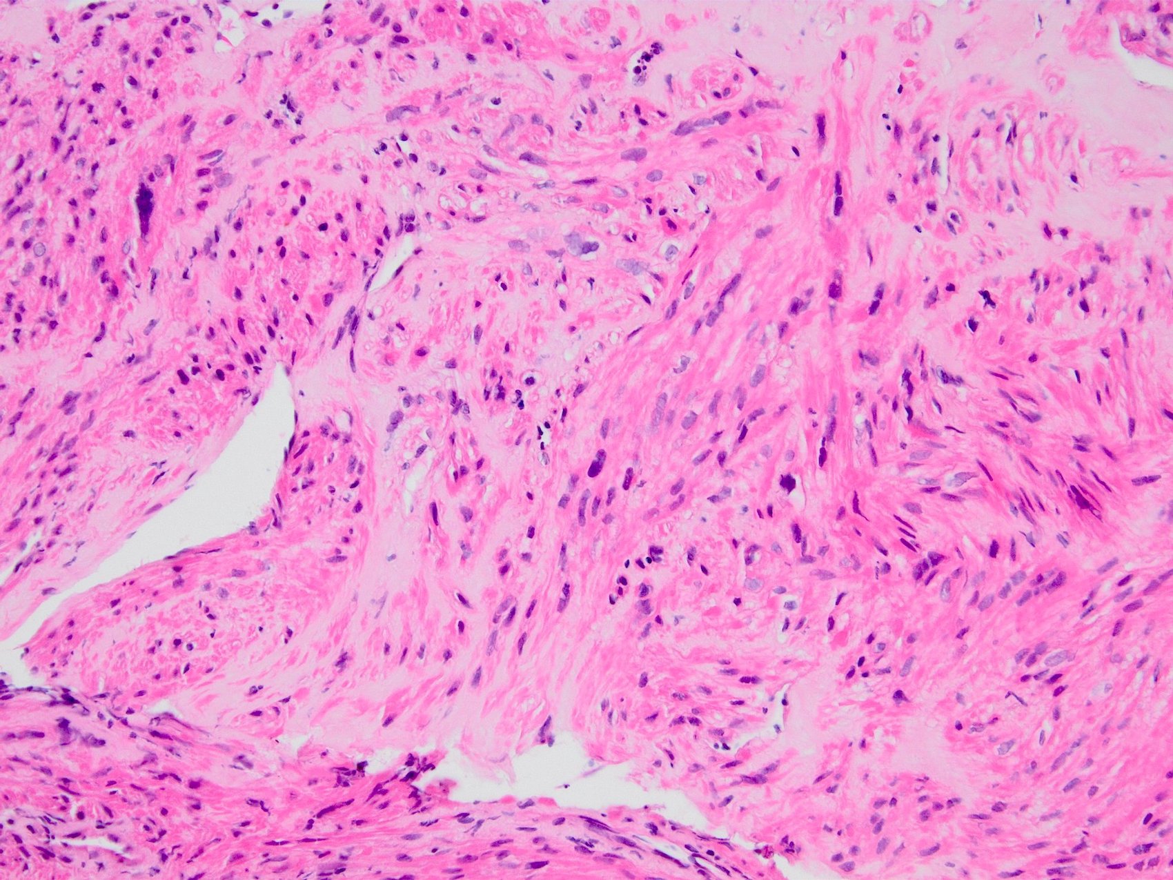
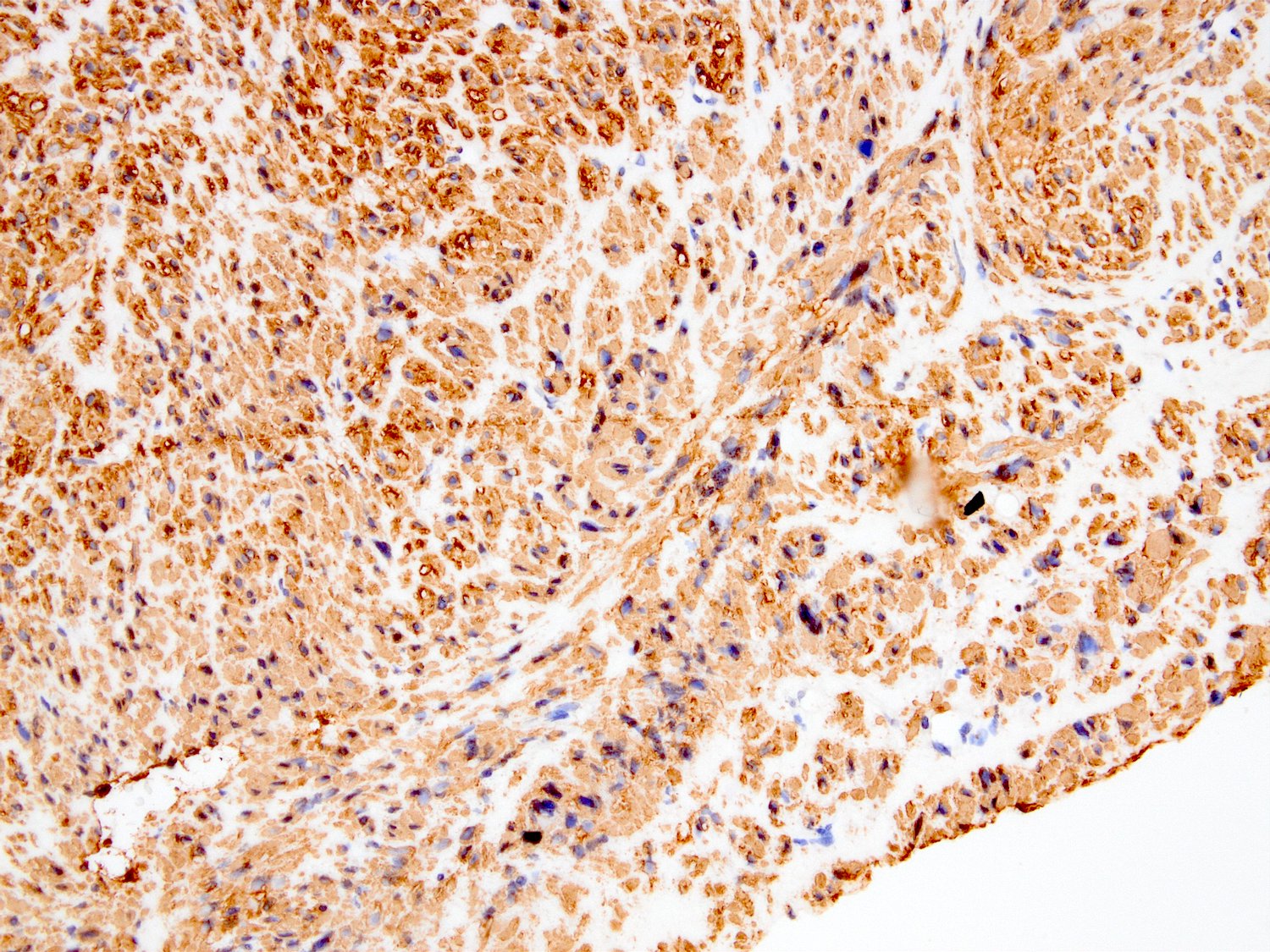
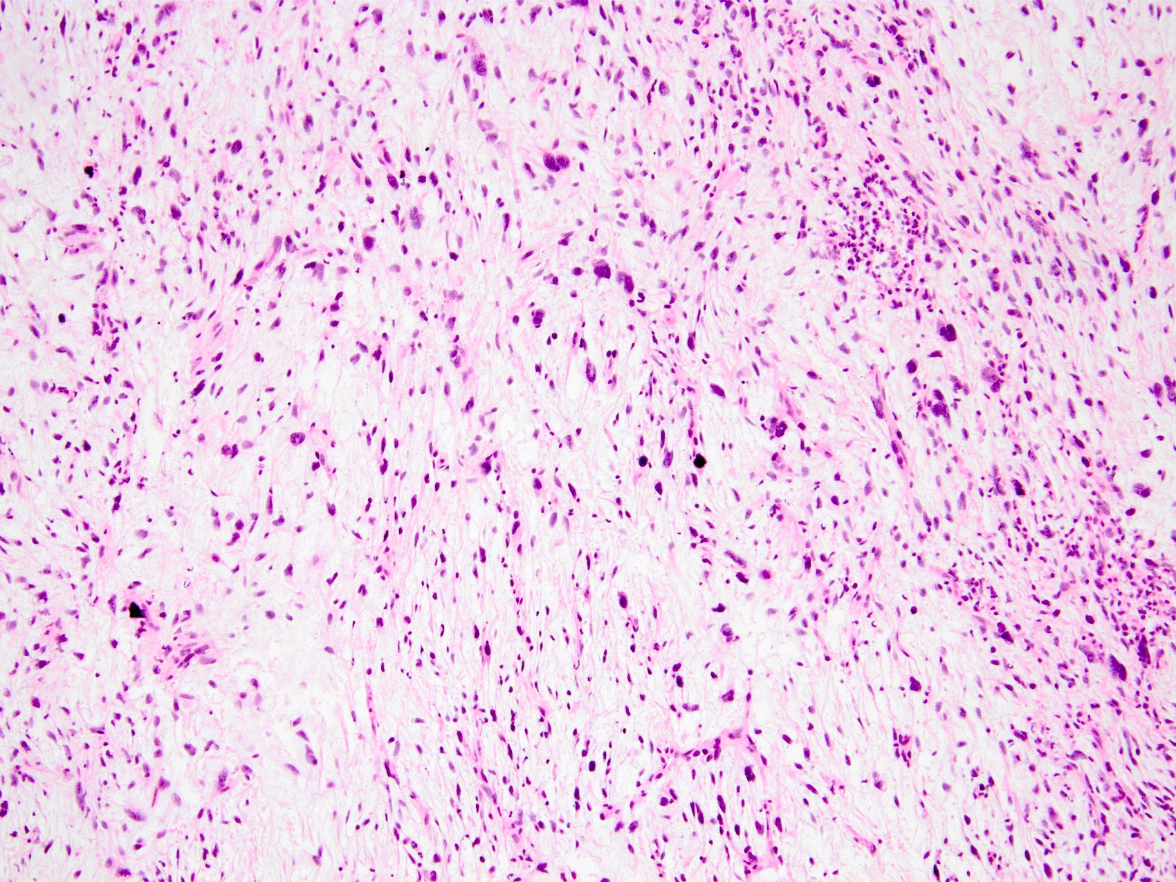
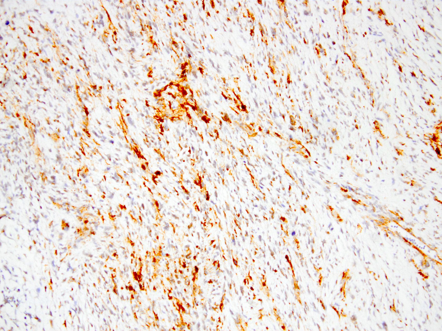
Nodular fasciitis
SMA in nodular fasciitis
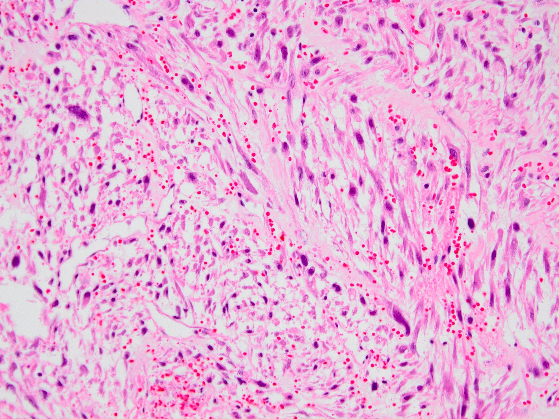
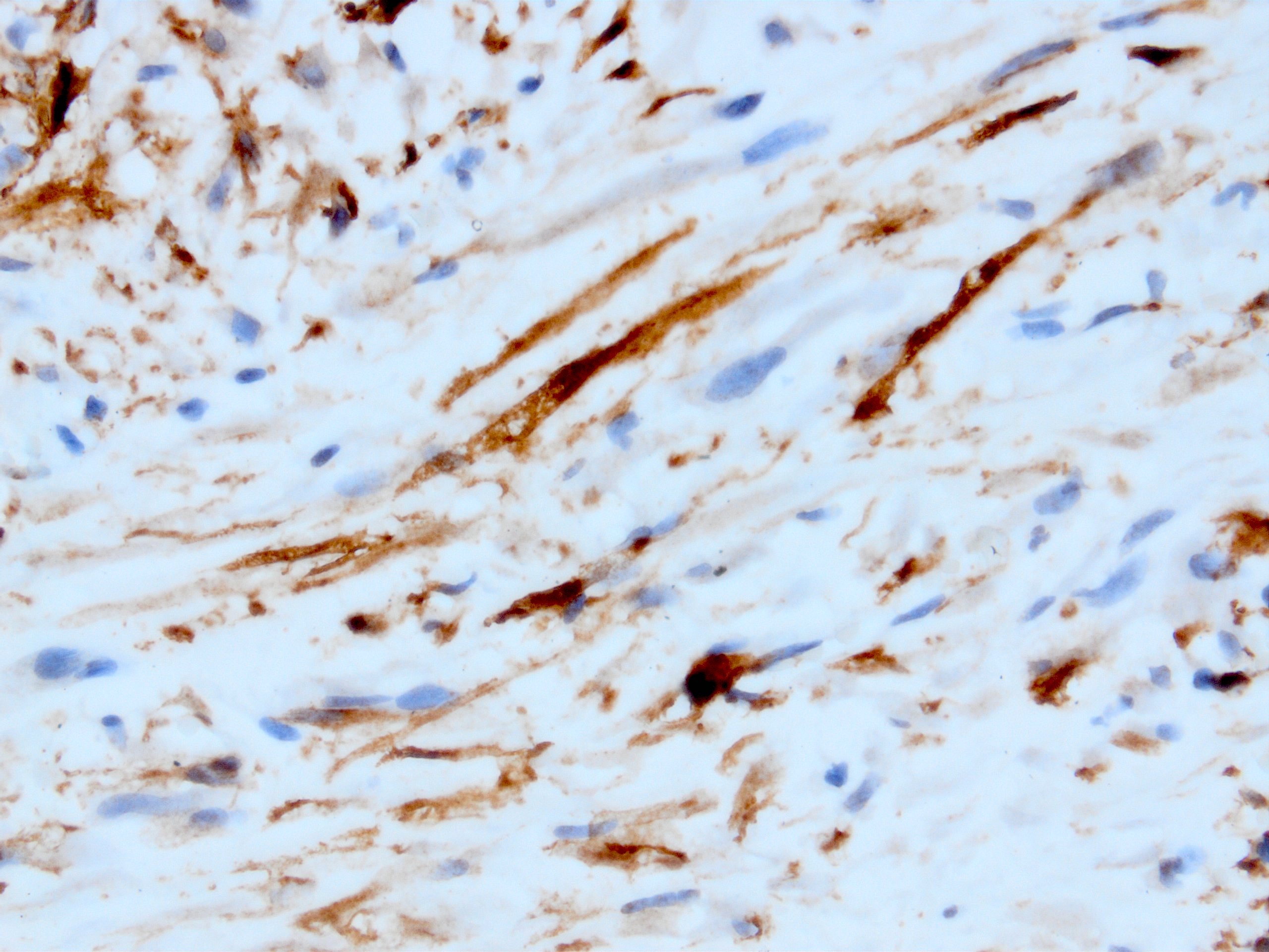
Myxofibrosarcoma
Focal SMA expression in myxofibrosarcoma

Glomus tumor

SMA in glomus tumor
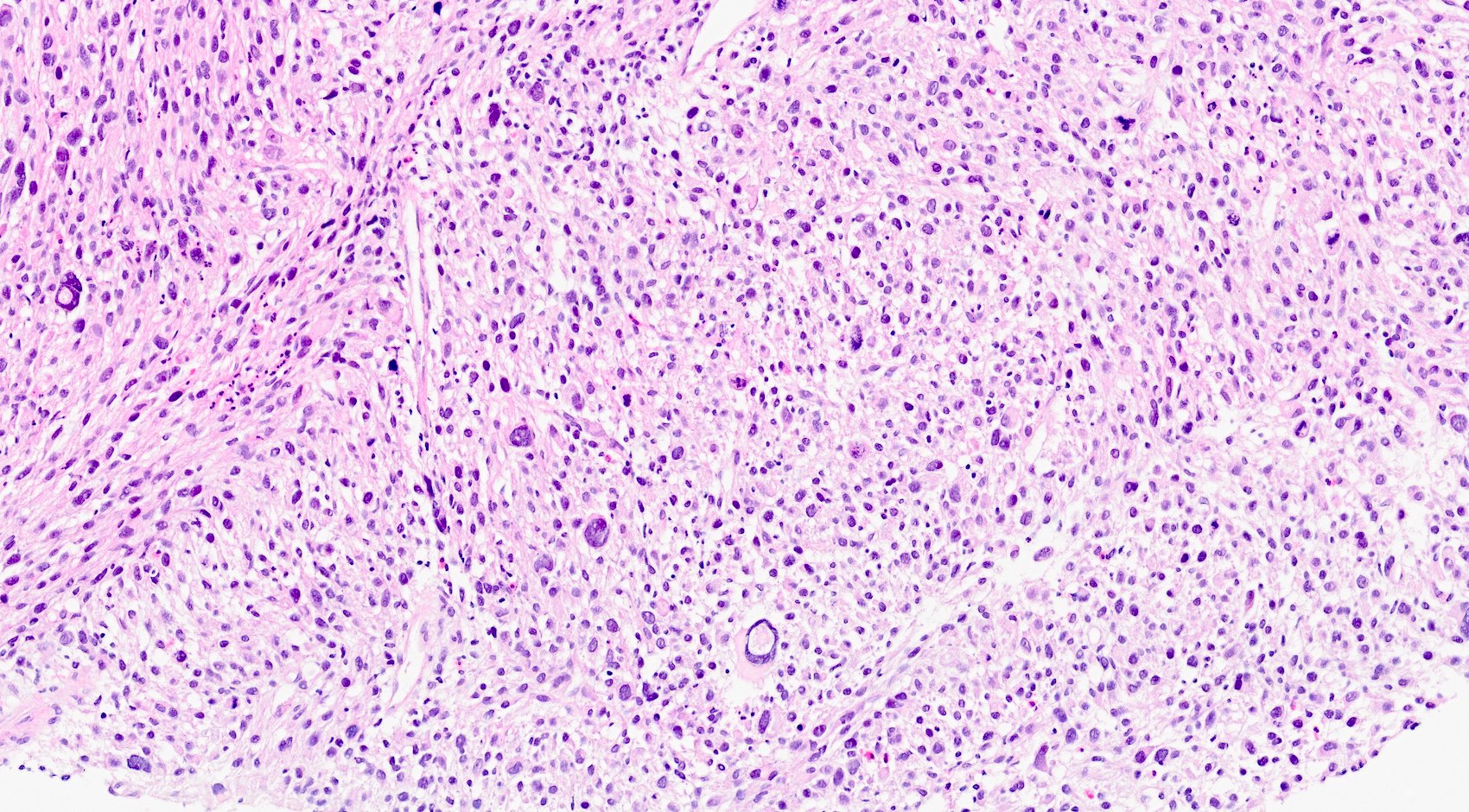
Undifferentiated pleomorphic sarcoma
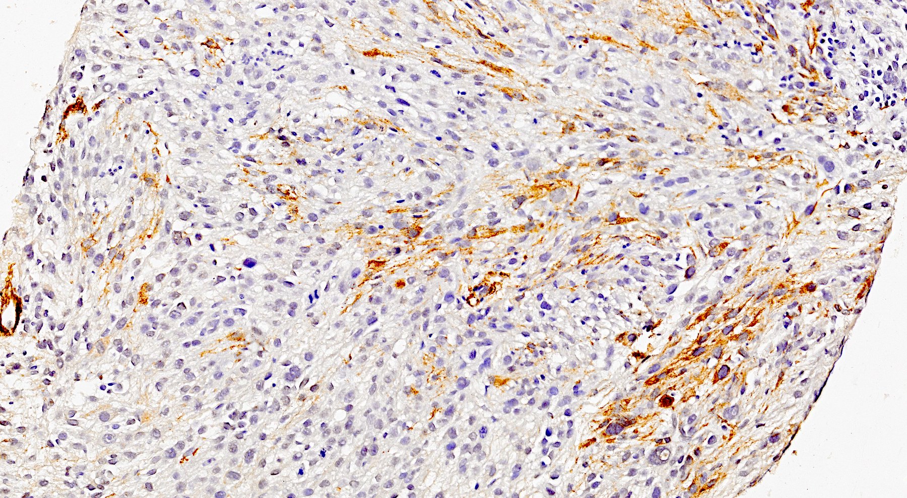
Focal SMA in undifferentiated pleomorphic sarcoma
Inflammatory myofibroblastic tumor
Myofibroblastic type SMA in IMT

Atypical apocrine adenosis
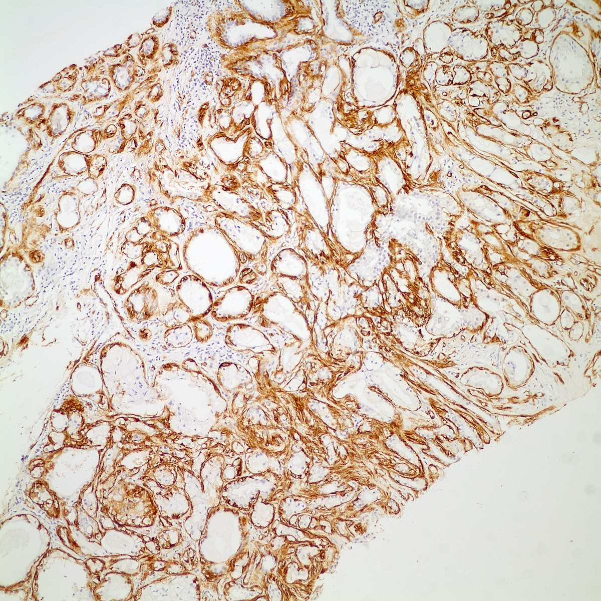
SMA in myoepithelial layer of breast adenosis

Radial scar / complex sclerosing lesion
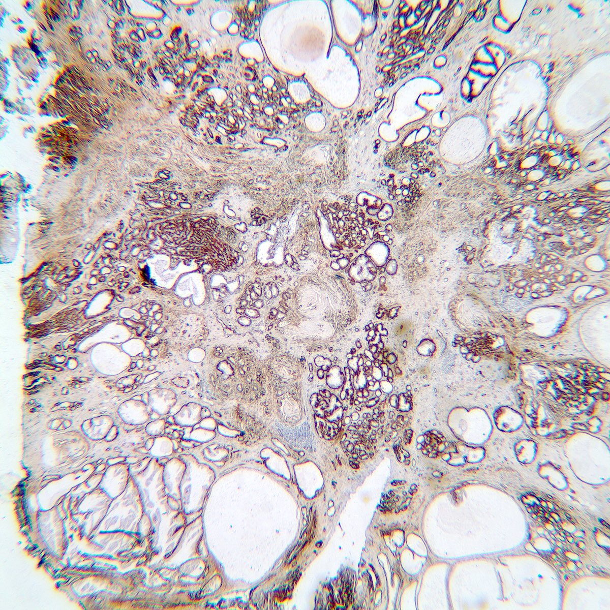
SMA in radial scar / complex sclerosing lesion
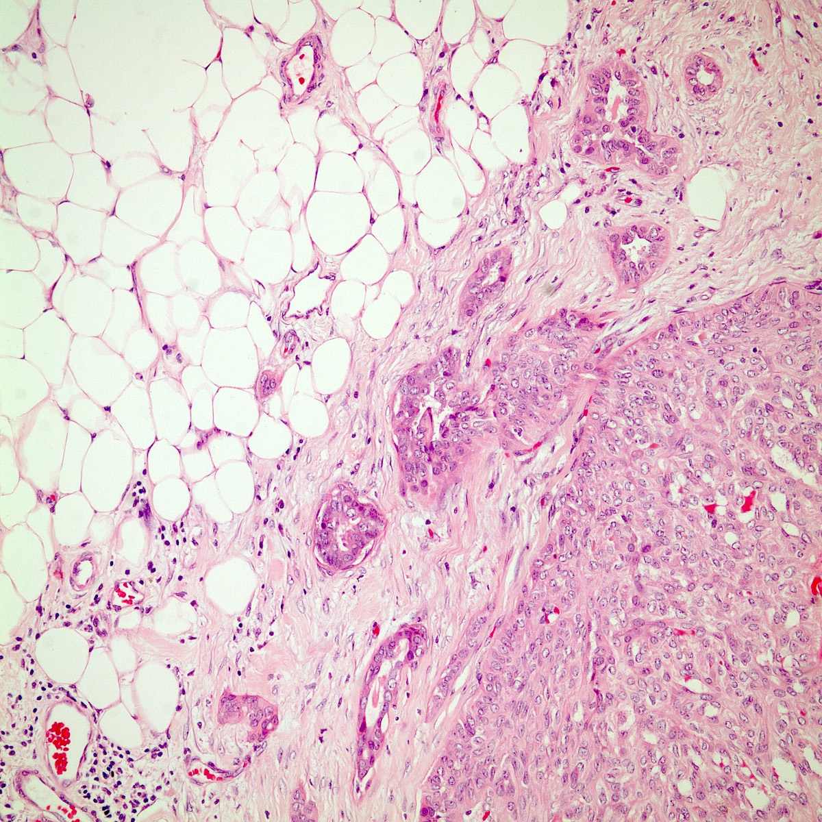
Sclerosing papilloma

Pseudoinvasion in sclerosing papilloma
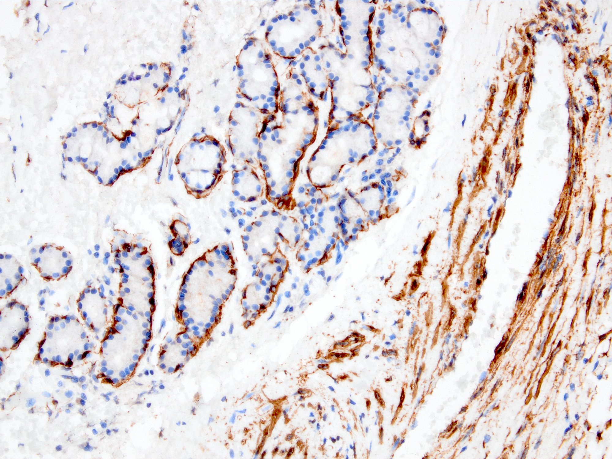
SMA positive myoepithelial layer
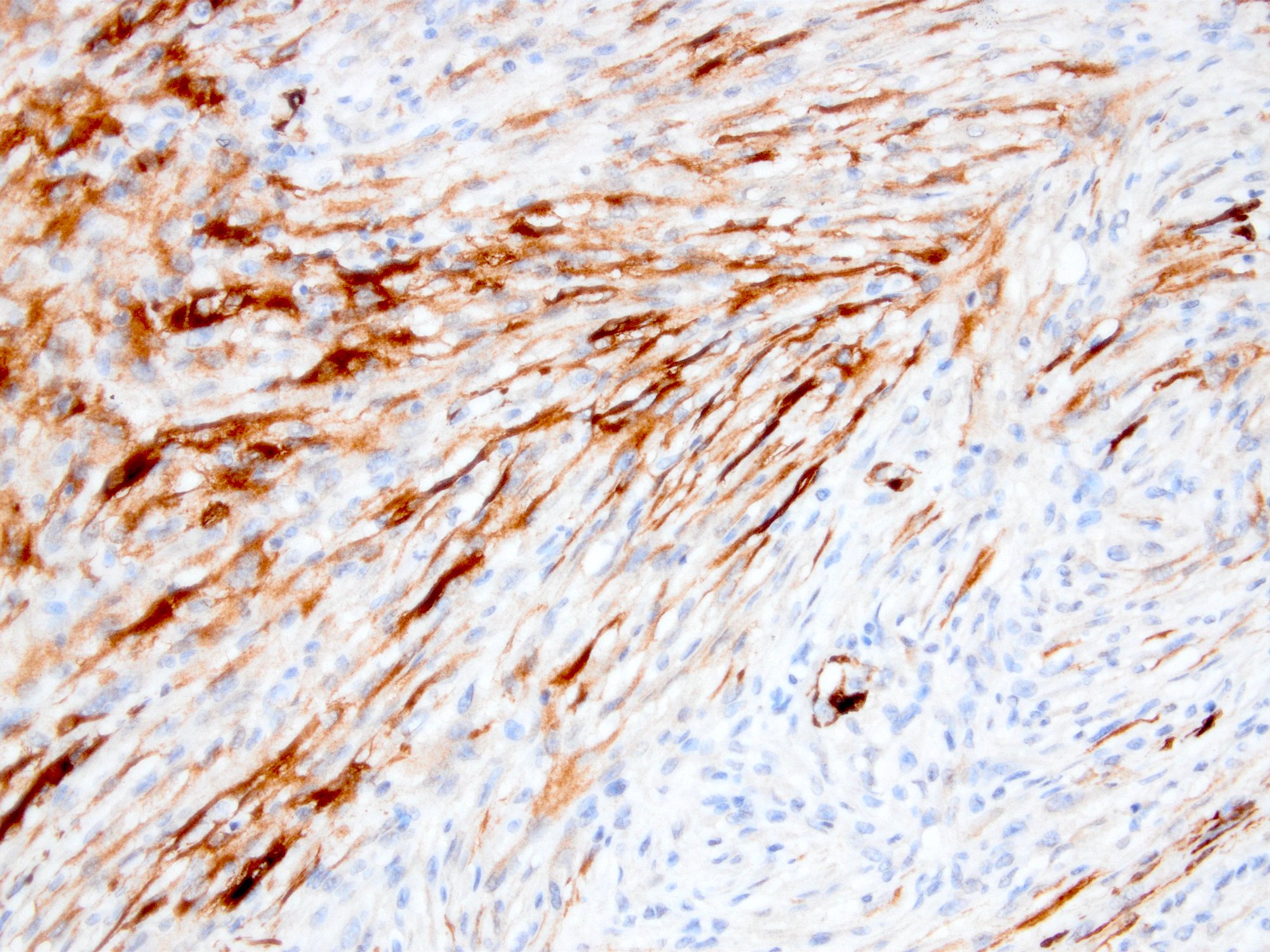
Tram track (myofibroblastic) staining pattern
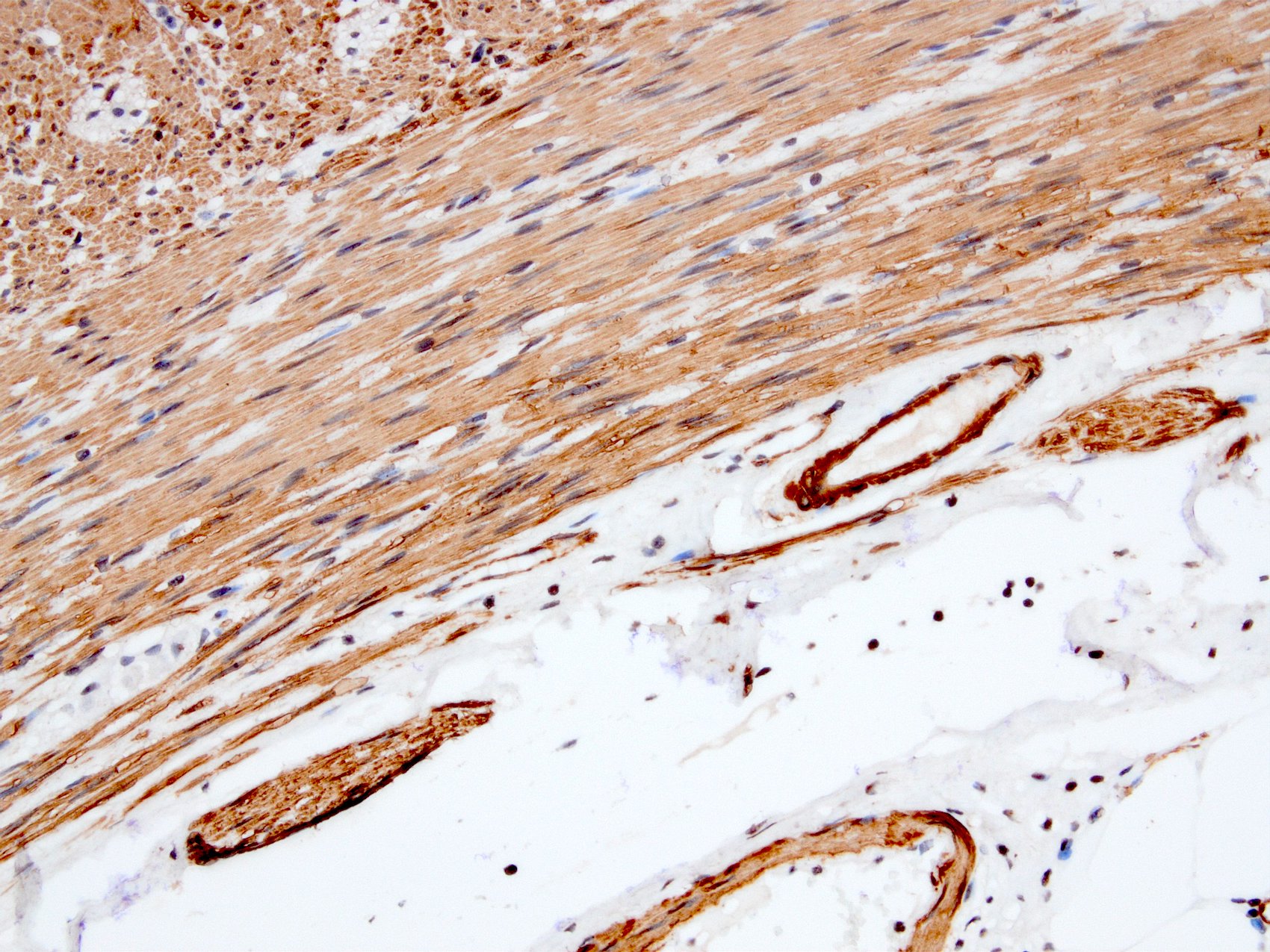
Block (smooth muscle-like) staining pattern
Images hosted on other servers:

SMA decorating vessels of tufted hemangioma

Glomangioma with diffuse and strong SMA expression

SMA expression in embryonal rhabdomyosarcoma
- Breast myoepithelial cells (most) (Breast Cancer Res 2003;5:R151, Proc Natl Acad Sci U S A 1993;90:999)
- Chondrocytes, choroidal nonvascular smooth muscle cells (Folia Biol (Praha) 2006;52:167, J Anat 2005;207:381)
- Decidual stromal cells, fibroblastic reticulum cells (Hum Reprod 1999;14:1599, J Cancer Res Clin Oncol 1981;101:149)
- Glomus coccygeum, hepatic stellate cells (Arch Pathol Lab Med 1999;123:905, Virchows Arch 1997;430:195)
- Myofibroblasts (except alveolar and some granulation tissue / scars) (J Histochem Cytochem 1992;40:1955, Lab Invest 1989;60:275, Int J Legal Med 1992;105:99)
- Osteoblasts (J Orthop Res 2002;20:622)
- Pericytes (J Histochem Cytochem 1989;37:315)
- Salivary glands (APMIS 1991;99:405)
- Smooth muscle and vascular smooth muscle (Proc Natl Acad Sci U S A 1981;78:298)
- Sweat glands and tracheobronchial glands (J Histochem Cytochem 1988;36:659)
- Adenoid cystic carcinoma (Arch Pathol Lab Med 1999;123:801)
- Atypical teratoid / rhabdoid tumor (J Neurosurg 1996;85:56, Brain Tumor Pathol 2008;25:79)
- Benign fibrous histiocytoma (in deep form 38%) (Am J Surg Pathol 2008;32:354)
- Biphenotypic sinonasal sarcoma (Am J Surg Pathol 2012;36:517)
- Cellular angiofibroma (focal, 41%) (Mod Pathol 2011;24:82)
- Cellular neurothekeoma (at least focal in 57%) (Am J Surg Pathol 2007;31:329)
- Collagenous spherulosis (Mod Pathol 2006;19:1351)
- Chronic obstructive pulmonary disease (COPD): large airways have increased expression of SMA (Respir Res 2011;12:48)
- Epstein-Barr virus associated smooth muscle tumour (EBV SMT) (Am J Surg Pathol 2006;30:75)
- Endometrial stromal sarcoma (65%) (Gynecol Oncol 2004;92:71)
- Epithelial myoepithelial carcinoma (Am J Surg Pathol 2007;31:44)
- Fibromatosis (56%) (Am J Surg Pathol 2002;26:1296)
- Gastrointestinal stromal tumor (GIST) (45%) (Am J Surg Pathol 2002;26:1296, Am J Pathol 1990;136:771)
- Glomus tumor (Hum Pathol 1999;30:1259, Am J Pathol 1990;136:771)
- Granulosa cell tumors of ovary, both adult and juvenile (variable) (Mod Pathol 1995;8:25)
- Inflammatory myofibroblastic tumor (Am J Surg Pathol 1991;15:1146, Ann Diagn Pathol 2001;5:335, Am J Surg Pathol 1992;16:896, Turk J Gastroenterol 2012;23:399)
- Leiomyoma (Am J Dermatopathol 2006;28:105, Am J Pathol 1987;128:91)
- Leiomyosarcoma (Int J Gynecol Pathol 2011;30:236, Anticancer Res 2005;25:1559)
- Liposarcoma, pleomorphic (focal in 40 - 50%), dedifferentiated (50%), well differentiated (in the form of pericytic mimicry) (Am J Surg Pathol 2002;26:601, Am J Surg Pathol 2004;28:1257, Am J Surg Pathol 2020;44:799, Hum Pathol 2016;54:92)
- Melanoma, desmoplastic / spindle cell (Am J Dermatopathol 1999;21:537, Am J Surg Pathol 2006;30:75, Am J Surg Pathol 1996;20:1489)
- Mesothelioma, sarcomatoid (60%) (Histopathology 2003;42:270)
- Myoepithelioma (Hum Pathol 2004;35:14, Am J Surg Pathol 2003;27:1183)
- Myofibroma / myopericytoma (Am J Pathol 1987;128:91)
- Myofibroblastic sarcoma (Chin Med J (Engl) 2007;120:363, Int J Oral Sci 2012;4:170, Am J Dermatopathol 2006;28:105)
- Neurothekeoma (40% focal) (Am J Pathol 1987;128:91)
- Nodular fasciitis (Ann Diagn Pathol 2002;6:94, Am J Dermatopathol 2006;28:105)
- PEComas (angiomyolipoma, pulmonary lymphangioleiomyomatosis) (J Egypt Natl Canc Inst 2013;25:125, J Clin Pathol 1993;46:479, Tohoku J Exp Med 2003;199:119)
- Plexiform fibrohistiocytic tumor (Am J Surg Pathol 1994;18:668, Histopathology 1991;19:503)
- Plexiform fibromyxoma (Am J Surg Pathol 2009;33:1624)
- Renal mixed epithelial and stromal tumor (Arch Pathol Lab Med 2006;130:80, Beijing Da Xue Xue Bao 2008;40:415)
- Rhabdomyoma (focal / rare) (Hum Pathol 1993;24:754, Hum Pathol 1993;24:608)
- Rhabdomyosarcoma embryonal, alveolar and sclerosing / spindle cell (Pediatr Dev Pathol 2005;8:427, Korean J Ophthalmol 2006;20:70, Virchows Arch 2006;449:554)
- Soft tissue perineurioma (21%) (Am J Surg Pathol 2005;29:845)
- Synovial sarcoma (25%) Mod Pathol 2007;20:760)
- Undifferentiated pleomorphic sarcoma (focal) (J Clin Pathol 2003;56:666, Histopathology 2006;48:453)
- Normal tissue:
- Cardiac muscle (positive during development) (J Cell Sci 2007;120:229)
- Skeletal muscle (J Cell Biol 1985;100:807)
- Basal cells of prostate glands (Am J Surg Pathol 1996;20:1489)
- Disease:
- Angiomyofibroblastoma (rarely focal) (Hum Pathol 1997;28:1046)
- Carcinomas (usually)
- Cellular benign fibrous histiocytoma (Am J Surg Pathol 1994;18:668)
- Clear cell sarcoma (J Clin Pathol 2010;63:416)
- Epithelioid sarcoma proximal type (15 - 33%) (Am J Surg Pathol 1997;21:130, Mod Pathol 2001;14:655)
- Fibrosarcoma, infantile and adult type (rare / focal; expression does not exclude diagnosis) (Am J Clin Pathol 2001;115:348)
- Hemosiderotic fibrolipomatous tumor (Histopathology 2006;48:453)
- Liposarcoma, myxoid type (rarely focal) (Am J Clin Pathol 1995;103:20)
- Low grade fibromyxoid sarcoma (LGFMS) (rare / focal) (Lab Invest 2005;85:408)
- Myofibroblastoma (occasionally focally positive) (Pathology 2005;37:144, Am J Surg Pathol 2001;25:1022)
- Ossifying fibromyxoid tumor (weak); 6% (J Laryngol Otol 1993;107:75, Am J Surg Pathol 2011;35:1615)
- Thecoma / fibrothecoma (Mod Pathol 1995;8:25)
- Schwannoma, solitary fibrous tumor (Arch Pathol Lab Med 2006;130:1503, Diagn Pathol 2021;16:32)
- Sclerosing epithelioid fibrosarcoma (Am J Surg Pathol 1995;19:979)
- Right 3rd intercostal space, wide excision:
- Leiomyosarcoma, grade 3 (see comment)
- Comment: Immunohistochemically, neoplastic cells showed diffuse strong cytoplasmic staining for SMA, desmin and h-caldesmon.
- Right thigh, excisional biopsy:
- Nodular fasciitis (see comment)
- Comment: Immunohistochemically, neoplastic cells were positive for SMA in myofibroblastic pattern (tram track staining) and negative for desmin.
Which statement is correct about the SMA immunostaining shown above?
- Consistent with the diagnosis of nodular fasciitis
- Demonstrates smooth muscle type of SMA staining
- Demonstrates tram track SMA staining
- Excludes the diagnosis of spindle cell rhabdomyosarcoma
Comment Here
Reference: Actin, alpha smooth muscle type