Stains & CD markers
Beta catenin

Copyright: 2003-2025, PathologyOutlines.com, Inc.
PubMed Search: Beta catenin
Beta catenin
Author: Pamela Wirth, Ph.D.
Editorial Board Member: Christian M. Schürch, M.D., Ph.D.
Deputy Editor-in-Chief: Borislav A. Alexiev, M.D.
Last author update: 25 June 2024
Last staff update: 25 June 2024
Copyright: 2003-2025, PathologyOutlines.com, Inc.
PubMed Search: Beta catenin
Table of Contents
Definition / general | Essential features | Terminology | Pathophysiology | Diagrams / tables | Clinical features | Interpretation | Uses by pathologists | Prognostic factors | Microscopic (histologic) images | Positive staining - normal | Positive staining - disease | Negative staining | Molecular / cytogenetics description | Sample pathology report | Practice question #1 | Practice answer #1 | Practice question #2 | Practice answer #2Cite this page: Wirth P. Beta catenin. PathologyOutlines.com website. https://www.pathologyoutlines.com/topic/stainsbetacatenin.html. Accessed August 24th, 2025.
Definition / general
- Protein encoded by the CTNNB1 gene with dual functions in cell adhesion and Wnt signaling (Am J Clin Pathol 2019;151:529)
Essential features
- Responsible for maintaining structural integrity of many epithelial tissues
- Mutations in CTNNB1 relevant to certain types of cancer
- In the nucleus, beta catenin can act as cofactor in the upregulation of oncogenes like cyclin D1 and MYC
- Increase in protein expression is considered a hallmark of Wnt driven cancers (J Hematol Oncol 2020;13:165)
- Belongs to armadillo protein family
- Discovered in late 1980s as an E-cadherin associated protein (Biol Res 2020;53:33)
Terminology
- Beta catenin (β catenin)
- CTNNB1
- Catenin (cadherin associated protein), beta 1
Pathophysiology
- In normal tissues, beta catenin levels are regulated (targeted and degraded) by a complex of cytoplasmic proteins including adenomatous polyposis coli (APC), glycogen synthase kinase 3 (GSK3) and casein kinase 1 (CK1) (Cold Spring Harb Perspect Biol 2013;5:a007898)
- Without activation of the Wnt pathway, most beta catenin localizes at the cellular membrane
- Upon Wnt activation, β catenin is translocated from the cell membrane (where it interacts with E-cadherin) to the cytoplasm and then nucleus, where it interacts with transcriptional activators, leading to cellular proliferation (Dev Cell 2009;17:9)
- Alterations in either beta catenin or APC can result in the accumulation of beta catenin within the nucleus
- Nuclear localization of beta catenin is known to associate with malignant transformation in many cancers (Oncotarget 2017;8:33972)
Diagrams / tables
Images hosted on other servers:

Cadherins and catenins in signal transduction

Role of beta catenin in cell
Clinical features
- Loss of beta catenin membrane expression with overexpression in the cytoplasm and nucleus is associated with various carcinomas, including breast, colon and pancreas
- Breast
- Nuclear and cytoplasmic accumulation in basal-like breast cancers
- Reduced membrane expression observed with tumor progression (Am J Pathol 2010;176:2911)
- Colon
- Beta catenin plays a critical role in tumorigenesis (activating Wnt signaling mutations present in 90% of colon cancers) (Sci Rep 2020;10:2957)
- Membrane expression in normal colorectal epithelium
- Staining pattern shifts to cytoplasmic / nuclear in tumor tissue (World J Gastroenterol 2008;14:3866)
- Thyroid
- Wnt pathway important for anaplastic and possibly papillary thyroid carcinomas
- Shifts from membrane location to cytoplasmic in malignant tumors (Front Endocrinol (Lausanne) 2012;3:31)
- Uterus
- Endometrial carcinoma is associated with beta catenin mutations (exon 3 in CTNNB1) (Front Oncol 2022;12:1009345)
- Liver
- Beta catenin expression is membranous for normal hepatocytes
- Mutations in CTNNB1 occur in about a third of human hepatocellular carcinomas (HCC) (J Clin Invest 2022;132:e154515)
- Pancreas
- Strong membrane staining in primary tumors of pancreatic neuroendocrine tumors (PanNETs); nuclear staining present in advanced stages including metastasis (World J Gastrointest Oncol 2016;8:615)
- Low grade sinonasal adenocarcinoma (nuclear localization) (Case Rep Pathol 2018;2018:8741017)
- Lung
- Reduced membrane expression of beta catenin with increased expression in cytoplasm and nucleus for non-small cell lung cancer (NSCLC) (Transl Lung Cancer Res 2017;6:97)
- Skin (melanoma)
- Loss of membrane expression as melanoma progresses from primary to metastatic
- Low frequency of CTNNB1 genetic mutations in melanomas, which does not correlate with nuclear expression (Mod Pathol 2002;15:454)
- Breast
Interpretation
- Membranous / cytoplasmic staining in normal tissues
- Increased cytoplasmic and nuclear expression in various cancers (see Clinical features)
- Weak nuclear expression of beta catenin may be observed in normal endometrium during proliferative phase
- Therefore, some authors only consider moderate levels of nuclear staining to be significant (Dev Cell 2009;17:9)
Uses by pathologists
- Beta catenin may be useful in the differential diagnosis of breast ductal carcinoma (membrane expression) versus lobular carcinoma (cytoplasmic expression)
- Nuclear beta catenin expression may be a helpful screening tool to distinguish ambiguous neoplasms like deep penetrating nevus (positive) from similar entities like blue melanocytic tumors, spitzoid tumors, nevoid and superficial spreading melanomas (all negative for nuclear expression) (Virchows Arch 2019;474:535)
- Increased nuclear expression of beta catenin may be useful as a diagnostic marker for desmoid type fibromatosis, as only a very limited subset (solitary fibrous tumor, synovial sarcoma and endometrial stromal sarcoma) of other mesenchymal neoplasms are positive for this marker (Mod Pathol 2005;18:68)
- Can help with classification of hepatocellular adenoma subtypes and identification of focal nodular hyperplasia (Am J Surg Pathol 2012;36:1691, Int J Hepatol 2013;2013:268625, Hum Pathol 2013;44:750)
- Beta catenin may be mutated or positive in fibromatosis (Mod Pathol 2012;25:1551)
- May help distinguish mesenteric fibromatosis (positive with nuclear staining due to mutations in APC / beta catenin pathway causing nuclear accumulation) from gastrointestinal stromal tumors (GISTs) (negative) and sclerosing mesenteritis (negative) (Am J Surg Pathol 2002;26:1296)
- Distinguish fibromatosis (diffuse or rarely focal nuclear staining for deep tumors) from low grade fibromyxoid sarcoma and other myofibroblastic or fibroblastic tumors / sarcomas (negative for nuclear staining) (Am J Surg Pathol 2005;29:653)
- Identify isolated tumor cells in neuroblastoma (Am J Clin Pathol 2009;131:49)
Prognostic factors
- Nuclear staining of beta catenin can indicate exon 3 mutations in CTNNB1, correlating with decreased survival in endometrial cancer (Am J Clin Pathol 2019;151:529)
- Reduced or negative membranous staining in gastric tumors associated with advanced stage and shorter survival (Br J Cancer 1999;81:1392, Int J Clin Exp Pathol 2017;10:8980)
- Poor prognostic factor for nasopharyngeal carcinoma (Hum Pathol 2013;44:1357)
- Reduced membrane expression and increased cytoplasmic / nuclear immunohistochemical staining in NSCLC is correlated with poor prognosis (Transl Lung Cancer Res 2017;6:97)
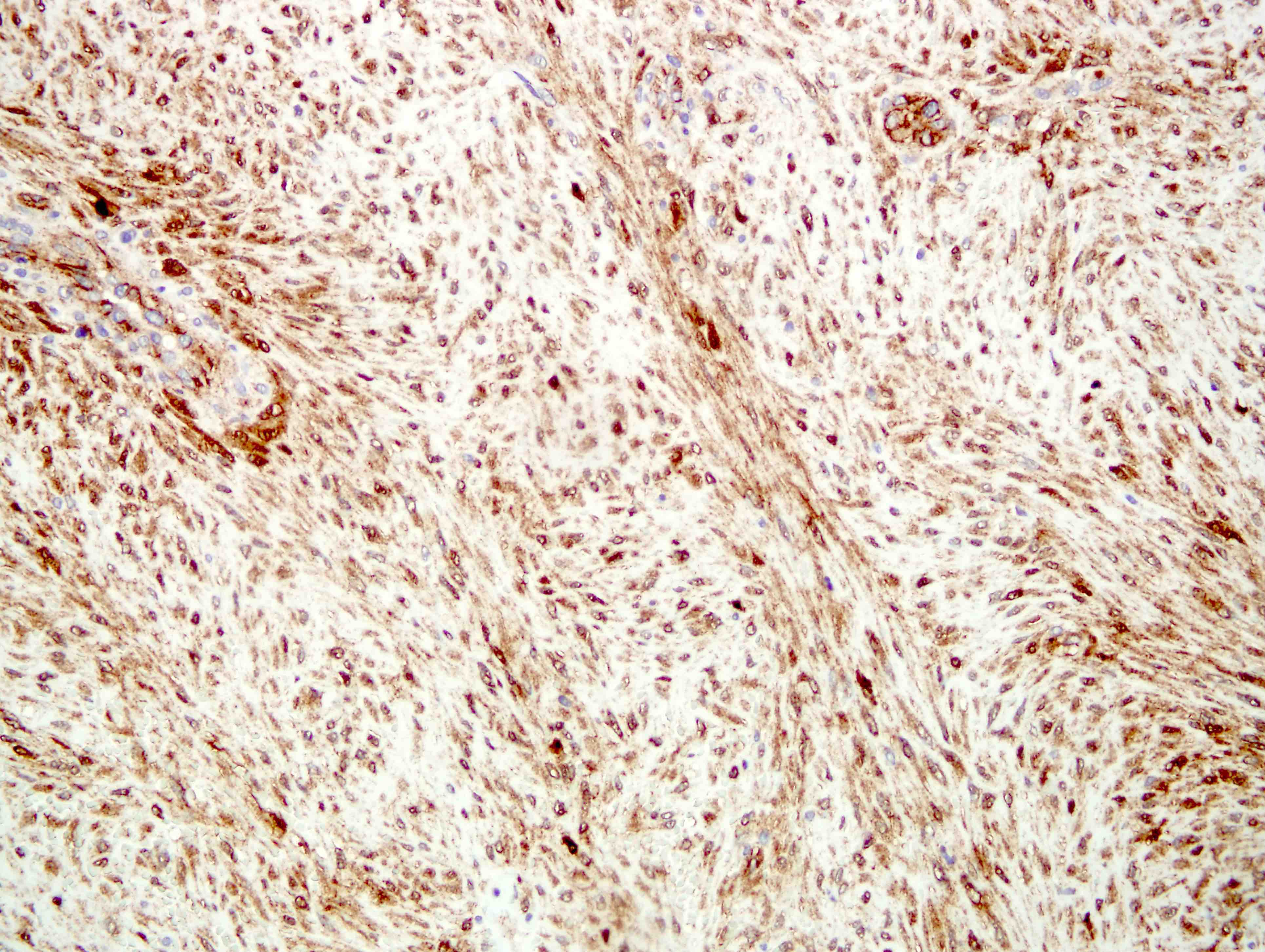
Microscopic (histologic) images
Contributed by Pamela Wirth, Ph.D. (source: University of Toronto and Human Protein Atlas),
Farres Obeidin, M.D., Christian M. Schürch, M.D., Ph.D. and Hyunkyu Shin, M.D.

Normal pancreatic tissue

Cervix

Breast carcinoma

Colorectal carcinoma

Lung cancer

Normal kidney

Nuclear beta catenin


Desmoid tumor


Desmoid tumor with positivity for beta catenin


Hepatocellular adenoma


Beta catenin activated hepatocellular adenoma with beta catenin positivity


Solid pseudopapillary neoplasm


Solid pseudopapillary neoplasm with positivity for beta catenin
Positive staining - normal
- Membrane expression of beta catenin can be found in many normal tissues and some benign tumors (Med Pharm Rep 2019;92:246, J Hepatocell Carcinoma 2018;5:61)
- Fibroblasts and endothelial cells (cytoplasmic or membranous staining)
Positive staining - disease
- Gastric cancer
- Nuclear expression (58%) (Int J Clin Exp Pathol 2010;3:782)
- Most gastric cancers show reduced or weak membrane expression (51.7%) (Int J Clin Exp Pathol 2017;10:8980, Br J Cancer 1999;81:1392)
- Invasive ductal carcinoma of the breast
- Membrane expression (70%) (Asian Pac J Cancer Prev 2021;22:3493)
- In basal-like breast cancer, nuclear (72%) and cytoplasmic (88%) expression is increased; membrane expression may also be present (Am J Pathol 2010;176:2911)
- Liver cancer
- Reduced membrane expression with an increase in cytoplasmic and nuclear expression in cirrhosis and hepatocellular carcinoma (73%) (J Hepatocell Carcinoma 2018;5:61)
- Bone cancer
- Osteoid producing primary tumors of bone exhibit cytoplasmic or membrane expression of beta catenin (73%) (Virchows Arch 2021;479:529)
- Endometrioid carcinoma of endometrium and ovary, particularly squamous morules (Hum Pathol 2005;36:605)
- Neuroblastoma (Am J Clin Pathol 2009;131:49)
- Ovarian microcystic stromal tumor (Am J Surg Pathol 2011;35:1429)
- Primitive neuroectodermal tumors (PNET), rhabdomyosarcoma, Wilms tumor (Am J Clin Pathol 2009;131:49)
- Pancreas: solid papillary neoplasm (Am J Clin Pathol 2009;132:831)
- Pulmonary lymphangioleiomyomatosis (Am J Clin Pathol 2011;135:776)
- Renal angioleiomyoma (Am J Clin Pathol 2011;135:776)
- Solitary fibrous tumors (nuclear staining in 33%, remainder had membranous or membranous / cytoplasmic staining) (Arch Pathol Lab Med 2005;129:776)
- Desmoid fibromatosis: 80% nuclear beta catenin staining (PLoS One 2021;16:e0250619)
Negative staining
- GIST, sclerosing mesenteritis, low grade fibromyxoid sarcoma, myofibroblastic or fibroblastic tumors (low grades may show some nuclear staining) (Mod Pathol 2014;27:S47)
Molecular / cytogenetics description
- Mutations in CTNNB1 (exon 3) have been detected in 30% of intestinal and diffuse type gastric cancers (World J Exp Med 2015;5:84)
- Deletions or missense mutations in exon 3 of CTNNB1 are the most common genetic abnormality leading to hepatocellular carcinoma (J Hepatocell Carcinoma 2018;5:61)
Sample pathology report
- Pancreas, distal pancreatectomy:
- Solid pseudopapillary neoplasm (SPPN) (see comment)
- Comment: A 28 year old female presents with abdominal pain and fever without jaundice, change in stool color or diarrhea. CT and ultrasound imaging of the abdomen exhibited a solid and cystic tumor measuring 8 cm near the tail of the pancreas. Tumor cells were positive for beta catenin, CD10, CD56, CD117 and cyclin D1. Beta catenin was expressed in the nucleus of tumor cells.
Practice question #1
Which exon mutation in beta catenin is most commonly detected in endometrial carcinomas, resulting in increased nuclear expression?
- Exon 3
- Exon 7
- Exon 8
- Exon 13
Practice answer #1
A. Exon 3. Studies have demonstrated that 30 - 60% of endometrial carcinoma cases exhibit nuclear accumulation of beta catenin in tumor cells because of mutations in exon 3 of CTNNB1. Answers B and C and are incorrect because these mutations are not commonly found in endometrial carcinoma and only weakly activate beta catenin in some other cancers like hepatocellular carcinomas. Answer D is incorrect because mutations in exon 13 are not associated with endometrial carcinomas but are more consistent with CTNNB1 syndrome with symptoms of cognitive and motor impairment.
Comment Here
Reference: Beta catenin
Comment Here
Reference: Beta catenin
Practice question #2
Which of the following staining patterns for beta catenin would be most common in pancreatic tissue?
- Increased membranous expression of beta catenin in stages III - IV of pancreatic tumors
- Loss of membrane expression in ductal cells of normal pancreatic tissue
- Increased nuclear expression of beta catenin in solid pseudopapillary neoplasm
- Normal pancreatic tissue with increased nuclear beta catenin expression
Practice answer #2
C. Increased nuclear expression of beta catenin in solid pseudopapillary neoplasm. Solid pseudopapillary neoplasms of the pancreas frequently harbor genetic mutations within the beta catenin gene, resulting in aberrant nuclear expression of the protein by IHC. Answer B is incorrect because beta catenin is predominantly located and positively expressed on the membrane of ductal cells in normal pancreatic tissue. Answer A is incorrect because beta catenin expression on the membrane is lost in later stages of pancreatic tumors. Answer D is incorrect because nuclear expression of beta catenin is associated with more advanced stages of pancreatic tumors.
Comment Here
Reference: Beta catenin
Comment Here
Reference: Beta catenin