Stains & CD markers
Cytokeratins (CK) - general

Copyright: 2003-2025, PathologyOutlines.com, Inc.
PubMed Search: Cytokeratins
Cytokeratins (CK) - general
Editorial Board Member: Christian M. Schürch, M.D., Ph.D.
Last author update: 30 December 2024
Last staff update: 30 December 2024
Copyright: 2003-2025, PathologyOutlines.com, Inc.
PubMed Search: Cytokeratins
Table of Contents
Definition / general | Essential features | Terminology | Pathophysiology | Diagrams / tables | Clinical features | Interpretation | Uses by pathologists | Microscopic (histologic) images | Positive staining - normal | Positive staining - disease | Negative staining | Sample pathology report | Additional references | Practice question #1 | Practice answer #1 | Practice question #2 | Practice answer #2 | Practice question #3 | Practice answer #3Cite this page: Maccio U. Cytokeratins (CK) - general. PathologyOutlines.com website. https://www.pathologyoutlines.com/topic/stainsckgeneral.html. Accessed August 25th, 2025.
Definition / general
- Cytoskeletal proteins that belong to the family of intermediate filaments (IFs) (Cell Biol Int 2018;42:132)
- Most fundamental markers of epithelial differentiation (Semin Cancer Biol 2022;86:816)
Essential features
- Cytokeratins (CKs) belong to the family of intermediate filaments
- Different types numbered from 1 to 20
- Classification based on molecular weight and isoelectric pH
- Different expression patterns in different epithelia and tumors
- Used as antibodies against single cytokeratin(s) or as cocktails containing combination of antibodies (most important AE1 / AE3, which is broad spectrum)
- Used as / for
- Markers of epithelial differentiation
- Analysis of sentinel lymph nodes or tumor bed to screen for isolated carcinoma cells
- Aberrant expression in some neoplasms other than carcinomas (melanoma, some sarcomas and lymphomas)
Terminology
Pathophysiology
- Cytoskeleton of eukaryotic cells is responsible for mechanical integrity of cells and plays a role in cell division, motility and cell to cell contact (Cell Biol Int 2018;42:132)
- Cytoskeleton consists of 3 different types of filamentous structures (Nature 2010;463:485)
- Microfilaments
- Microtubules
- Intermediate filaments (IFs)
- Intermediate filament family includes (Curr Opin Cell Biol 2024;87:102325)
- Cytokeratins (CKs) → marker for epithelial differentiation
- Vimentin → marker for mesenchymal differentiation
- Glial fibrillary acidic proteins (GFAP) → marker for glial differentiation
- Desmin → marker for muscular differentiation
- Over 25 subtypes of CKs are defined based on molecular weight (40 to 68 kDa) and isoelectric pH (5 to 8) (Acta Histochem Suppl 1991;41:117)
- Moll catalog number ranges from 1 (highest molecular weight) to 19 (lowest molecular weight) (Cell 1982;31:11)
- New nomenclature has been proposed (J Cell Biol 2006;174:169)
- CKs are grouped into 2 main categories for diagnostic purposes
- Acidic CKs (type A / class I)
- Basic CKs (type B / class II)
- These 2 groups are further designated based on their migration pattern in a 2 dimensional (2D) gel electrophoresis, which separates them based on size (Semin Cancer Biol 2022;86:816)
- For diagnostic purposes, CKs are divided into low molecular weight cytokeratins (LMWCK) and high molecular weight cytokeratins (HMWCK)
- 28 genes coding for acidic CKs clustered at long arm of chromosome 17 (17q21.2) and 26 genes coding for basic CKs at long arm of chromosome 12 (12q13.13) (Hum Genomics 2022;16:1)
- CKs assemble into obligate noncovalent heterodimers containing 1 cytokeratin protein of type A / class I and 1 cytokeratin protein of type B / class II (e.g., CK8/18, CK5/14, etc.) (PLoS One 2015;10:e0132706)
- All CKs dimers have the same tridomain structure: a head, a central rod and a tail domain (Semin Cell Dev Biol 2022;128:80)
- During the transformation of normal cells into malignant cells, the cytokeratin patterns are usually maintained: this property allows cytokeratins to be applied as reliable markers for epithelial differentiation (Cold Spring Harb Perspect Biol 2018;10:a018275)
Diagrams / tables
Contributed by Umberto Maccio, M.D., M.Sc., M.B.A.
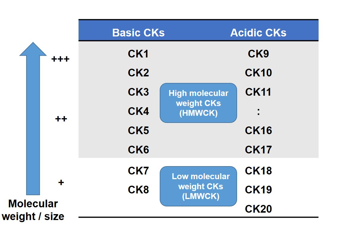
Cytokeratin nomenclature
Images hosted on other servers:

Pancytokeratin positivity in different tumors

Structural model of keratin
Clinical features
- Genetic mutations in keratin genes may cause various keratin disorders known as keratinopathies (Curr Opin Cell Biol 2023;85:102264)
- Some examples are
- Epidermolysis bullosa simplex (mutations in KRT5 and KRT14)
- Keratinopathic ichthyosis (KRT1, KRT2 and KRT10)
- Cryptogenic liver disease (KRT8 and KRT18)
- Steatocystoma multiplex (KRT17)
- Epidermolytic icthyosis and epidermolytic nevi (KRT1 and KRT10)
Interpretation
- Diffuse cytoplasmic reactivity (Mod Pathol 2000;13:962)
- Cytoplasmic dot-like reactivity in neuroendocrine small cell carcinomas and Merkel cell carcinoma (Arch Pathol Lab Med 2010;134:1711)
- Distribution of most common CKs in normal epithelia (Histopathology 2002;40:403)
- Low molecular weight CKs (LMW CKs)
- CK8 / CK18: first to appear during embryogenesis and present in hepatocytes, pancreatic acini, proximal renal tubules and the adrenal cortex
- Secondary CKs (e.g., CK19, CK7 and CK20) expressed alongside CK8 / 18 in most simple and complex epithelia (including urothelium, mesothelium and nonkeratinizing squamous epithelium
- CK17 prominent in basal cells of complex epithelia, urothelium and pilosebaceous units)
- High molecular weight CKs (HMW CKs)
- CK5 / 14 primary CKs in basal cells of squamous and complex epithelia
- CK4 / CK13 appear during maturation in nonkeratinizing squamous epithelium
- CK5 pairs with CK13 in urothelium
- Low molecular weight CKs (LMW CKs)
Uses by pathologists
- Demonstration or exclusion of epithelial differentiation in tumors with unclear morphological differentiation, metastasis or cancer of unknown primary (Hum Pathol 2013;44:1195)
- Depending on the diagnostic question, pathologists can use
- Cocktails of CKs (see below) for screening purposes or to prove the epithelial differentiation of a neoplasia (sensitive but not specific)
- Single antibodies for specific CK (commonly in combination with other markers) are used to identify a possible tissue of origin of a carcinoma (e.g., CK20+ / CK7- for colorectal carcinoma)
- In sentinel lymph node biopsy to screen for the presence of carcinoma cells (Appl Immunohistochem Mol Morphol 2001;9:297)
- In tumor bed area after neoadjuvant treatment to screen for isolated carcinoma cells (Arch Pathol Lab Med 2009;133:633)
- Monitor tumor progression (Int J Biol Markers 1994;9:63)
- Many cocktails or antibody combinations are available for routine diagnosis (Actas Dermosifiliogr 2013;104:181)
- AE1: all acidic CKs (except CK9, CK12, CK17, CK18)
- AE3: all basic CKs (CK1 - CK8)
- AE1 / AE3 (pancytokeratin): AE1 + AE3, broad spectrum CK cocktail
- MNF116 (PANK), OSCAR: similar to AE1 / AE3
- CK22: CK1 - CK19
- 34βE12 (CK903): CK1, CK5, CK10, CK14 (high molecular weight CK, HMWCK)
- CAM 5.2: CK8 and CK18
- CK5/6: HMWCK
Microscopic (histologic) images
Contributed by Umberto Maccio, M.D., M.Sc., M.B.A.
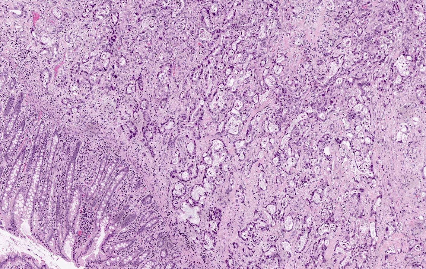

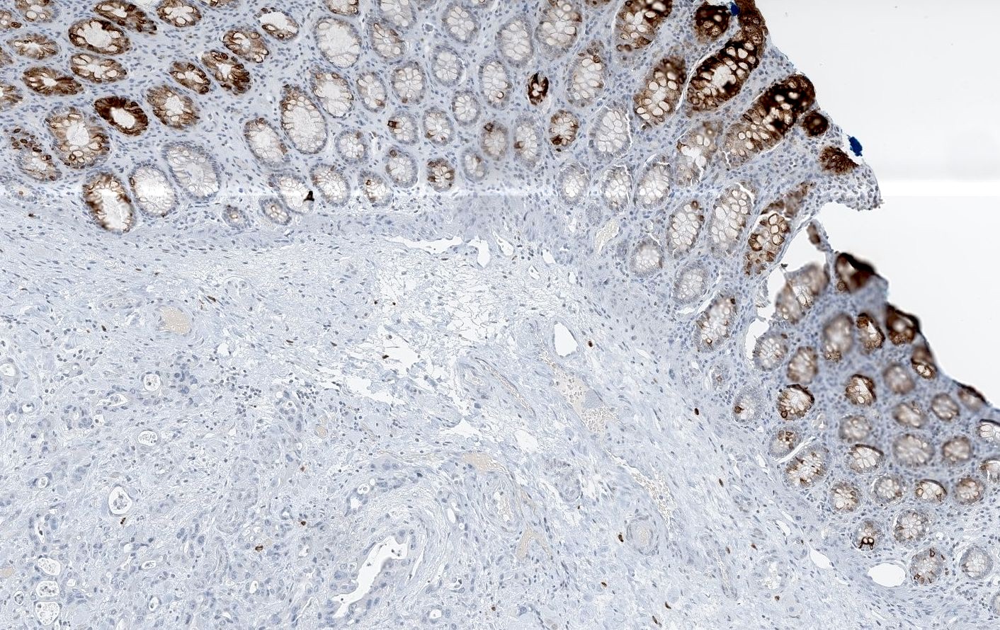
Pancreatic adenocarcinoma
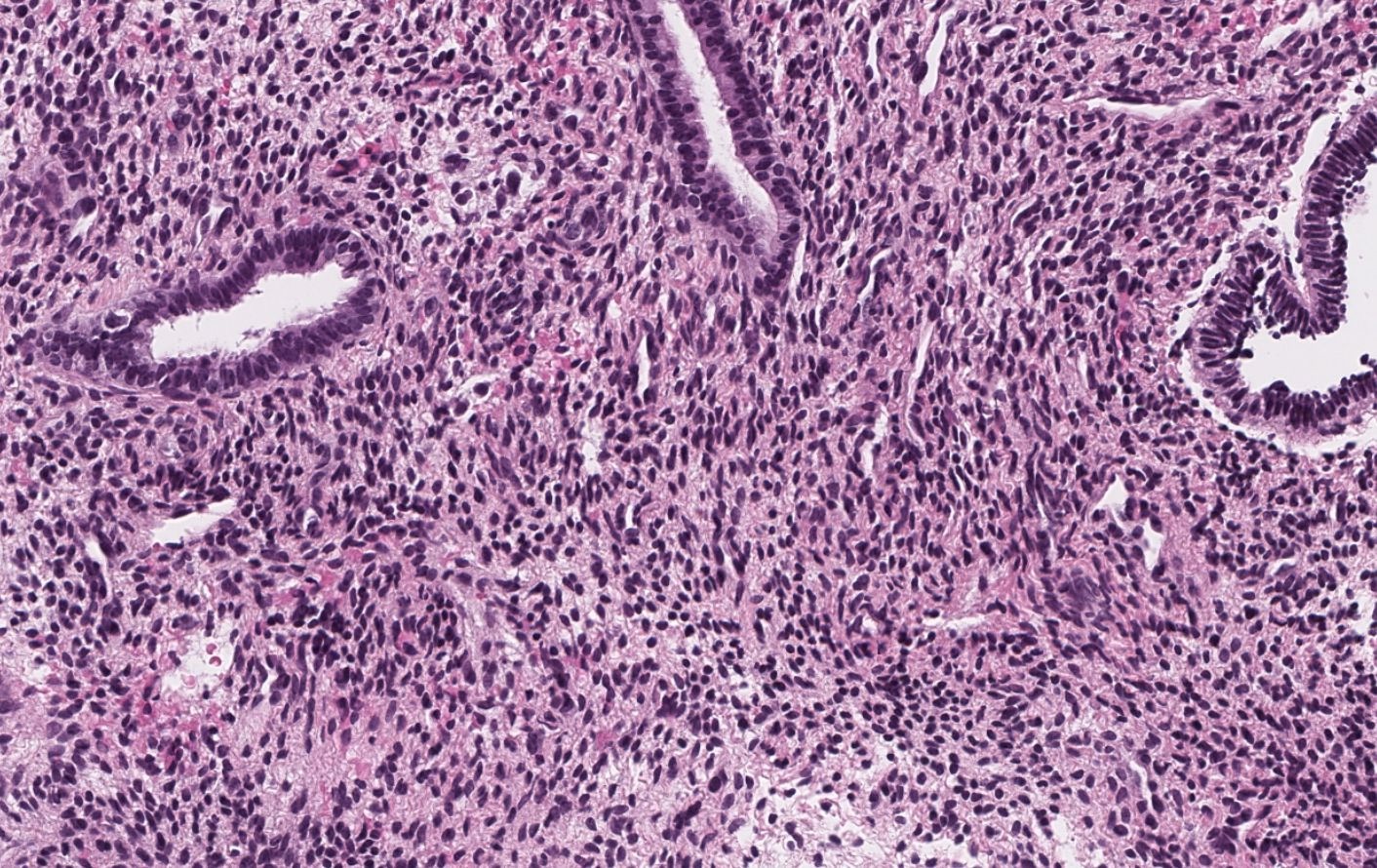
Endometrium metastasis of lobular carcinoma
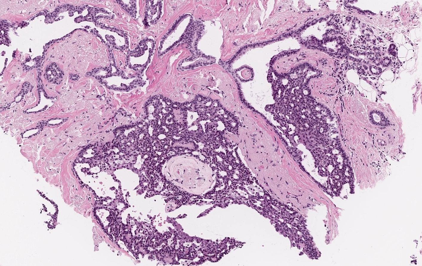
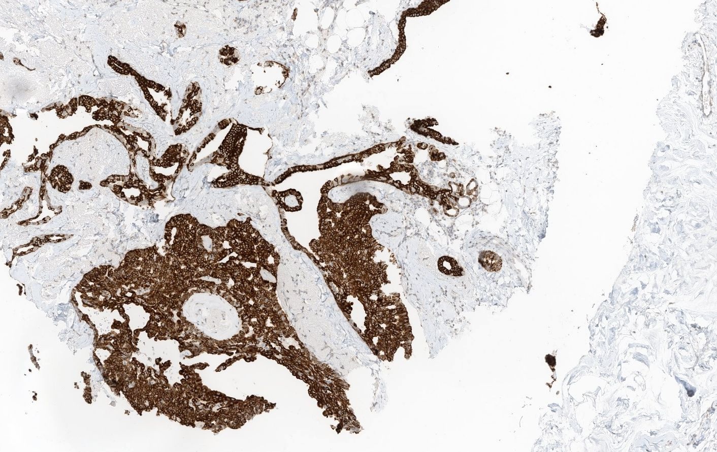
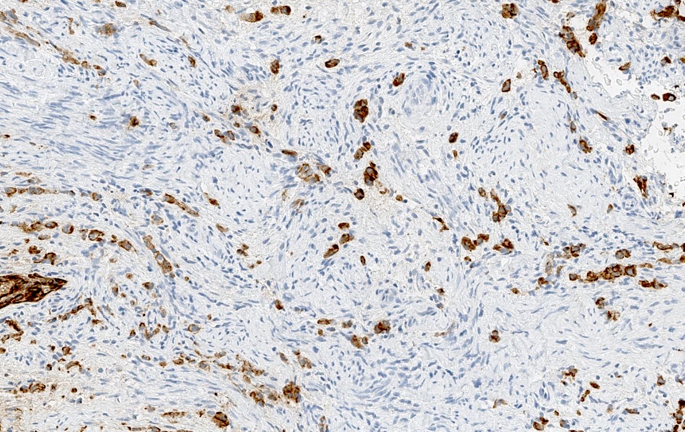
Usual ductal hyperplasia (UDH)
Positive staining - normal
- Epithelial cells (J Cell Sci 2022;135:jcs260594)
- Mesothelium (Int J Clin Exp Pathol 2011;4:631)
- Occasional pancytokeratin positive spindle shaped myofibroblasts in multiple organs, especially in case of degenerative or chronic inflammatory conditions (e.g., media of the aorta, muscular wall of the gallbladder, placental stroma, ovary in the vicinity of a corpus luteum) (Int J Surg Pathol 2023;31:927)
Positive staining - disease
- Carcinomas (Cancers (Basel) 2018;10:108)
- Positive in tumors with epithelial differentiation, which can be identified with electron microscopy, even in tumors that are not classified strictly as carcinomas) (Histopathology 2014;64:101)
- Synovial sarcoma
- Epithelioid sarcoma
- Epithelioid angiosarcoma
- Rhabdomyosarcoma (more common in alveolar type)
- Leiomyosarcoma
- Mesothelioma
- Nonseminoma germ cell tumors (embryonal carcinoma, yolk sac tumor)
- Chordoma
- Thymoma
- Some gliomas and reactive astrocytes (due to crossreactivity between AE3 and GFAP, as in schwannoma) (Virchows Arch 1997;431:139, Mod Pathol 2006;19:115)
Negative staining
- Some carcinomas (Cancers (Basel) 2018;10:108)
- Poorly differentiated carcinomas
- Spindle cell squamous cell carcinomas
- Adrenocortical carcinoma (negative in 84% of cases) (Hum Pathol 2015;46:1799)
- Hepatocellular carcinoma (negative in 72% of cases) for AE1 / AE3 (as hepatocytes are CK18+, which is omitted in AE1 / AE3) (Arch Pathol Lab Med 2014;138:1583)
- Typically nonepithelial tumors
- Melanoma (caveat: aberrant expression in 1 - 40% of cases depending on cocktail, especially in cases with epithelioid morphology) (Dermatopathology (Basel) 2021;8:359)
- Hematological neoplasms (caveat: aberrant expression in 1.5% in many lymphomas and leukemia, with up to 26% of positivity in mantle cell lymphoma) (Pathol Res Pract 2008;204:569)
- Sarcomas (CK positivity in some types) (see Positive staining - disease)
Sample pathology report
- Sentinel lymph node, left axilla, biopsy:
- Micrometastasis of invasive breast carcinoma (see comment)
- Comment: Lymph node tissue contains atypical cells, which demonstrate immunopositivity for the cytokeratin cocktail AE1 / AE3. These cells are arranged in a cluster measuring up to 1.5 mm, consistent with a diagnosis of micrometastasis. For definitive tumor staging, please refer to the report on the segmentectomy performed concurrently.
Additional references
Practice question #1
An endometrial biopsy showed atypical isolated cells in endometrial stroma. Immunohistochemistry for AE1 / AE3 is shown in the picture above. Which of the following statements is correct?
- AE1 / AE3 positivity in the atypical cells is diagnostic for carcinoma
- AE1 / AE3 positivity in the atypical cells is suggestive of carcinoma, although other markers and correlation with the clinical context are required for a definitive diagnosis
- AE1 / AE3 positivity rules out a hematological neoplasia
- AE1 / AE3 positivity rules out a sarcoma
Practice answer #1
B. AE1 / AE3 positivity in the atypical cells is suggestive of carcinoma, although other markers and correlation with the clinical context are required for a definitive diagnosis. AE1 / AE3 positivity, especially when combined with morphological features indicative of carcinoma, is suggestive for this diagnosis but this must be interpreted within the appropriate clinical context (e.g., the image shown above is a metastatic lobular breast carcinoma to the endometrium; therefore, the AE1 / AE3 positive cells could support a diagnosis consistent with metastasis). Answer A is incorrect because AE1 / AE3 positivity can be observed in many other tumors (e.g., some sarcomas or melanomas) so the diagnosis of carcinoma can only be made if the morphology and the clinical context are appropriate and should be done with great caution in cases of cancer of unknown primary. Answer C is incorrect because AE1 / AE3 can be aberrantly expressed in some lymphomas. Answer D is incorrect because AE1 / AE3 can be expressed in various sarcomas.
Comment Here
Reference: Cytokeratins (CK) - general
Comment Here
Reference: Cytokeratins (CK) - general
Practice question #2
Which of the following best describes the structure of cytokeratins?
- Cytokeratins are composed of microfilaments and are responsible for cell motility
- Cytokeratins are intermediate filaments and each cytokeratin dimer has a tridomain structure consisting of a head, a central rod and a tail domain
- Cytokeratins belong to the family of microtubules and provide tensile strength to cells
- Cytokeratins are essential components of the extracellular matrix, playing a role in cell adhesion
Practice answer #2
B. Cytokeratins are intermediate filaments and each cytokeratin dimer has a tridomain structure consisting of a head, a central rod and a tail domain. Cytokeratins are classified as intermediate filaments, not microfilaments or microtubules. They have a characteristic dimer structure which is essential for their function in providing structural support and maintaining cellular integrity. Unlike microflaments, which are involved in cell motility, cytokeratins primarily contribute to the mechanical stability of cells, especially in epithelial tissues. Answer A is incorrect because microfilaments, not cytokeratins, are primarily responsible for cell motility. Answer C is incorrect because cytokeratins are not microtubules but intermediate filaments. Answer D is incorrect because cytokeratins are cytoskeletal components, not extracellular matrix proteins.
Comment Here
Reference: Cytokeratins (CK) - general
Comment Here
Reference: Cytokeratins (CK) - general
Practice question #3
Cytokeratins (CKs) are classified based on their molecular weight and isoelectric pH. Which of the following statements accurately describes the classification of CKs?
- CKs are divided into 2 groups: basic CKs (type B / class II) and acidic CKs (type A / class I) and they always form homodimers
- CKs are divided into 2 groups: basic CKs (type B / class II) and acidic CKs (type A / class I) and they form obligate heterodimers
- CKs are divided based on their expression in carcinomas versus sarcomas
- CKs are classified solely based on their tissue of origin
Practice answer #3
B. CKs are divided into 2 groups: basic CKs (type B / class II) and acidic CKs (type A / class I) and they form obligate heterodimers. CKs are categorized into basic and acidic groups according to their molecular weight and isoelectric points. Rather than forming homodimers, CKs form obligate heterodimers, with each pair consisting of one acidic CK and one basic CK. Answer A is incorrect because CKs do not form homodimers, as they are obligate heterodimers and homodimers of cytokeratins do not exist. Answer C is incorrect because CK classification is not based on their presence in carcinomas or sarcomas but on their molecular characteristics. Answer D is incorrect because CK classification involves molecular weight and pH, not just tissue of origin.
Comment Here
Reference: Cytokeratins (CK) - general
Comment Here
Reference: Cytokeratins (CK) - general