Stains & CD markers
SMAD4 / DPC4


Copyright: 2003-2025, PathologyOutlines.com, Inc.
PubMed Search: SMAD4 DPC4[title]
SMAD4 / DPC4
Editorial Board Member: Catherine E. Hagen, M.D.
Deputy Editor-in-Chief: Raul S. Gonzalez, M.D.
Last author update: 19 January 2021
Last staff update: 18 June 2021
Copyright: 2003-2025, PathologyOutlines.com, Inc.
PubMed Search: SMAD4 DPC4[title]
Table of Contents
Definition / general | Essential features | Terminology | Pathophysiology | Diagrams / tables | Clinical features | Interpretation | Uses by pathologists | Prognostic factors | Microscopic (histologic) description | Microscopic (histologic) images | Positive staining - normal | Positive staining - disease | Negative staining | Sample pathology report | Practice question #1 | Practice answer #1Cite this page: Liao X, Zhang D. SMAD4 / DPC4. PathologyOutlines.com website. https://www.pathologyoutlines.com/topic/stainsdpc4.html. Accessed August 19th, 2025.
Definition / general
- One of the SMAD family of transcription factor proteins, encoded by SMAD4 gene located on chromosome 18q21.1 (Cancer Res 1998;58:3700)
- 552 amino acid polypeptide with a molecular weight of 60.439 Da; has 2 functional domains known as MH1 and MH2
- Tumor suppressor, which inhibits the TGFβ signaling pathway suppressing epithelial cell growth (Cell 2009;136:13)
Essential features
- SMAD4 tumor suppressor gene is mutated in ~ 55% of pancreatic ductal adenocarcinoma, 25% of extrahepatic cholangiocarcinoma, 34% of ampullary carcinoma and 10 - 20% of colorectal carcinomas, including appendix (Cancer Res 2000;60:2002, PLoS One 2017;9:e115383, Mod Pathol 2003;16:272, Oncogene 1999;18:3098)
- Majority of SMAD4 gene mutations in human cancer are missense, nonsense and frameshift mutations at the mad homology 2 region (MH2), which interfere with the homo-oligomer formation of SMAD4 protein and the hetero-oligomer formation between SMAD4 and SMAD2 proteins, resulting in disruption of TGFβ signaling (Mol Cell 2004;15:813)
- Immunohistochemical staining for SMAD4 showing loss of nuclear stain is practically a surrogate for SMAD4 genetic mutation (Am J Pathol 2000;156:37)
Terminology
- SMAD family member 4 or DPC4 (deleted in pancreatic cancer 4)
- Mammalian SMAD4 is a homolog of the Drosophila protein, mothers against decapentaplegic, named Medea
Pathophysiology
- SMAD4 interacts with receptor-regulated SMADs (R-SMADs), such as SMAD2, SMAD3, SMAD1, SMAD5 and SMAD8 (also called SMAD9) to form heterotrimeric complexes (Cold Spring Harb Perspect Biol 2016;8:a022087)
- Once in the nucleus, the complex of SMAD4 and 2 R-SMADs binds to DNA and regulates the expression of different genes depending on the cellular context
Diagrams / tables
Contributed by Xiaoyan Liao, M.D., Ph.D. and Dongwei Zhang, M.D., Ph.D.
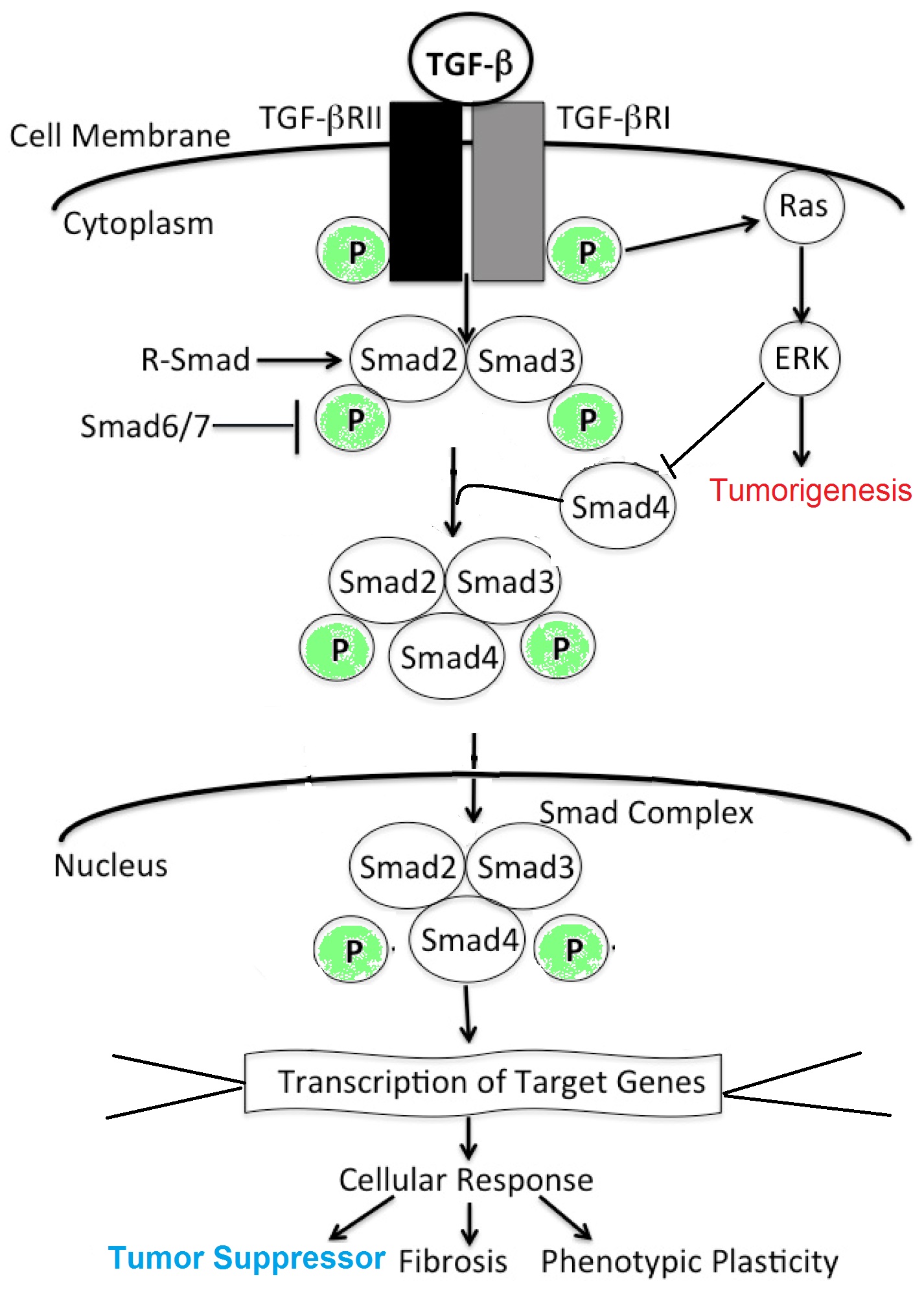
TGFβ / SMAD4
Clinical features
- SMAD4 is also found mutated in the autosomal dominant disease juvenile polyposis syndrome, which is characterized by hamartomatous polyps in the gastrointestinal tract (Science 1998;280:1086)
- Mutations in SMAD4 (mostly substitutions) can cause Myhre syndrome, a rare inherited disorder characterized by mental disabilities, short stature, unusual facial features and various bone abnormalities (Am J Hum Genet 2012;90:161)
Interpretation
- Nuclear stain if SMAD4 protein intact
- Loss of nuclear stain or weak cytoplasmic stain indicate SMAD4 loss and are a surrogate for SMAD4 gene mutation / deletion (Am J Clin Pathol 2001;116:831)
Uses by pathologists
- Loss of SMAD4 nuclear expression combining with other immunomarkers can be used to determine tumor origin in cases of metastasis of unknown primary
- In pancreatic tissue, loss of SMAD4 nuclear expression can distinguish malignancy (in situ or invasive) versus benign process; particularly helpful in biopsies (Am J Clin Pathol 2001;116:831)
- In mucinous carcinoma peritonei associated with a mucinous appendiceal neoplasm, SMAD4 loss can indicate high grade transformation (Am J Surg Pathol 2014;38:583)
- In patients with increased juvenile colon polyps, loss of SMAD4 in those polyps is helpful for making the diagnosis of juvenile polyposis syndrome
Prognostic factors
- In colorectal carcinoma, SMAD4 loss / SMAD4 mutation is associated with higher stage, mucinous differentiation, background Crohn's disease and concurrent RAS mutations (PLoS One 2019;14:e0212142)
Microscopic (histologic) description
- Loss of nuclear expression of SMAD4 in tumor cells in contrast to retained nuclear staining in surrounding stromal cells, lymphocytes or benign epithelial cells
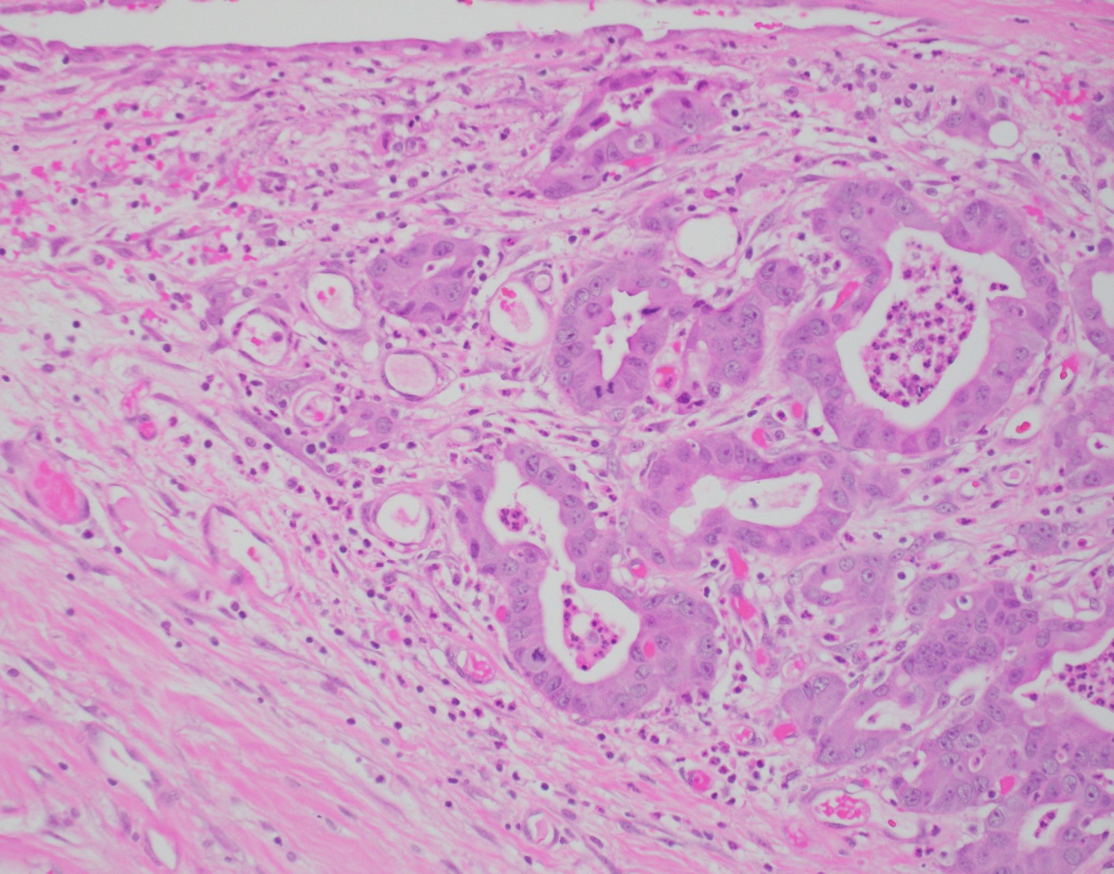
Microscopic (histologic) images
Contributed by Xiaoyan Liao, M.D., Ph.D. and Dongwei Zhang, M.D., Ph.D.
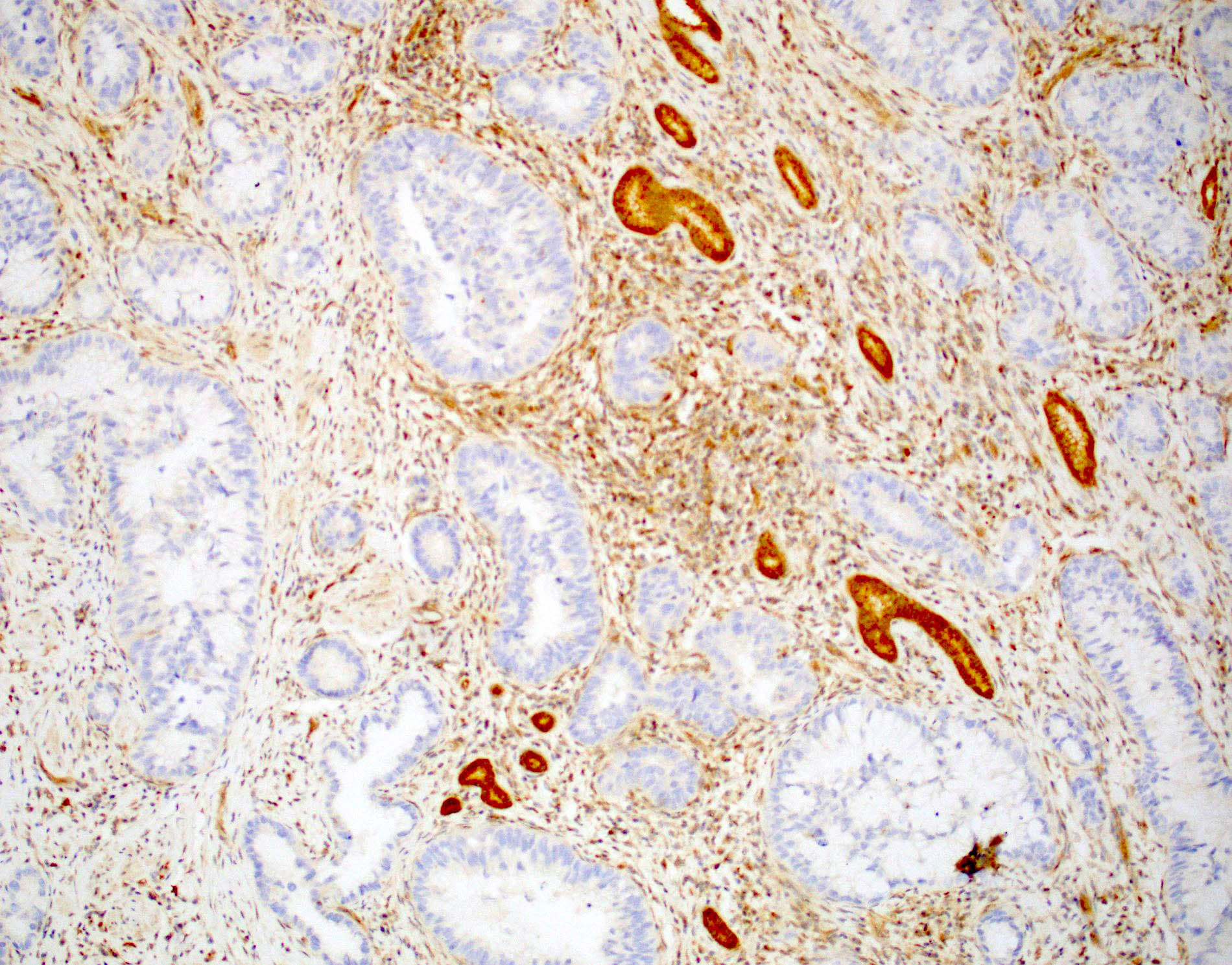
Extrahepatic cholangiocarcinoma
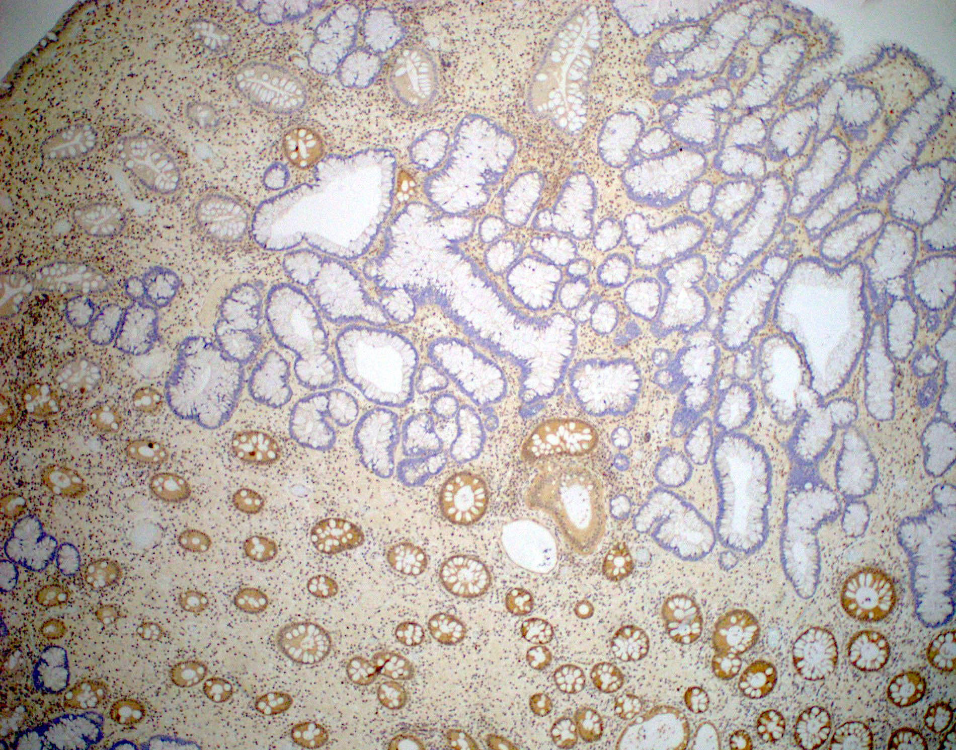
Juvenile polyposis syndrome
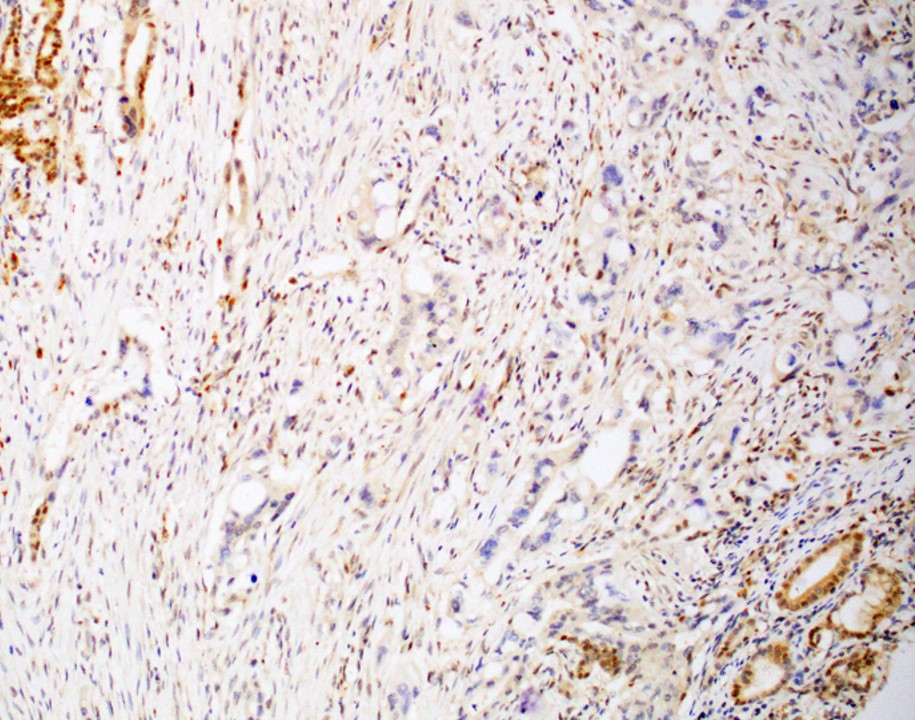
Pancreatic ductal adenocarcinoma
Positive staining - normal
- Nuclear stain in nearly all normal tissue from all organs
Positive staining - disease
- Nuclear stain indicates no SMAD4 protein loss or SMAD4 gene mutation
Negative staining
- Loss of nuclear stain in pancreatobiliary and colorectal carcinomas
Sample pathology report
- Pancreas, mass, biopsy:
- Well differentiated adenocarcinoma.
- SMAD4 immunohistochemistry shows loss of nuclear expression in tumor cells, confirming the diagnosis.
Practice question #1
A patient presents with hernia sac mass, resection of which showed moderately differentiated adenocarcinoma of unknown primary. The patient underwent abdominal CT examination, which showed a possible pancreatic mass. Which immunohistochemical stain result would be most helpful in determining the tumor cells are of pancreatic origin?
- CDX2 negative
- CDX2 positive
- Loss of SMAD4 nuclear stain
- Retained nuclear SMAD4 stain
Practice answer #1