

Testis & paratestis
Nonneoplastic lesions
Granulomatous lesions of testis and paratestis
Author: Sean R. Williamson, M.D.

Last author update: 1 December 2012
Last staff update: 13 February 2024
Copyright: 2002-2024, PathologyOutlines.com, Inc.
PubMed Search: Granulomatous lesions of testis and paratestis
Table of Contents
Definition / general | Infectious causes | BCG therapy | Sperm granuloma | Malakoplakia | Sarcoidosis | Iatrogenic (injectables) | Nonspecific / unknown cause | Gross images | Microscopic (histologic) images | Differential diagnosisCite this page: Williamson SR. Granulomatous lesions of testis and paratestis. PathologyOutlines.com website. https://www.pathologyoutlines.com/topic/testisorchitis.html. Accessed May 13th, 2024.
Definition / general
- Rare; usually men 40 - 59 years with sudden onset of tender testicular mass, variable fever
- May be a response to acid fast products of disintegrated sperm, postinfectious or due to trauma or sarcoidosis
- Resembles pyogenic epididymo-orchitis
- Benign, although granulomatous inflammation may be associated with seminoma
- Recommend cultures to rule out infectious process (brucellosis, leprosy, sarcoidosis, syphilis, TB)
- Granulomatous ischemic lesion
- Usually affects head of epididymis
- May be due to ischemia with secondary granulomatous reaction and scarring
- Gross description
- Solid, unilateral nodular enlargement of testis; resembles lymphoma
- Microscopic (histologic) description
- Lymphocytes and plasma cells infiltrate interstitium and surround seminiferous tubules
- Giant cells and histiocytes that resemble (but are not) actual granulomas
- Granulomatous ischemic lesion
- Zone of necrosis involving efferent ducts and interstitial connective tissue, with adjacent lymphocytes and macrophages
- Macrophages form large clusters with cholesterol crystals and foreign body type giant cells in duct lumen
- Also intratubular epithelial regeneration and proliferation of small ducts showing epithelial regeneration and numerous spermatozoa in their lumen
- Associated with ceroid granuloma, spermatic granuloma and epidermoid metaplasia of the efferent ducts
- Reference: Am J Surg Pathol 1997;21:951
Infectious causes
AIDS
Brucellosis
E. coli related pyogenic epididymo-orchitis
Gonorrhea
Histoplasma capsulatum
Leprosy
Mumps
Syphilis
Tuberculosis
- Associated with markedly reduced spermatogenesis, arrested maturation, germ cell aplasia, tubular hyalinization / thickening of basement membranes, interstitial inflammation and fibrosis, reduction in Leydig cells, Sertoli cell only pattern (J Pathol 1991;163:47, Urology 1999;53:203, Mod Pathol 1989;2:233, Hum Pathol 1989;20:210)
- Often other infections in testis or epididymis (Candida, CMV, Histoplasma, mycobacteria, toxoplasmosis)
- Testicular atrophy related findings do not appear to be immune mediated (Hum Pathol 1989;20:572)
- Testis is an uncommon location for Kaposi sarcoma in AIDS patients
Brucellosis
- Zoonotic infection acquired from sheep, camels, cattle, dogs, goats, reindeer, swine via skin / mucous membrane contact or contaminated animal products
- Affects testis and epididymis (epididymo-orchitis) in 2 - 20% of cases, causing scrotal pain, swelling, fever
- Often diagnosed by laboratory studies
- Case report: 32 year old man with painless testicular mass-brucellosis (Int J Urol 2004;11:683)
- Micro description: granulomatous or testicular abscess
- Treatment: antibiotics; orchiectomy if resembles a neoplasm or refractory to therapy
- Additional references: Urol Int 2009;82:158, BMC Res Notes 2011;4:286, Clin Infect Dis 2001;33:2017
E. coli related pyogenic epididymo-orchitis
- Usually due to E. coli
- Resembles granulomatous orchitis
- Complications: venous thrombosis, septic testicular infarct
Gonorrhea
- Usually spreads from posterior urethra to prostate, seminal vesicles and epididymis
- Testis involved only if untreated
Histoplasma capsulatum
- Rarely presents as testicular mass
- May resemble sperm granuloma (J Clin Pathol 1974;27:929)
- Caseating granulomatous inflammation with giant cells
- Small yeast forms (2 - 5 micrometers) are identifiable by silver stain (J Urol 2000;164:1652)
Leprosy
- Does not occur in U.S.
- Rarely presents with orchitis (Am J Clin Pathol 1980;73:712)
- Testicular involvement thought to be facilitated by lower temperature of scrotum
- 3 phases of testicular involvement
- Vascular phase: blood vessels show perivascular lymphocytic inflammation and interstitium is filled with macrophages containing mycobacteria
- Interstitial phase: endarteritis, Leydig cell clusters, interstitial fibrosis, histiocytes containing acid fast bacteria and reduced spermatogenesis
- Obliterative phase: dense fibrosis, no detectable tubules, reduced vessels, rare acid fast bacteria; associated with gynecomastia and infertility
Mumps
- Testicular infections rare in infected children (prepubertal) but occur in 15 - 40% of postpubertal men one week after parotiditis
- Usually unilateral (bilateral in 15 - 30%); epididymitis is also common (85%) and often precedes orchitis
- 33% of infected postpubertal men develop testicular atrophy, 2 - 10% become infertile
- Incidence increasing, due to reduced use of vaccine (BJU Int 2010;105:1060)
Syphilis
- Testis usually involved first
- Discrete gummas contribute to enlarged, irregular testis
- Gummas: diffuse interstitial inflammation with edema, lymphocytes and plasma cells, with obliterative endarteritis and perivascular cuffing
- Spirochetes usually identified in gummatous but not fibromatous stages
Tuberculosis
- Usually begins in epididymis and spreads to testis
- Prostate and seminal vesicles are usually also infected
BCG therapy
[Pending]
Sperm granuloma
[Pending]
Malakoplakia
[Pending]
Sarcoidosis
[Pending]
Iatrogenic (injectables)
[Pending]
Nonspecific / unknown cause
[Pending]
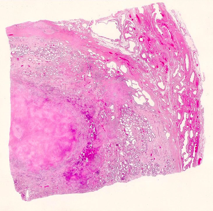
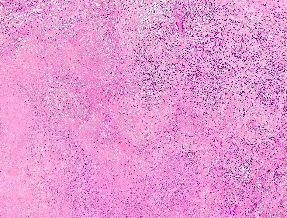
Gross images
Contributed by Yale Rosen, M.D. and @SueEPig on Twitter
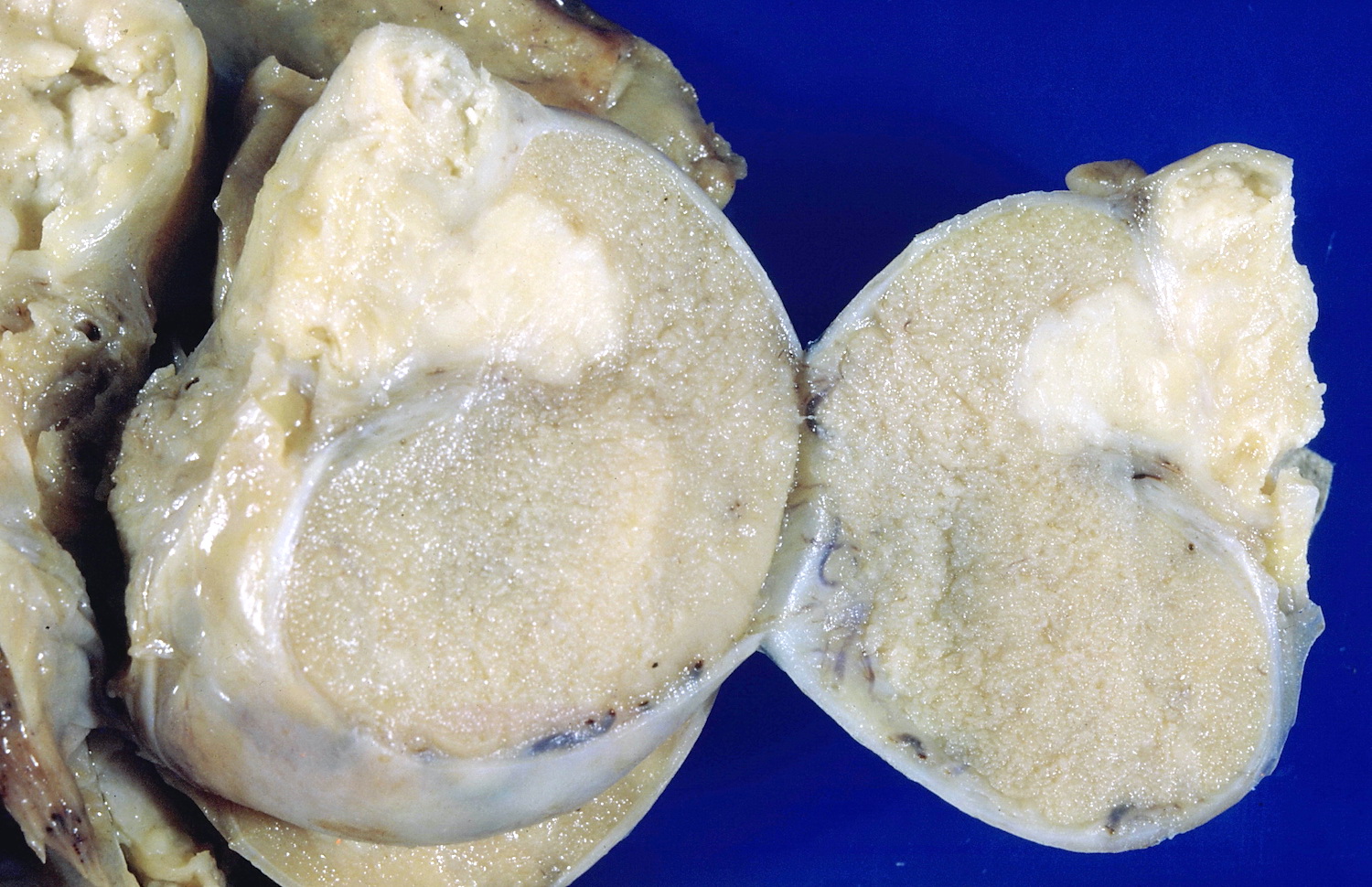
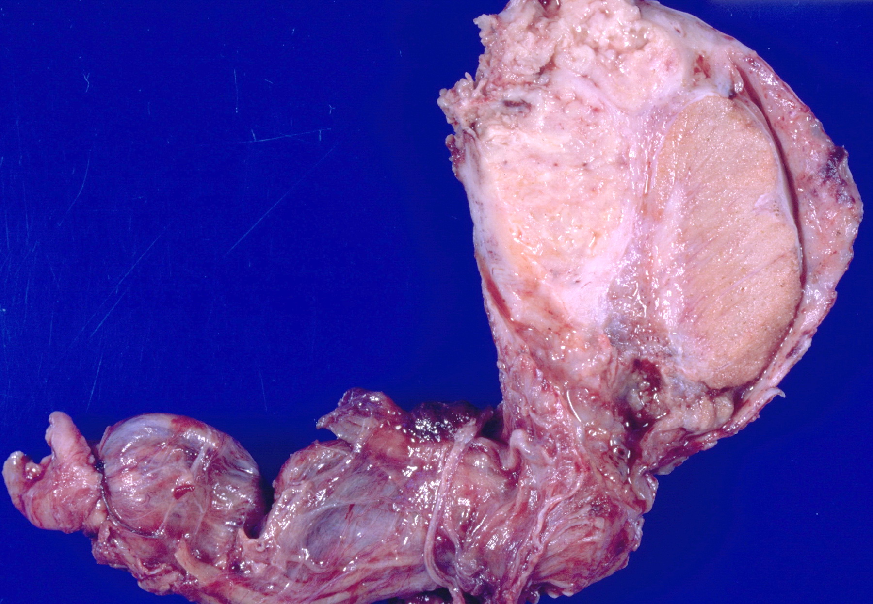
TB orchitis
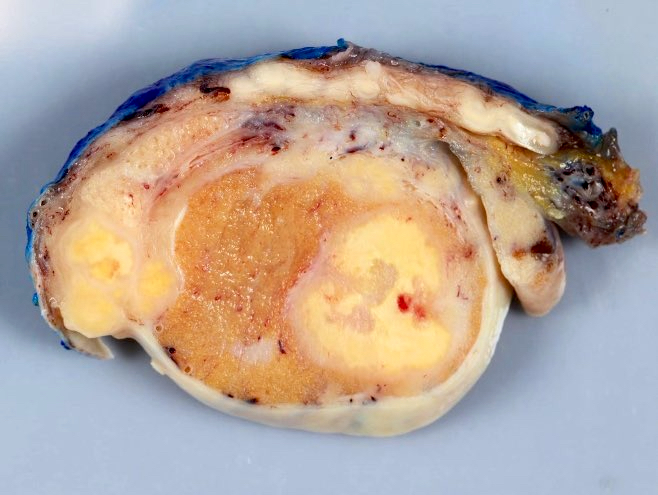
Granulomatous orchitis
Images hosted on other servers:

Brucellosis
Microscopic (histologic) images
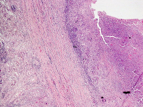
Contributed by Sean R. Williamson, M.D. and @SueEPig on Twitter
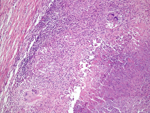
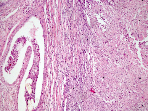
Tuberculosis involving testis and paratestis
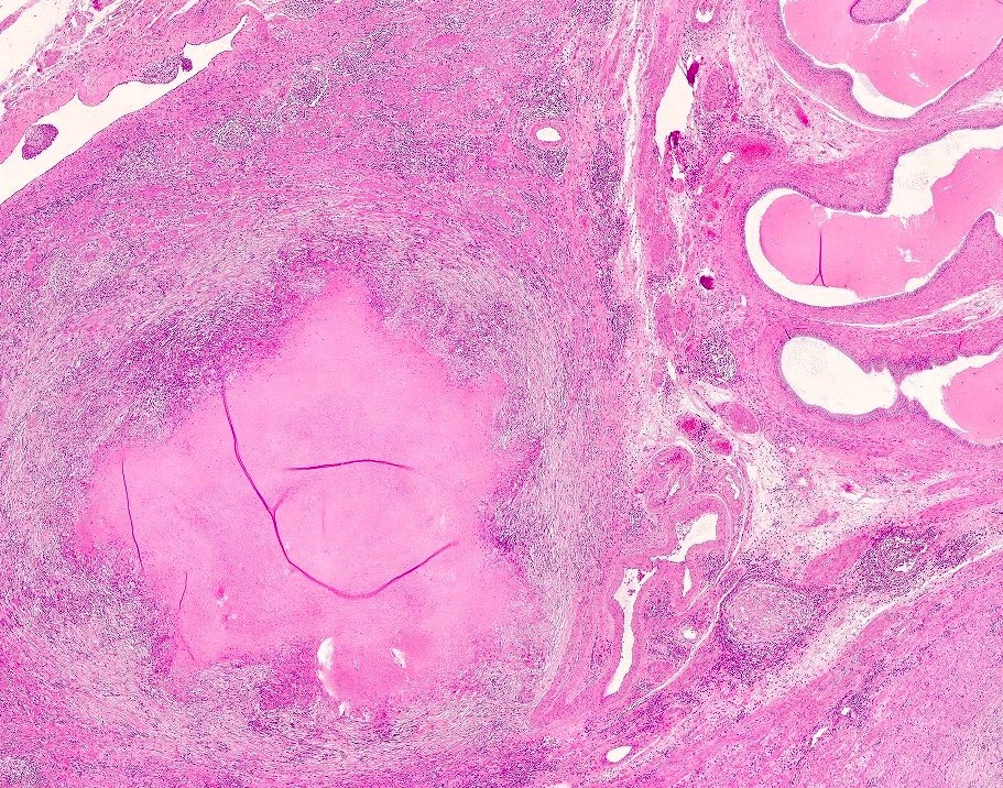
Granulomatous orchitis
Differential diagnosis
- Granulomatous inflammation associated with seminoma
- Infection or inflammation (Hum Pathol 1990;21:1080)
- Lymphoma
