Vulva & vagina
Nontumor
Vulvovaginal cysts
Authors: Morgan Hrones, M.D., Natalia Buza, M.D.
Editorial Board Member: C. Blake Gilks, M.D.
Deputy Editor-in-Chief: Jennifer A. Bennett, M.D.


Last author update: 3 January 2022
Last staff update: 25 April 2024
Copyright: 2002-2025, PathologyOutlines.com, Inc.
PubMed Search: Vulvovaginal cysts
Table of Contents
Definition / general | Essential features | ICD coding | Epidemiology | Sites | Pathophysiology | Clinical features | Diagnosis | Radiology description | Case reports | Treatment | Microscopic (histologic) description | Microscopic (histologic) images | Videos | Sample pathology report | Differential diagnosis | Practice question #1 | Practice answer #1 | Practice question #2 | Practice answer #2Cite this page: Hrones M, Buza N. Vulvovaginal cysts. PathologyOutlines.com website. https://www.pathologyoutlines.com/topic/vaginacysts.html. Accessed September 15th, 2025.
Definition / general
- Benign cysts located within the vulva or vagina
Essential features
- Vulvovaginal cysts may be congenital or acquired
- Congenitally derived vulvovaginal cysts may represent an embryological derivative, a result of an urological abnormality or ectopic tissue
- Cysts of the vagina are relatively common and represent generally benign conditions
- Bartholin gland cyst: often a product of chronic bacterial inflammation; nonsulfated sialomucin secretion product
- Epidermal / epithelial inclusion cyst: lined by squamous epithelium; may occur post surgery or trauma
- Gartner duct cyst: mesonephric origin (rare), non mucin secreting
- Müllerian cyst: lined by mucin secreting columnar cell
- Urothelial cyst: derived from periurethral and Skene glands
- Mammary-like gland cyst
- Cyst of canal of Nuck: mesothelial cyst
- Endometriotic cyst / cystic endometriosis
- Vaginitis emphysematosa: variably sized vaginal nodules that produce a characteristic popping sound
ICD coding
Epidemiology
- Müllerian cyst
- Comprises 30 - 44% of all vaginal cysts (Obstet Gynecol Surv 2021;76:101)
- Gartner duct cyst
- Most commonly identified vaginal cyst in children (Obstet Gynecol Surv 2021;76:101)
- Accounts for 4 - 21% of all vaginal cysts in adult females (Obstet Gynecol Surv 2021;76:101)
- Bartholin gland cyst
- Accounts for 7 - 21% of all vaginal cysts in reproductive age females (Obstet Gynecol Surv 2021;76:101)
- Vaginitis emphysematosa
- Rarely reported (< 200 cases recorded) (Arch Pathol Lab Med 1987;111:746)
- Average age ranges from 42 to 65 years
Sites
- Gartner duct cyst
- Most commonly located along the anterolateral vaginal wall at 11 o'clock and 1 o'clock regions
- May extend deep into the vaginal wall
- Bartholin gland cyst
- Found in the posterolateral inferior third of the vagina
- Urothelial cyst
- Usually situated low in the vagina, close to the urethra due to its urogenital sinus derivation
- Müllerian cyst
- May be found anywhere in the vagina but most commonly found in the vulvar vestibule
Pathophysiology
- Gartner duct cyst
- Mesonephric duct (Wolffian duct) normally forms genital organs in males and regresses in females; a remnant of mesonephric duct sometimes fails to regress and results in the formation of a Gartner duct cyst (JNMA J Nepal Med Assoc 2020;58:505)
- Bartholin gland cyst
- Often results from a blockage of the Bartholin gland due to inflammation, trauma, childbirth or infection (StatPearls: Bartholin Gland Cyst [Accessed 8 December 2021])
- Müllerian cyst
- Formation occurs when a portion of Müllerian epithelium fails to involute during the normal replacement of Müllerian epithelium with squamous epithelium of the urogenital sinus (Differentiation 2018;103:46)
- May show focal squamous metaplasia (Differentiation 2018;103:46)
- Epidermal / epithelial inclusion cyst
- May occur post surgery or trauma
Clinical features
- May present as a painful swollen cystic lesion
- Often asymptomatic
Diagnosis
- Most diagnosed clinically by position and appearance
- If clinical diagnosis remains unclear, further evaluation with transvaginal ultrasound (TVUS) may be employed (Aust N Z J Obstet Gynaecol 2018;58:388)
- Histopathological confirmation in surgically excised specimens
Radiology description
- Ultrasound findings: well defined, unilocular cyst
Case reports
- 29 year old woman with a recurrent cystic mass and an enlarged left labia majora (Case Rep Womens Health 2019;24:e00141)
- 32 year old woman with a chief complaint of pelvic organ prolapse (JNMA J Nepal Med Assoc 2020;58:505)
- 57 year old woman with a bulky appearing left labial mass (Case Rep Womens Health 2019;22:e00115)
Treatment
- Bartholin duct cyst: complete excision or marsupialization with antibiotic administration if necessary
Microscopic (histologic) description
- Bartholin duct cyst
- Lined by residual mucinous epithelium, low cuboidal or transitional epithelium
- May exhibit focal squamous metaplasia or denudation
- Müllerian cyst
- Lined by mucin secreting or ciliated (tubal type) columnar cells
- May show focal squamous metaplasia
- Gartner duct cyst
- Lined by a single layer of cuboidal to low columnar nonmucinous epithelium
- Urothelial cyst
- Lined by transitional or squamous epithelium
- Epidermal / epithelial inclusion cyst
- Lined by squamous epithelium (keratinizing or nonkeratinizing)
- Mammary-like gland cyst
- Ectopic mammary tissue may present as a persistent vulvar cyst (World J Surg Oncol 2009;7:70)
- Cyst of canal of Nuck
- Lined by mesothelial cells
- Endometriotic cyst / cystic endometriosis
- Cystic endometrial type glands surrounded by variable amount of endometrial stroma and hemosiderin laden macrophages
- Vaginitis emphysematosa
- Variably sized cysts with pink, hyaline-like material, foreign body giant cells and chronic inflammatory infiltrate (Arch Pathol Lab Med 1987;111:746)
Microscopic (histologic) images
Contributed by Morgan Hrones, M.D. and Natalia Buza, M.D.
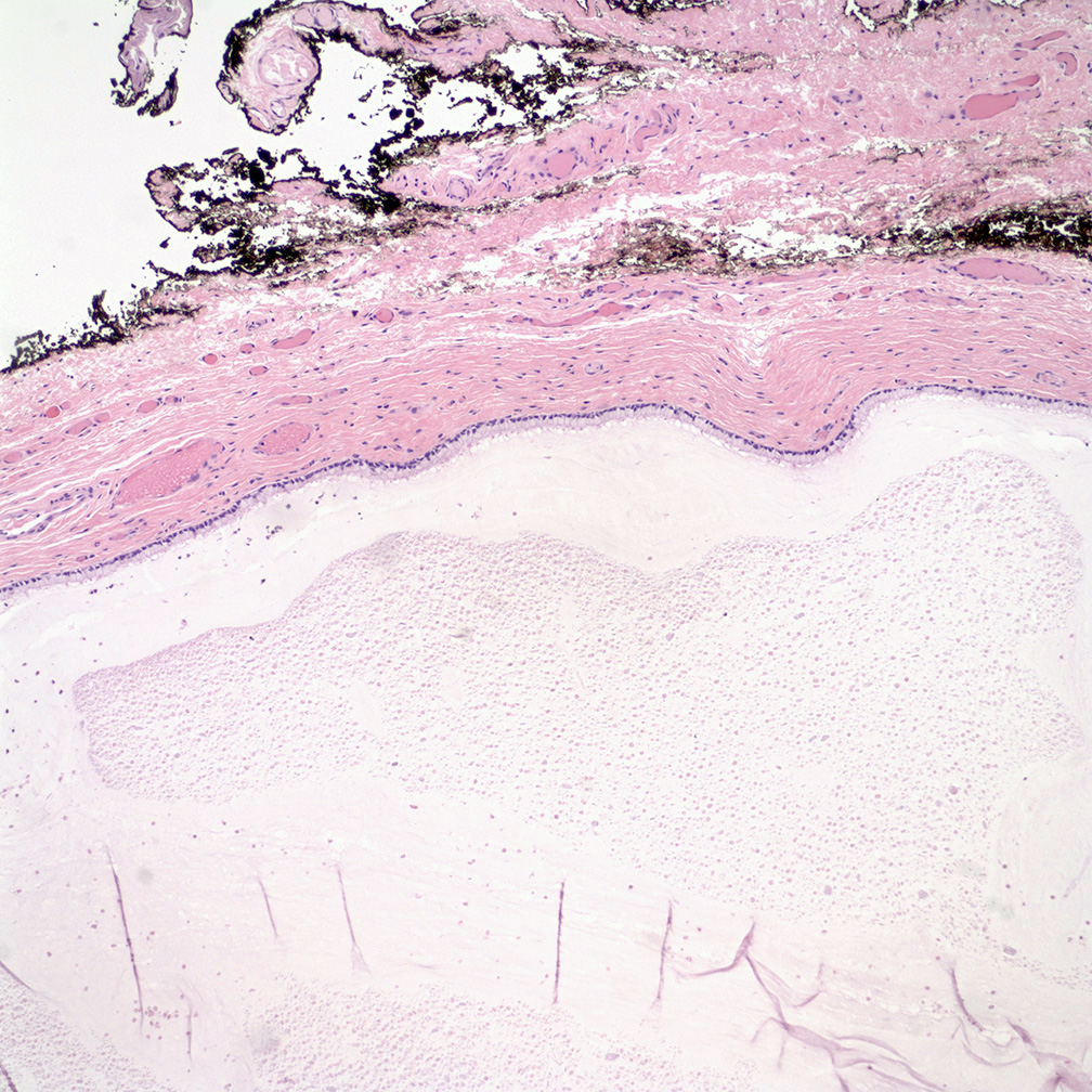
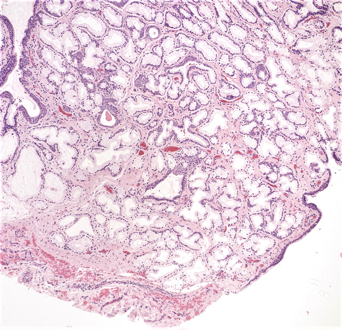
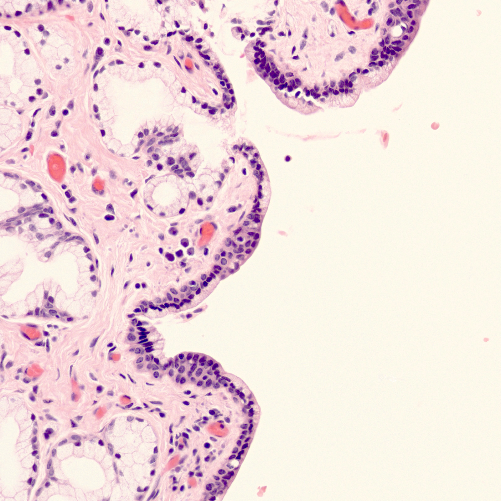
Mucinous cystic lining
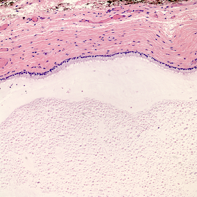
Bland appearing cyst lining
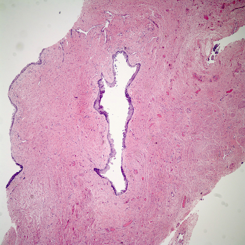
Benign urothelial cyst
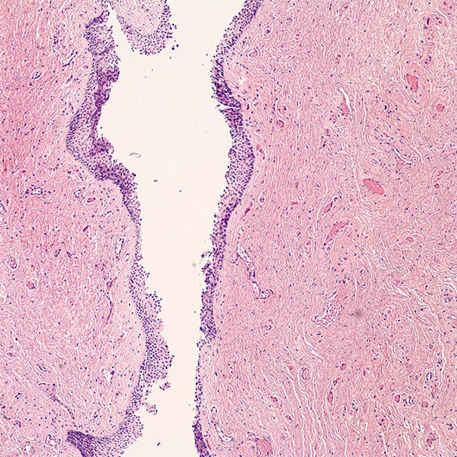
Benign urothelial lined cyst
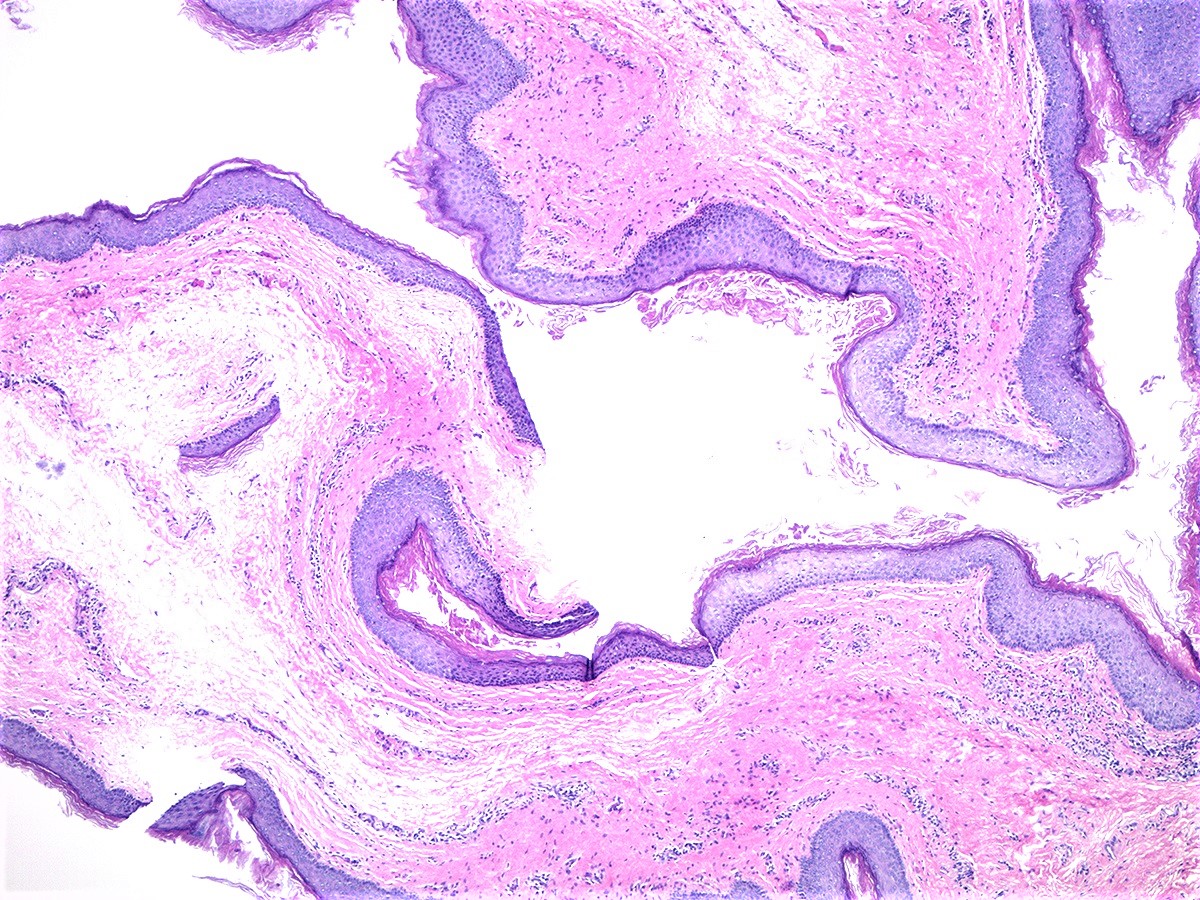
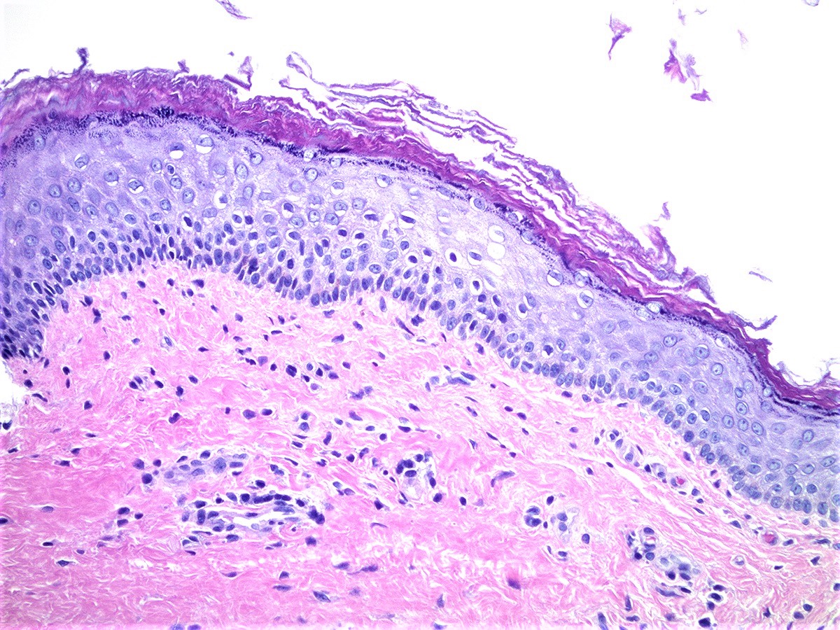
Benign epidermal inclusion cyst
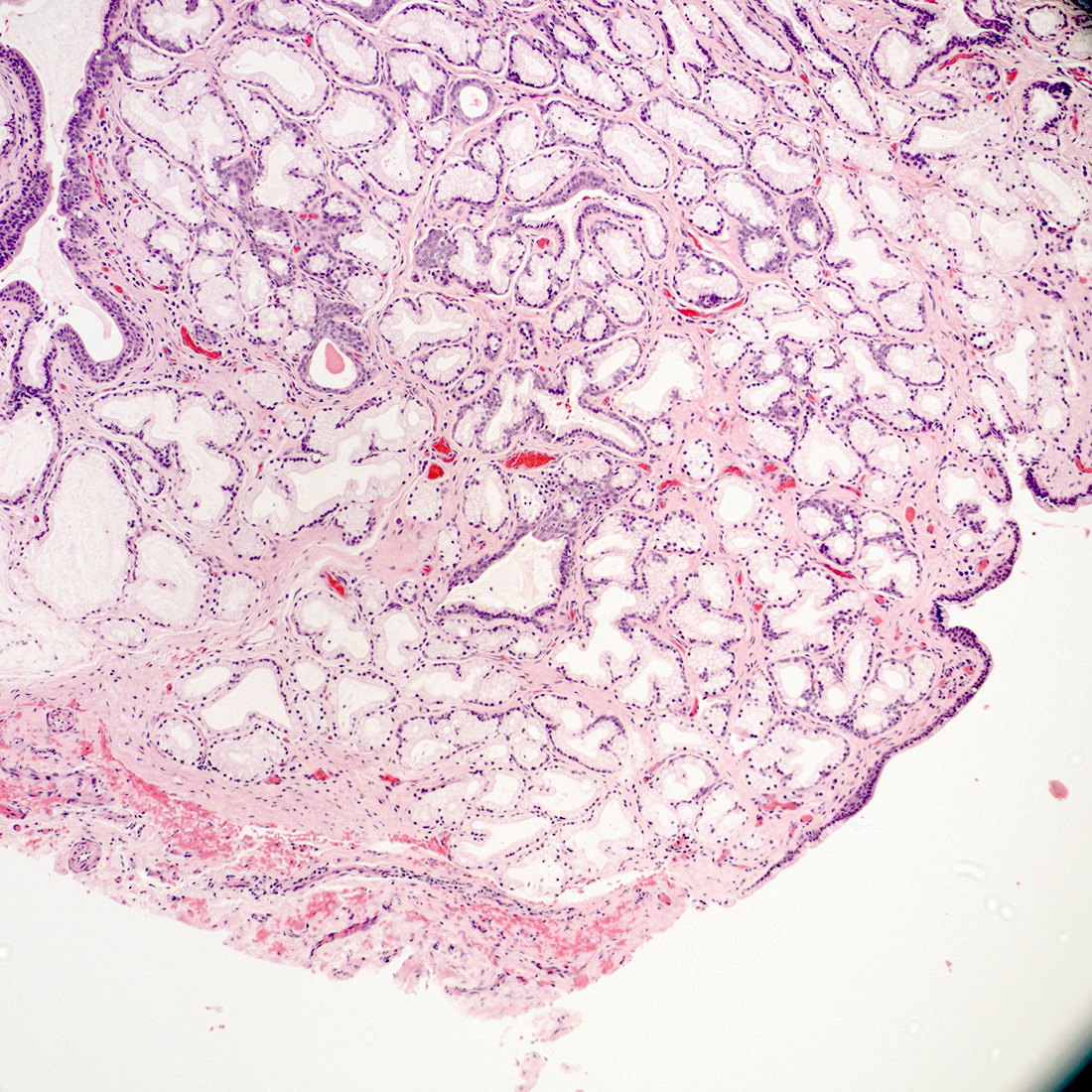
Bartholin cyst with adjacent Bartholin glands
Bartholin cyst
Videos
Bartholin gland cyst of the vulva - histopathology
Sample pathology report
- Vulva, right posterior, cystectomy:
- Bartholin gland cyst (2 cm)
Differential diagnosis
- Urethral diverticulum:
- Communication with urethra detected by MRI (AJR Am J Roentgenol 1993;161:809)
- Diagnostic confirmation at the time of surgery
Practice question #1
A 42 year old woman presents with a painful cystic swelling along the right posterolateral vulva, near the vaginal introitus. The cyst is excised and sent to pathology, where a benign appearing cystic lesion lined by simple cuboidal epithelium with areas of denudation and surrounding inflammation are seen. These findings, along with the image shown above, best describe which of the following vulvovaginal cysts?
- Bartholin cyst
- Epidermal inclusion cyst
- Gartner duct cyst
- Urothelial cyst
Practice answer #1
Practice question #2
Which of the following represents a physiological remnant of the mesonephric duct in adult females?
- Bartholin cyst
- Epidermal inclusion cyst
- Gartner duct cyst
- Urothelial cyst
Practice answer #2
