

Images hosted on other servers:

Left sided appendicitis

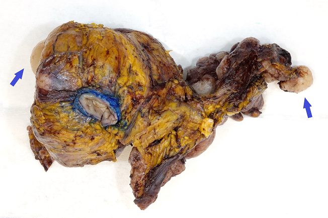
Acute appendicitis as inflammatory mass
Images hosted on other servers:

Laparoscopic appendectomy
Contributed by Elliot Weisenberg, M.D. and Andrey Bychkov, M.D., Ph.D.
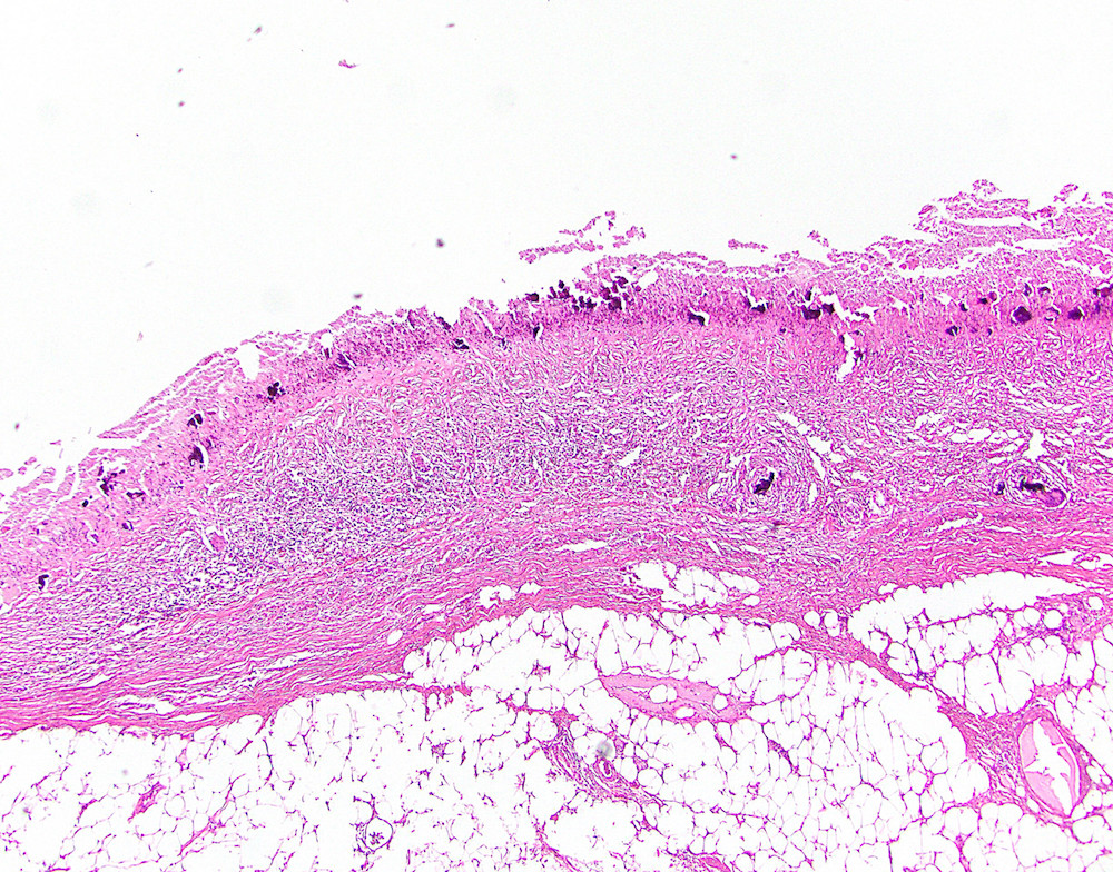
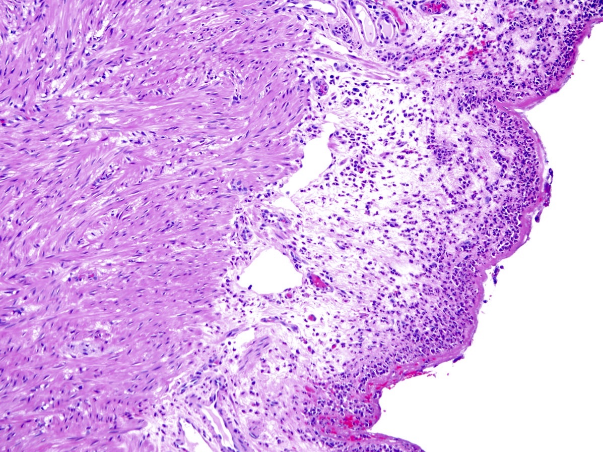
Fibrinopurulent exudate
Mucosal ulceration
Severe disease
Dark mucosa
Contributed by Qingqing Liu, M.D., Ph.D.
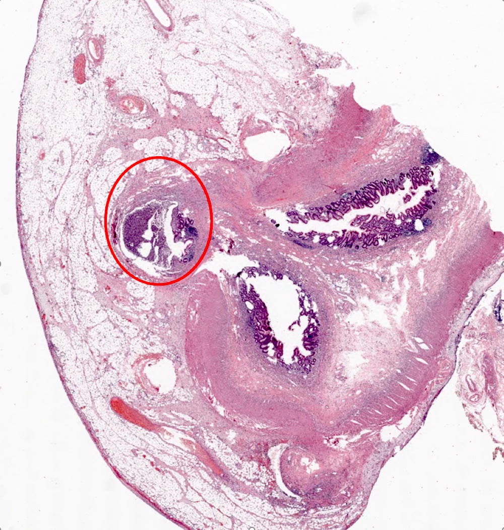
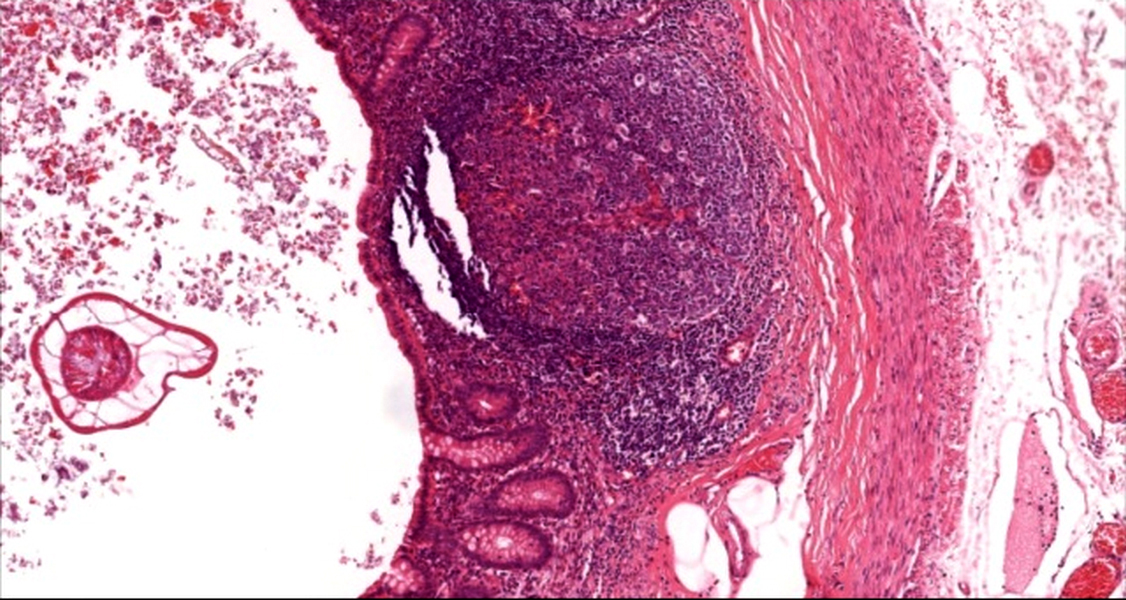
Appendiceal diverticulitis
Appendiceal endometriosis
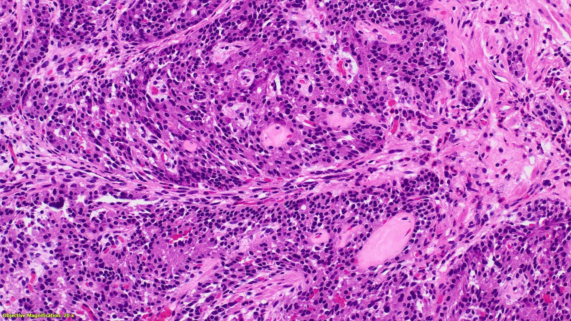
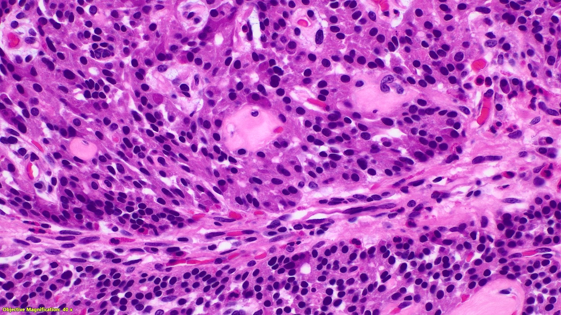
Contributed by Raul S. Gonzalez, M.D. and Elliot Weisenberg, M.D.
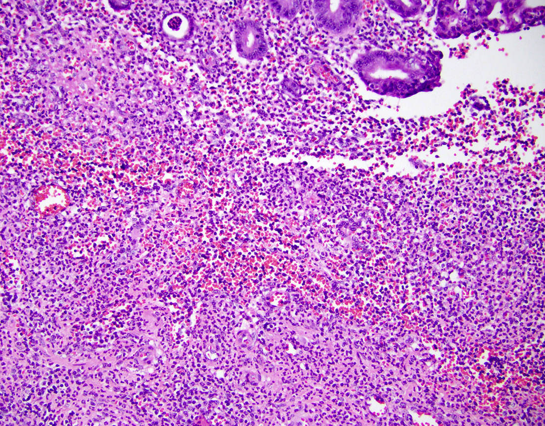
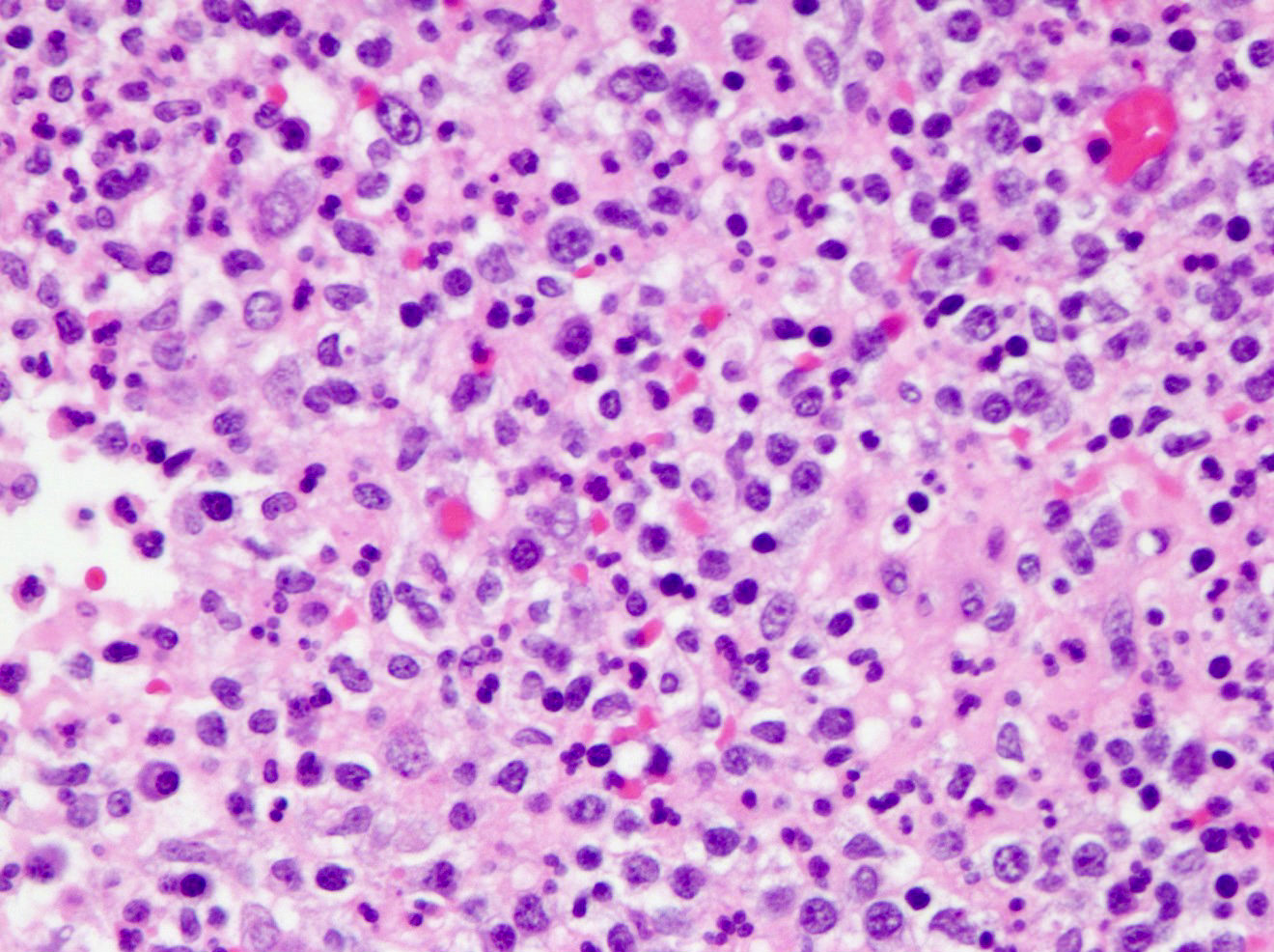
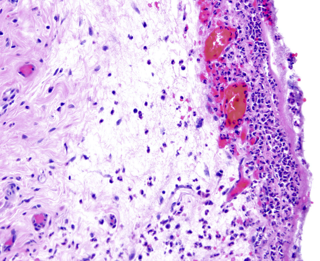
Marked neutrophilic infiltration
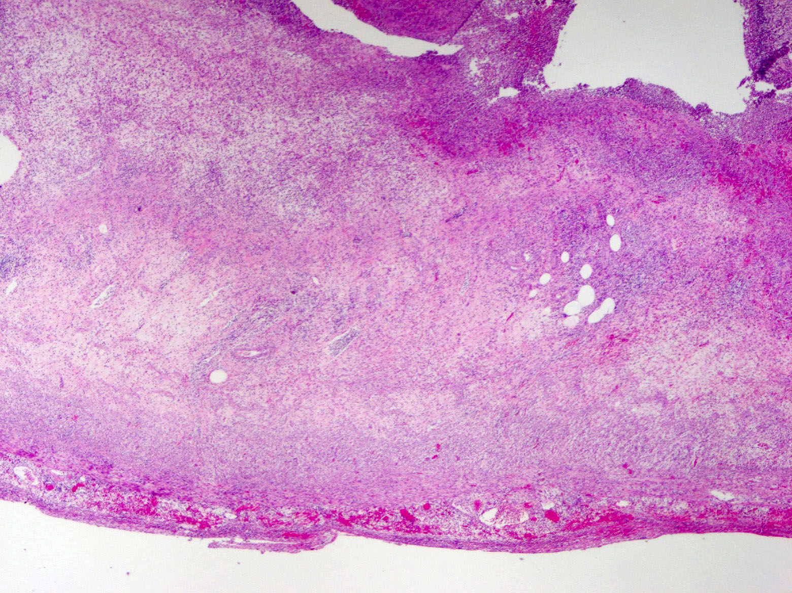
Transmural inflammation
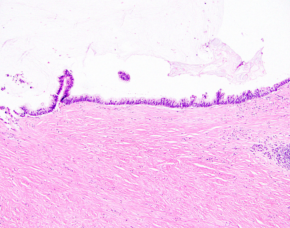
Intraluminal neutrophils
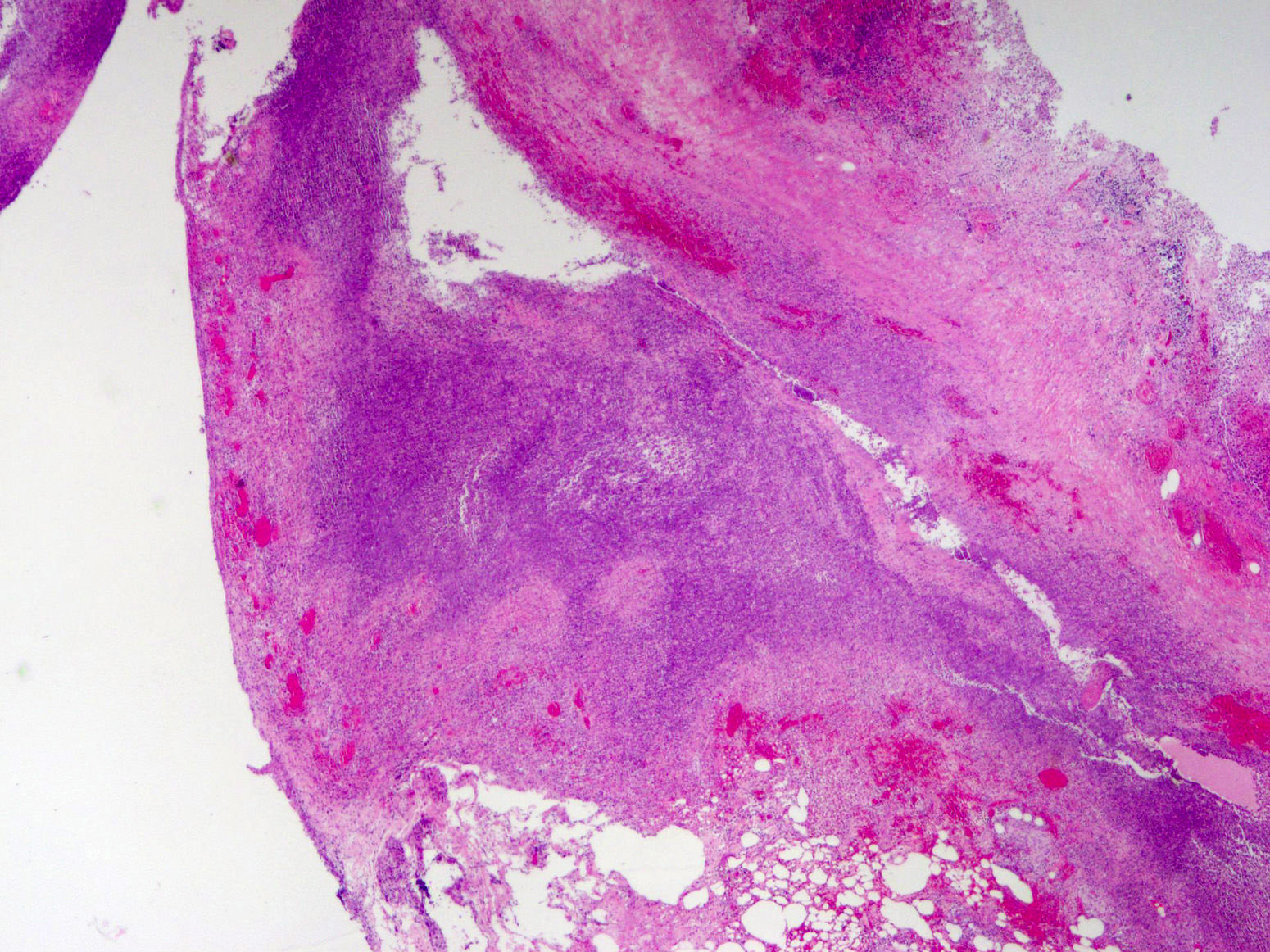
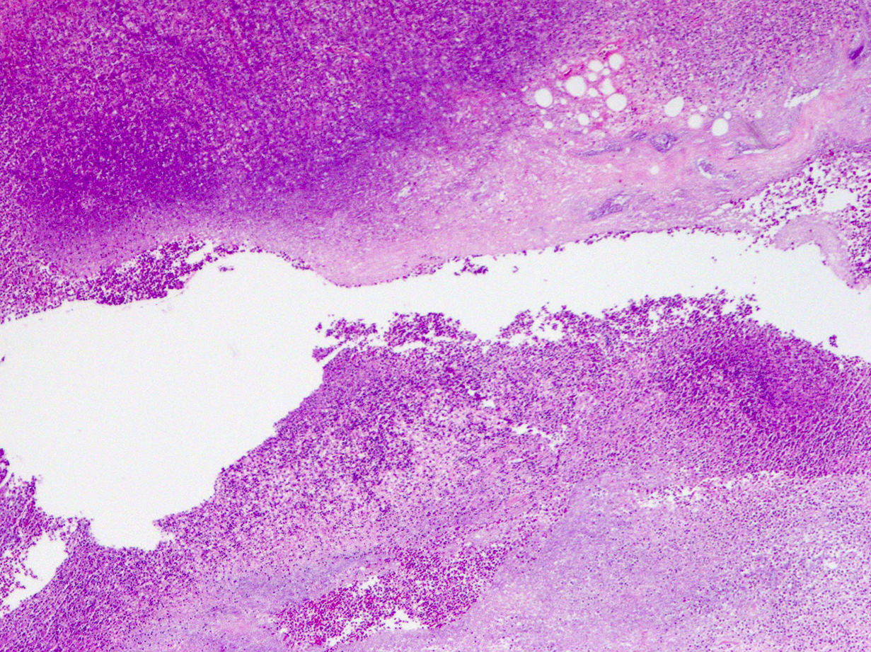
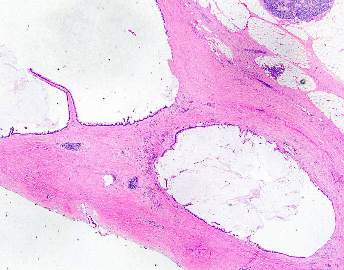
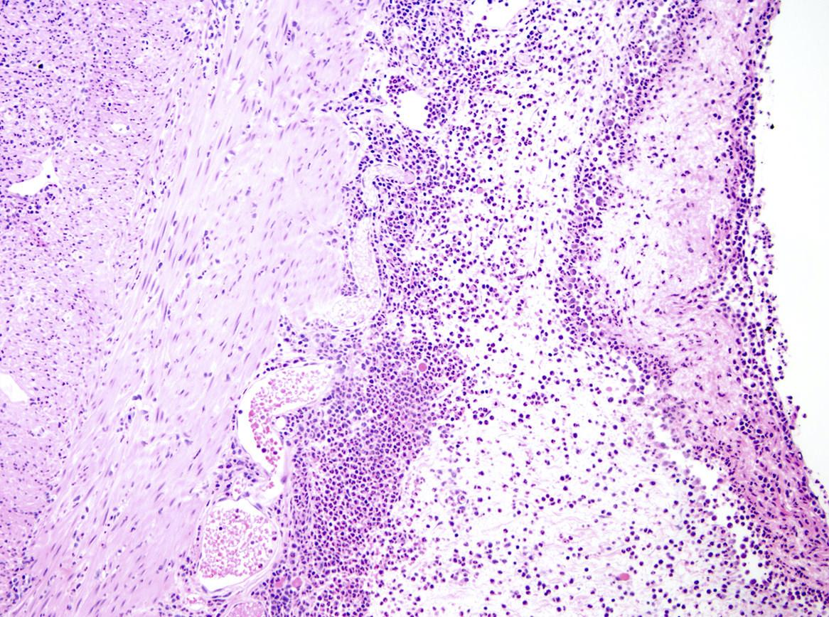
Periappendiceal abscess
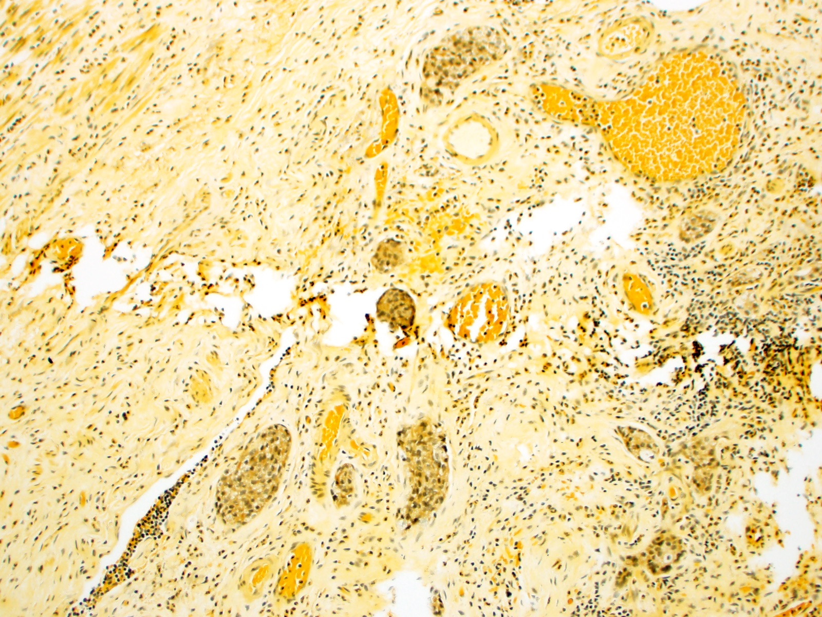
Xantho-granulomatous
inflammation
Contributed by Bella Lingjia Liu, M.D.
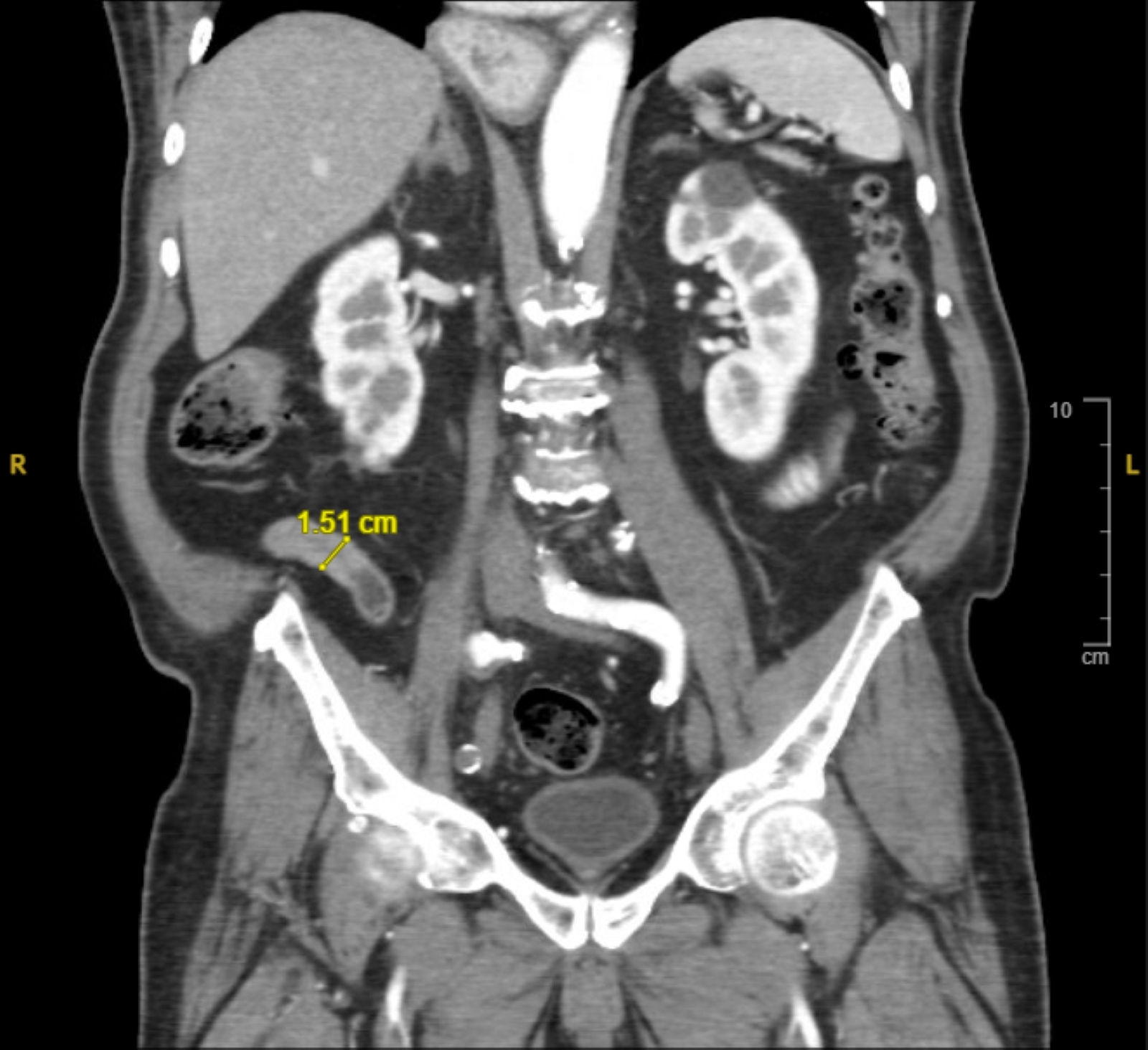
CT of abdomen
Images hosted on other servers:

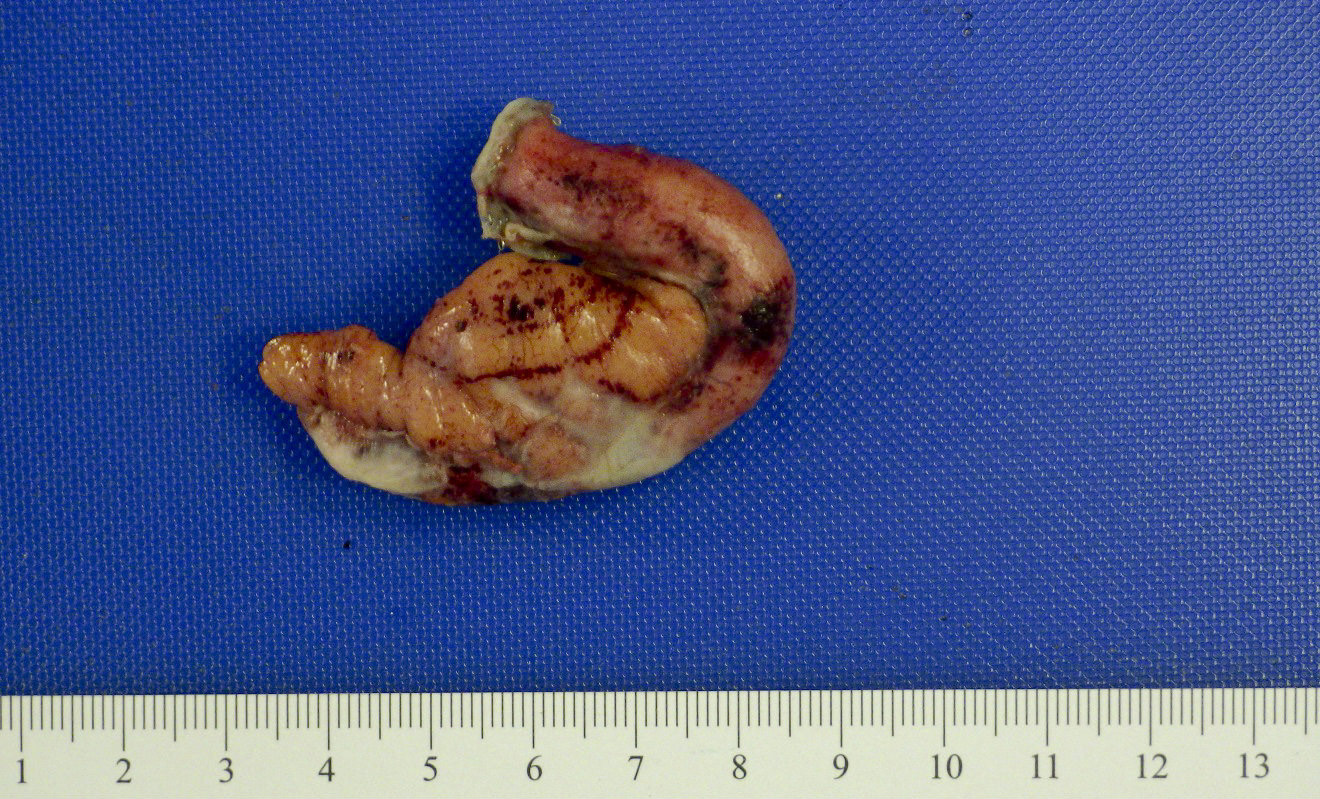
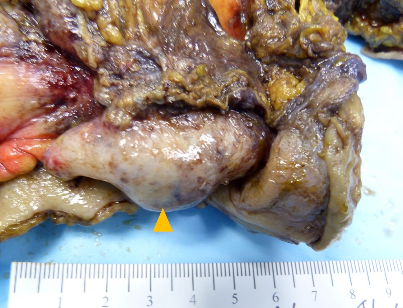
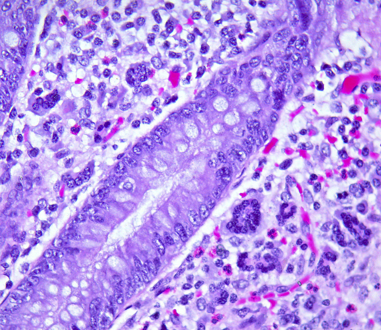
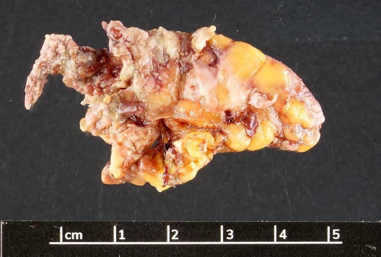
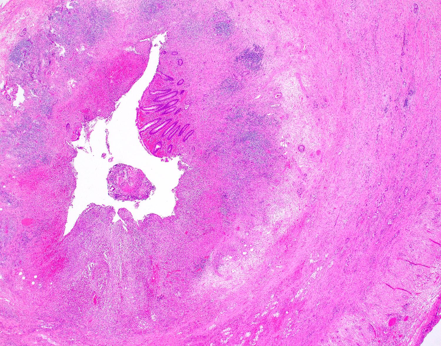
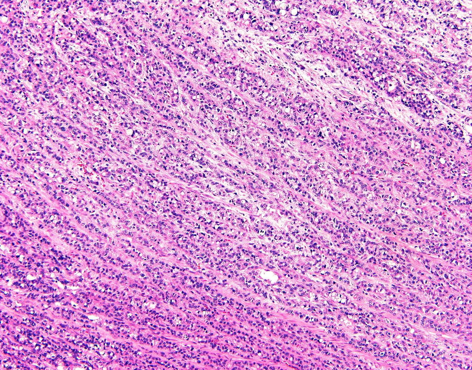
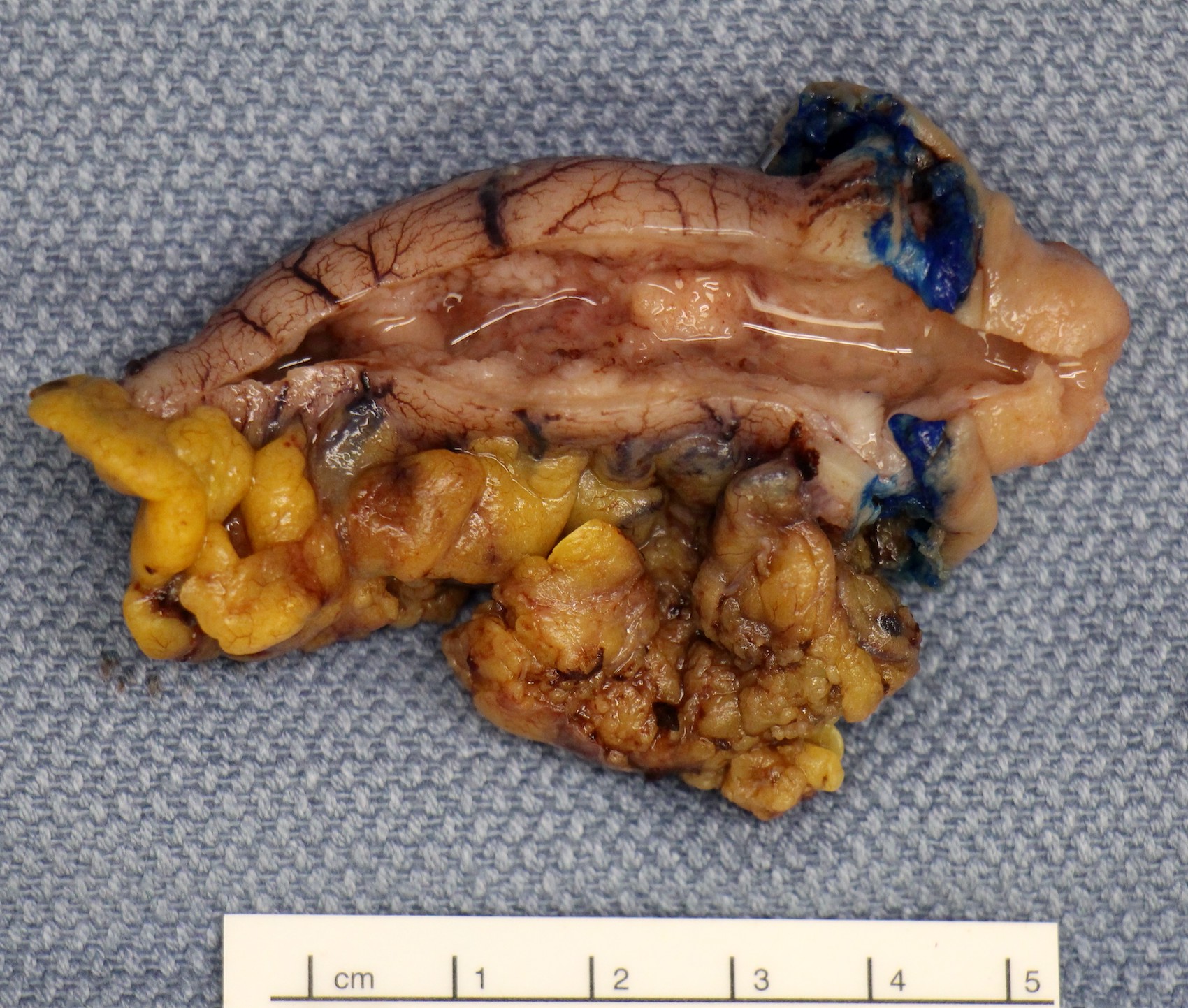
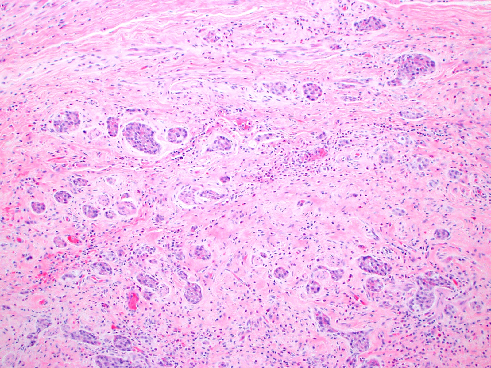
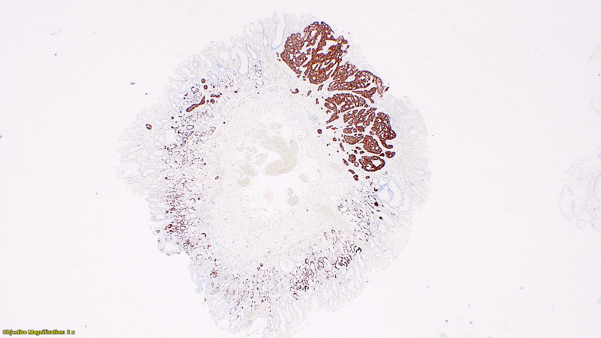
Adenocarcinoma involving appendiceal tip

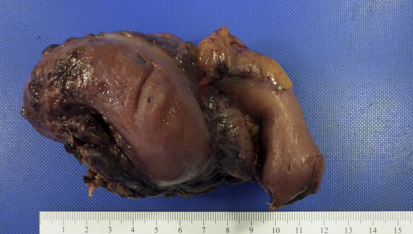
Adenocarcinoma involving appendix
Contributed by Bella Lingjia Liu, M.D.

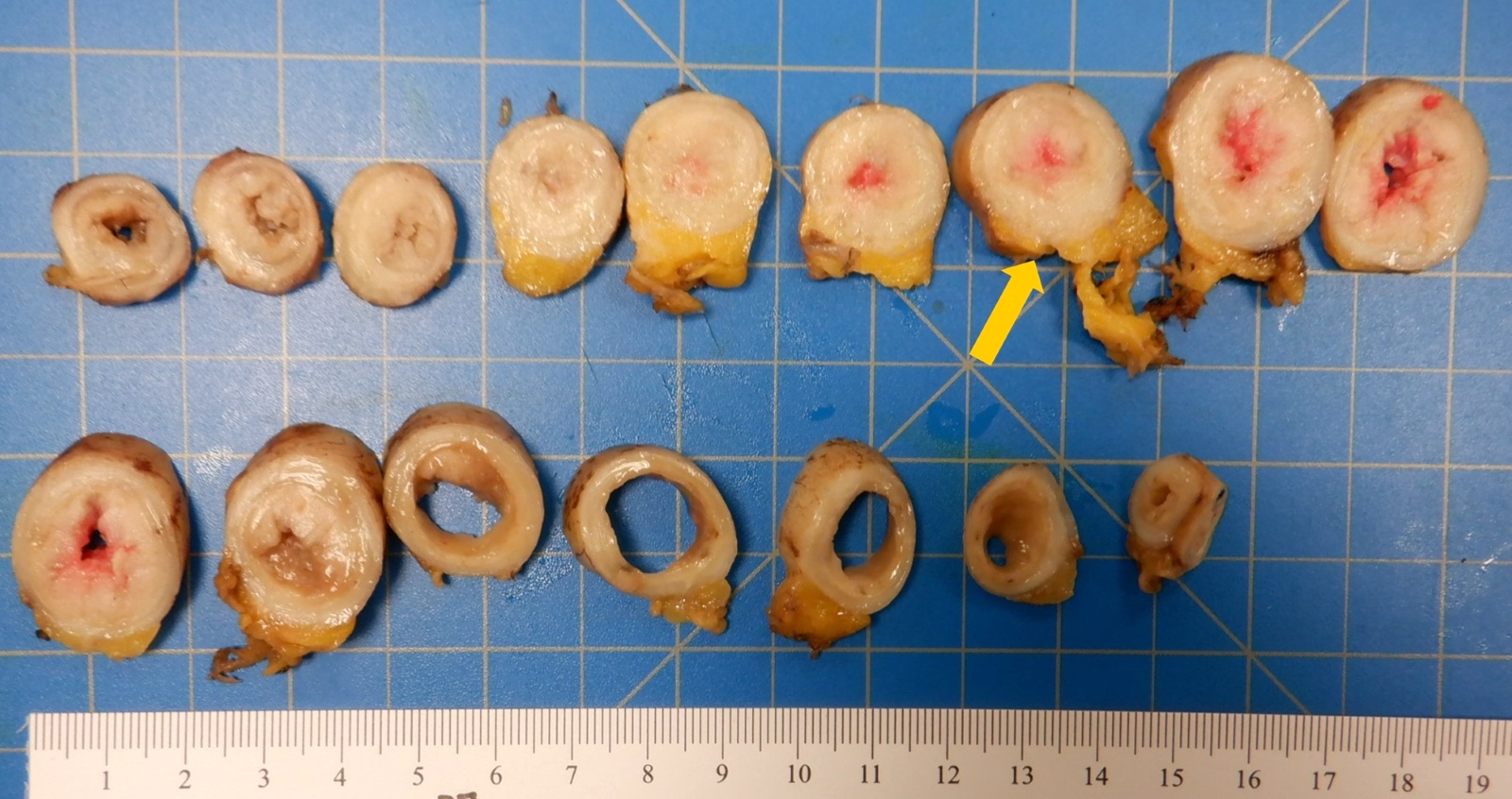
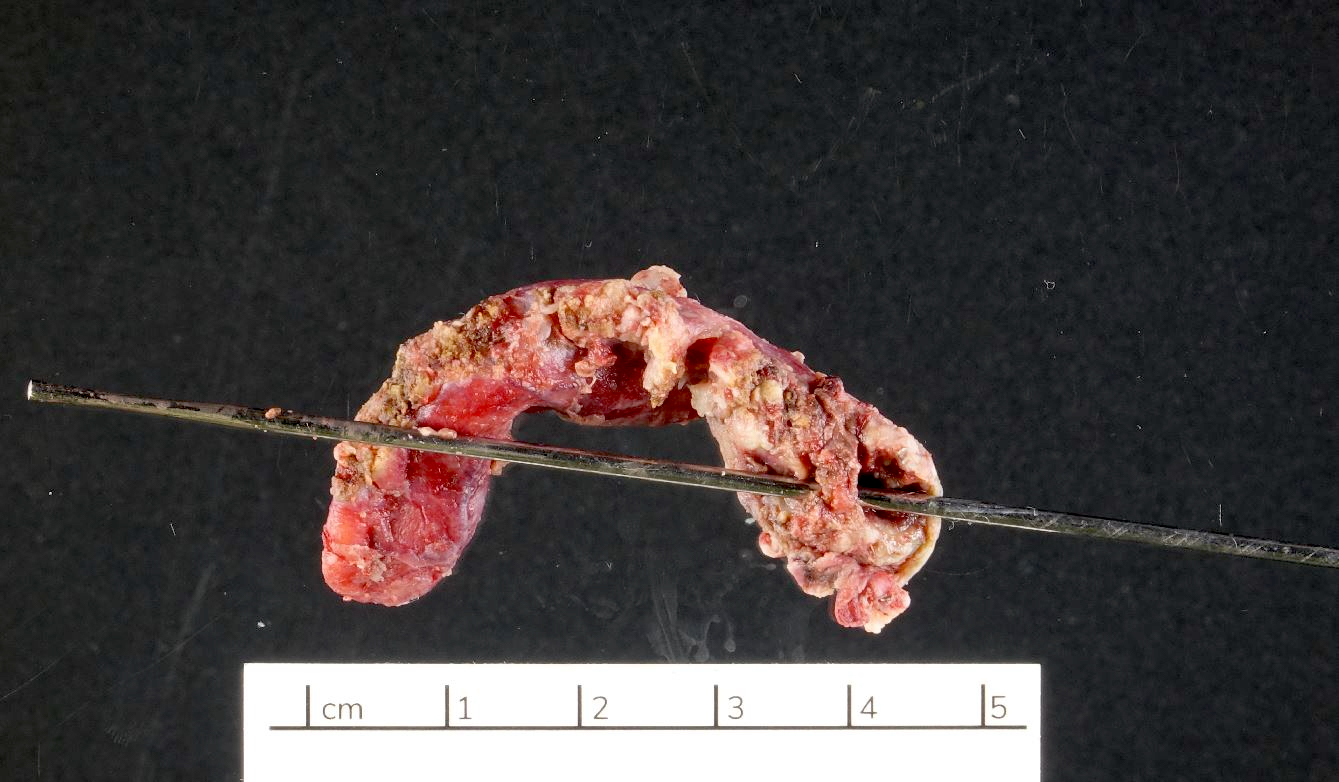
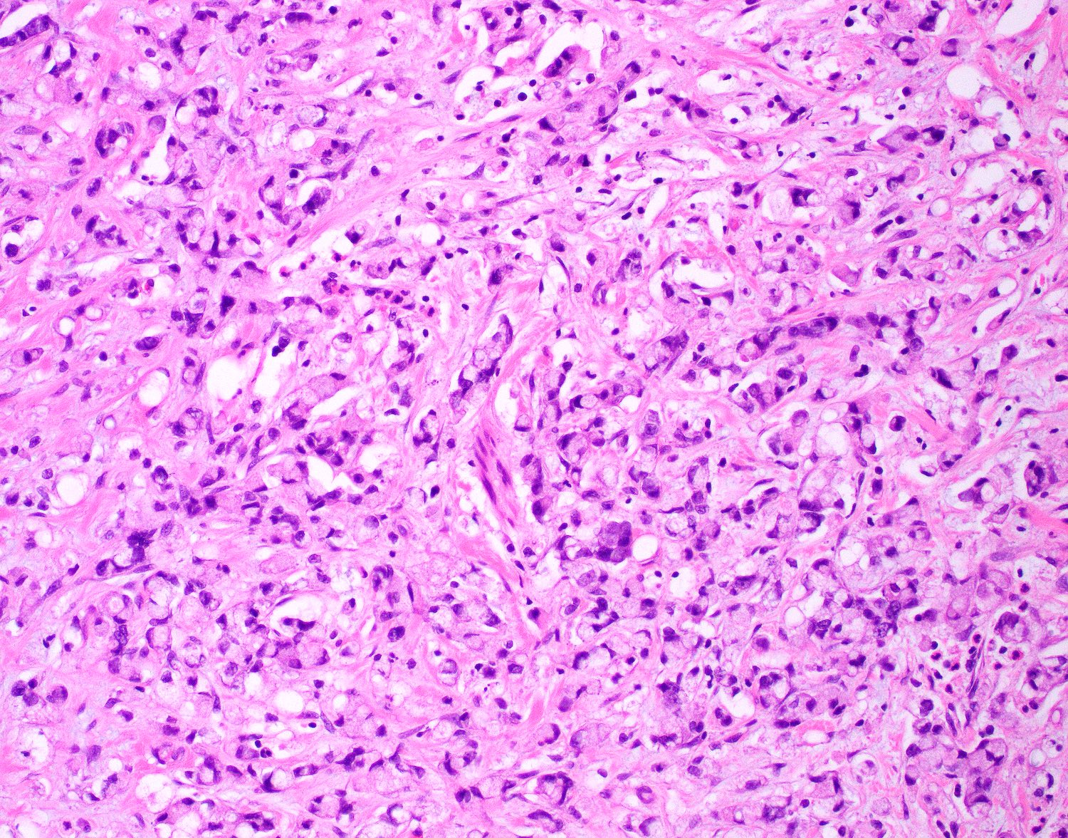
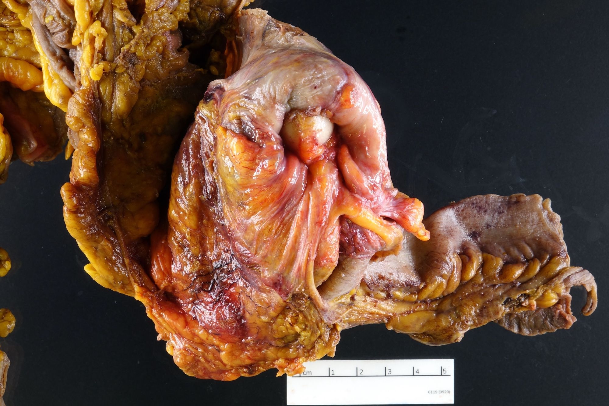
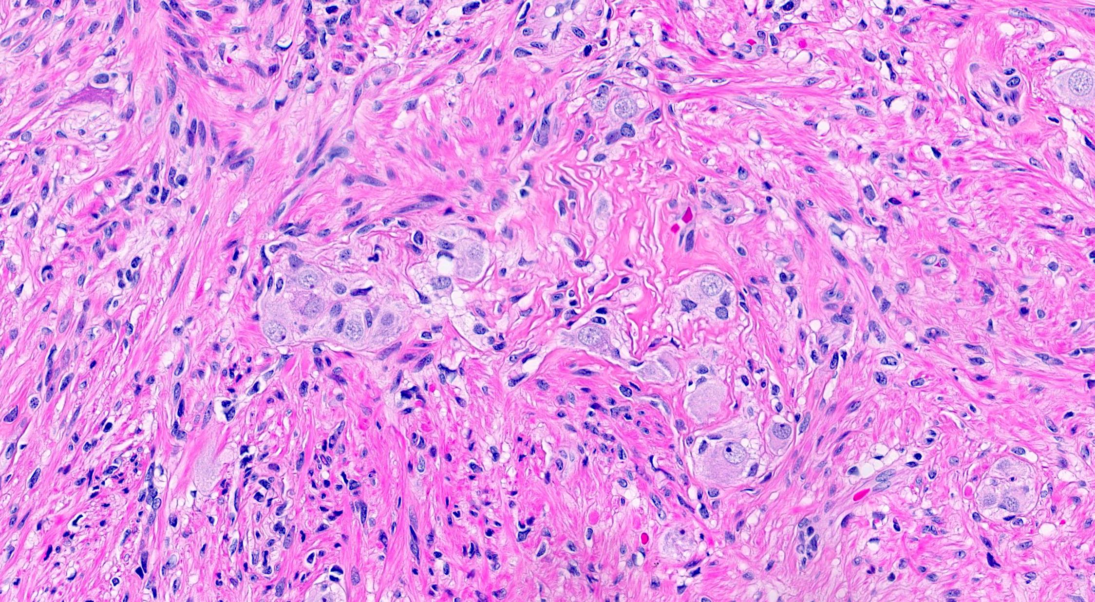
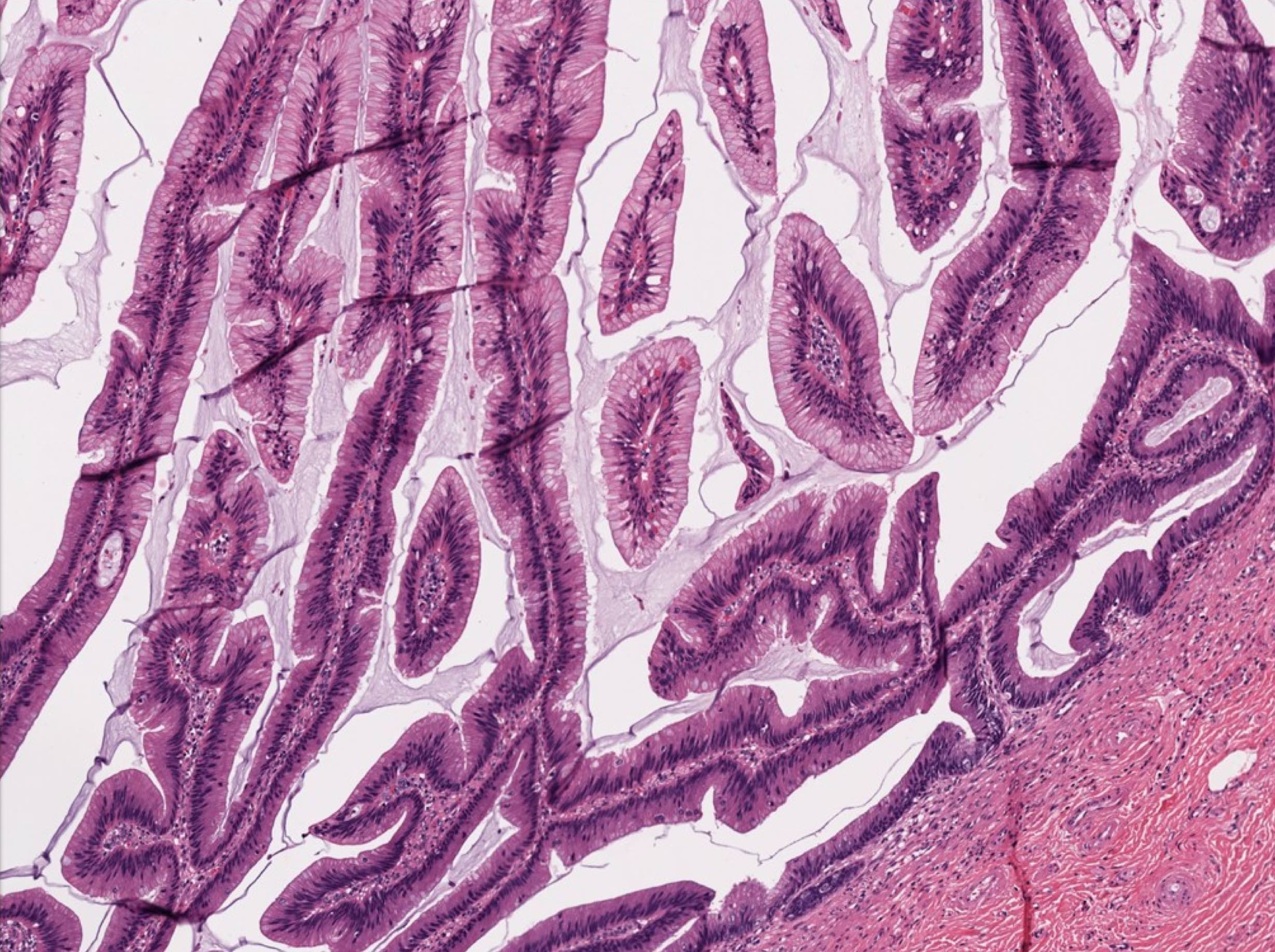
Mucinous adenocarcinoma
Appendiceal adenocarcinoma
Contributed by Bella Lingjia Liu, M.D.
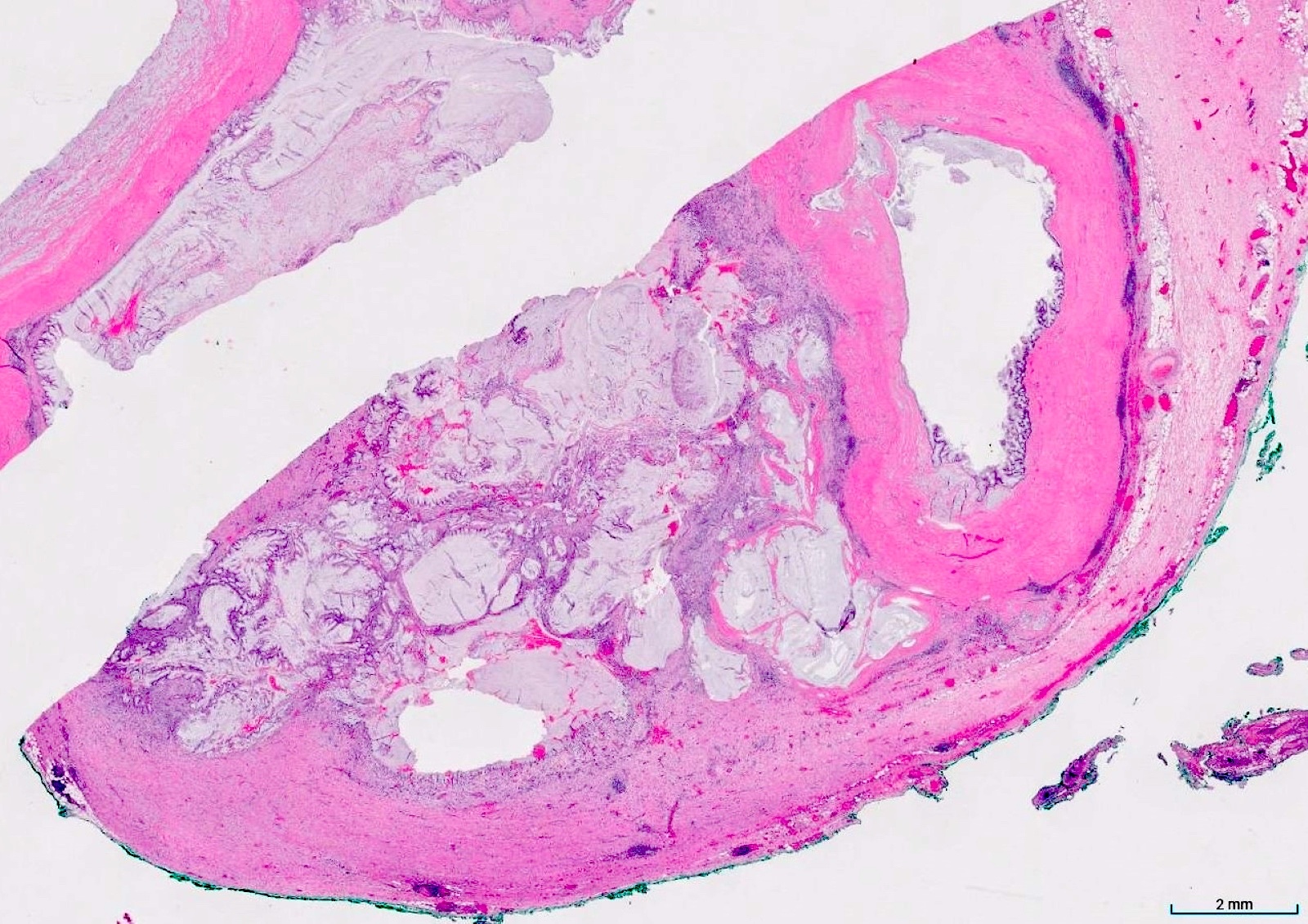
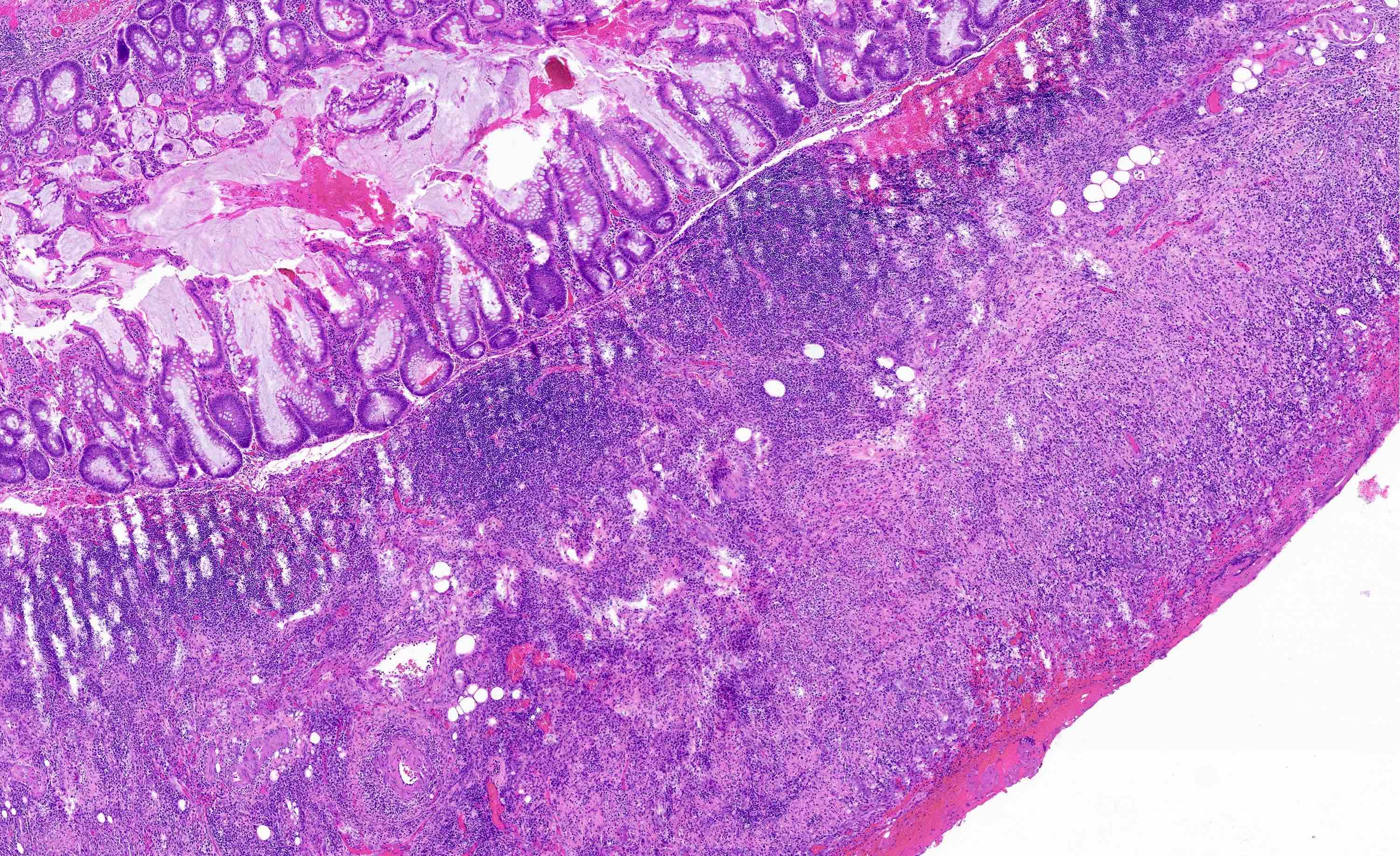
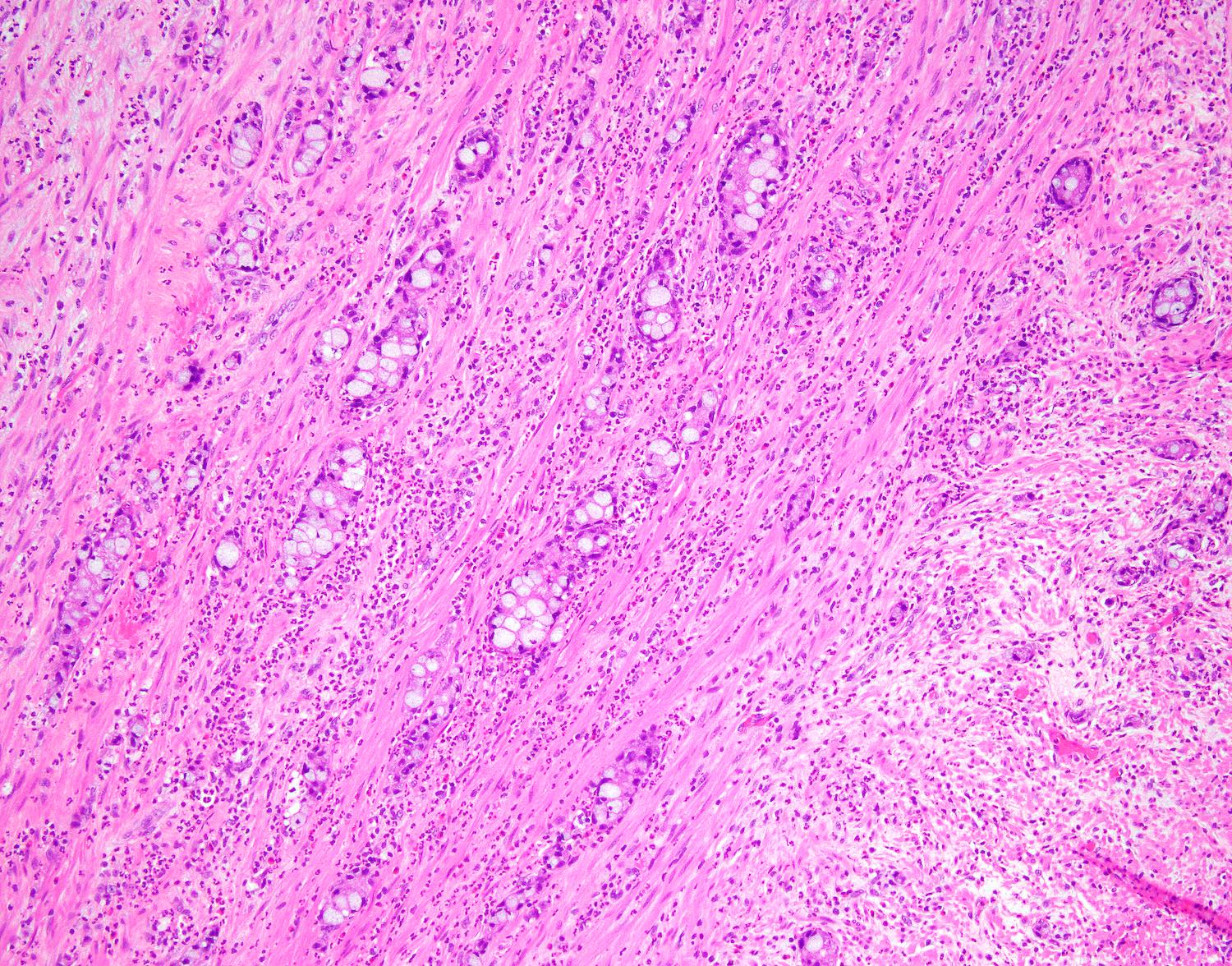
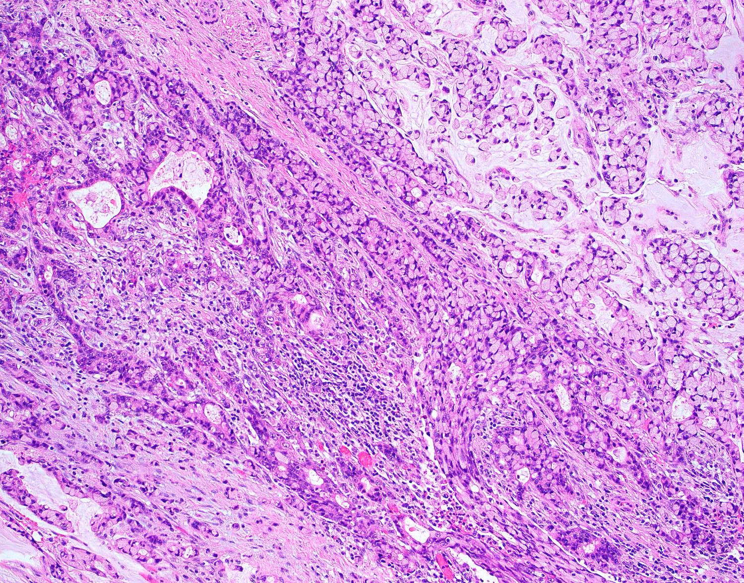
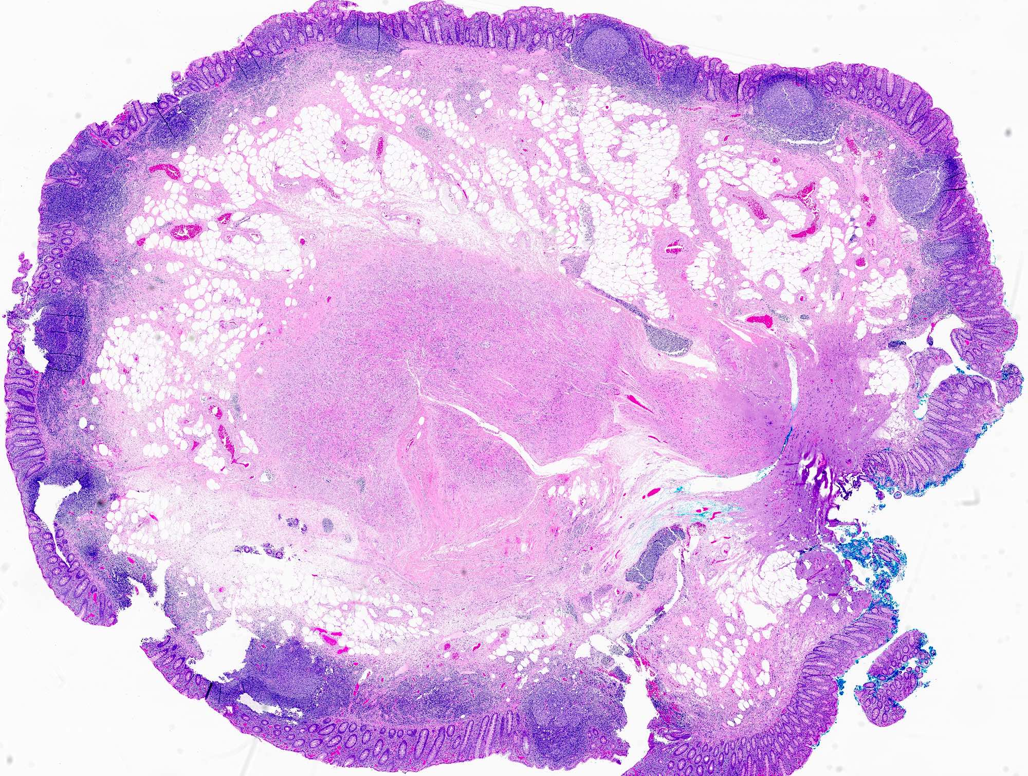
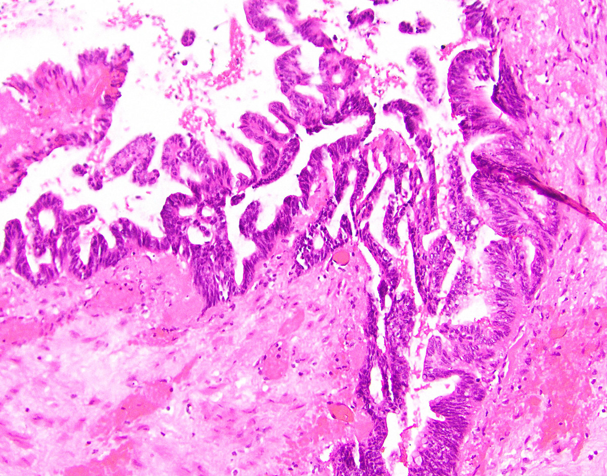
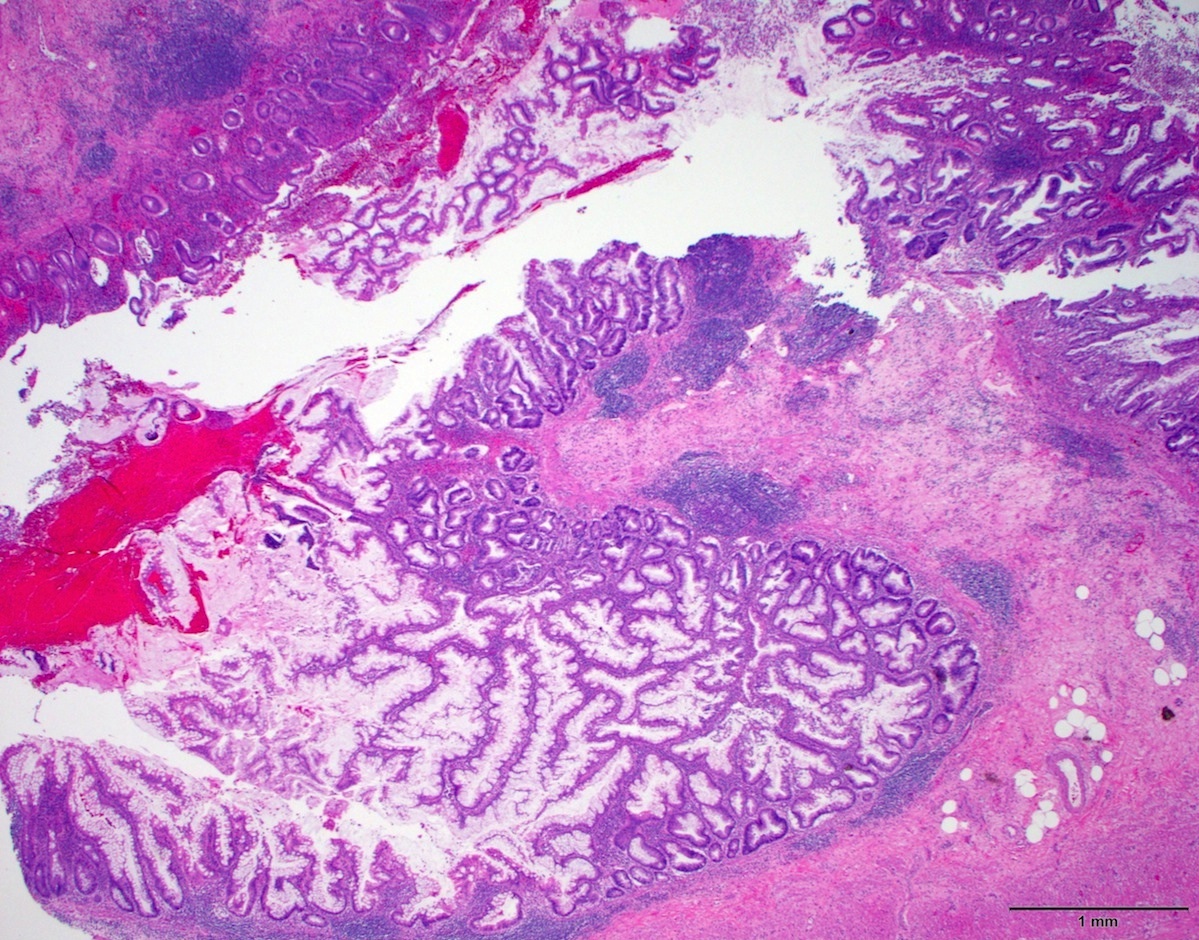
Adenocarcinoma with adjacent LAMN
Infiltrative glands with desmoplasia
Tumor cells in mucin
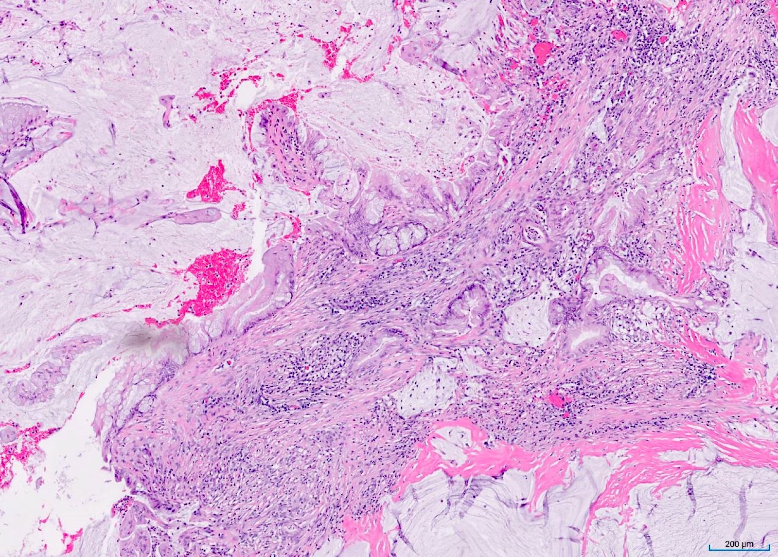
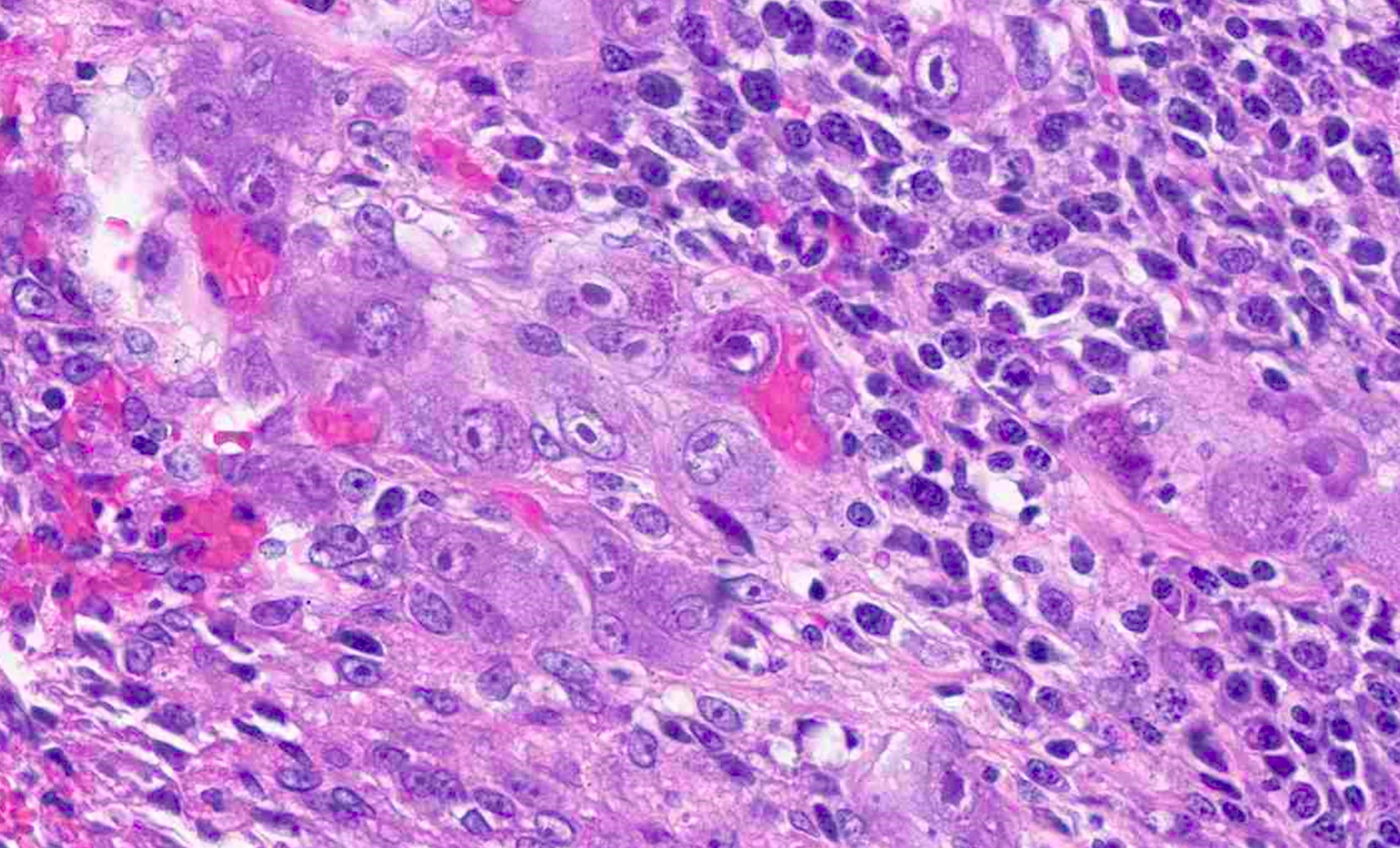
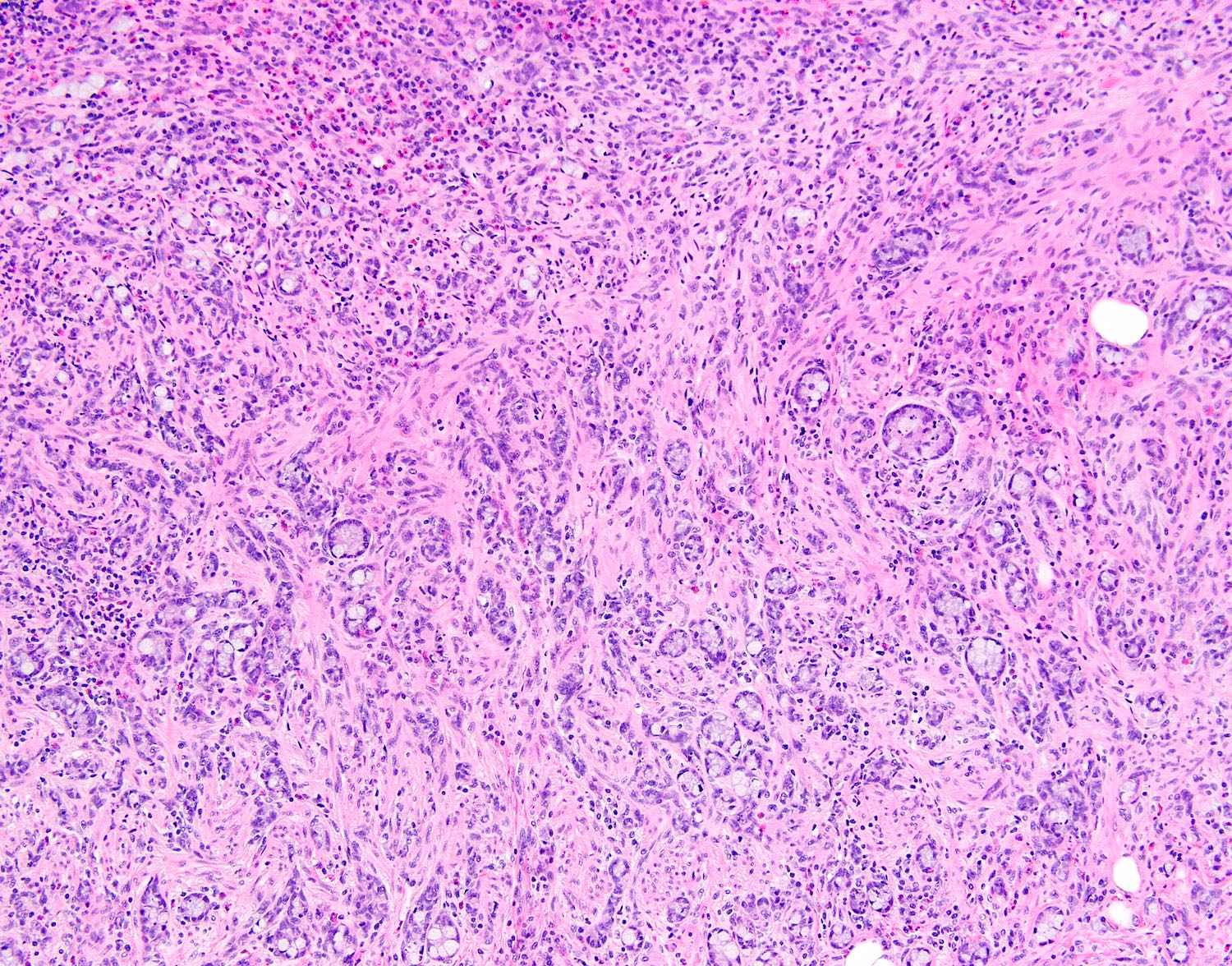
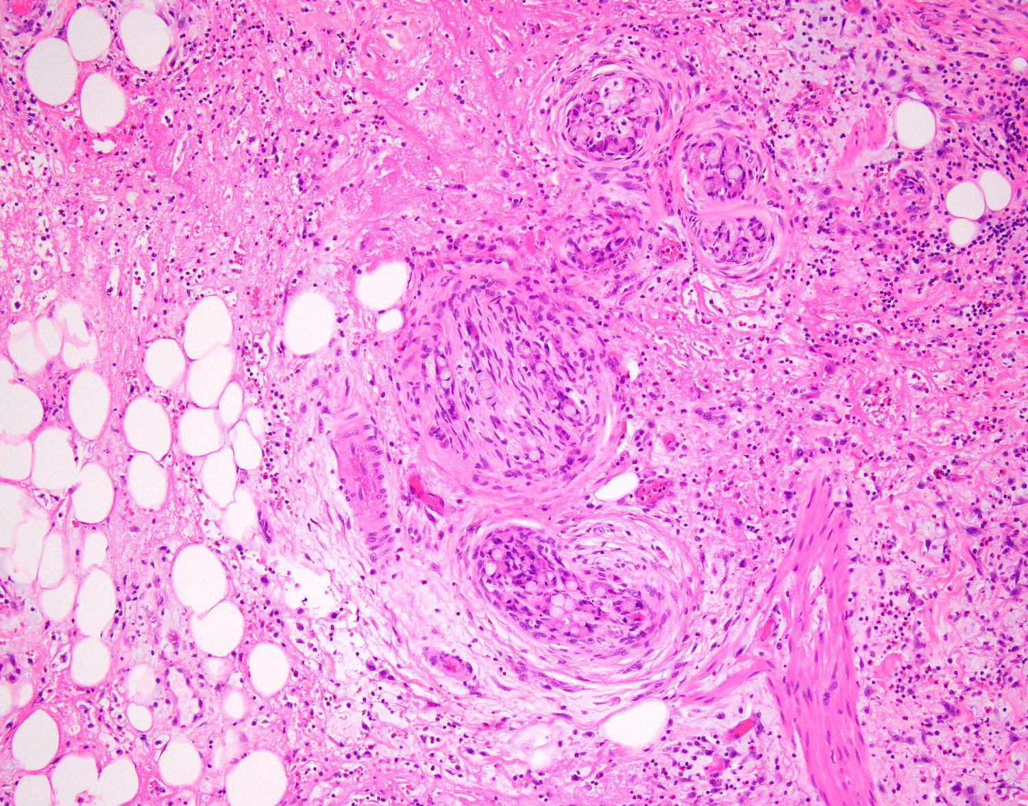
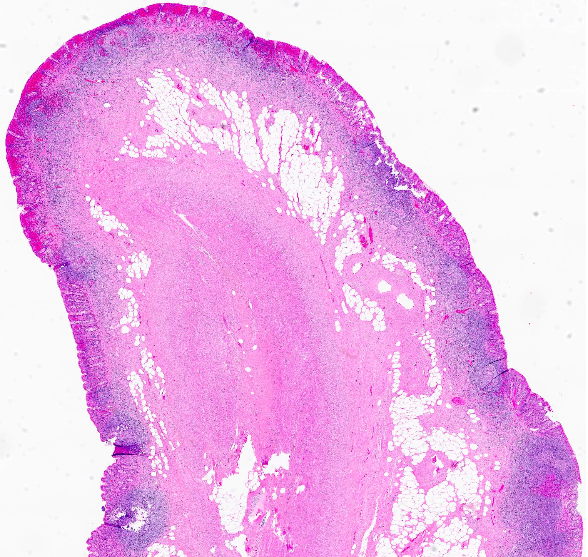
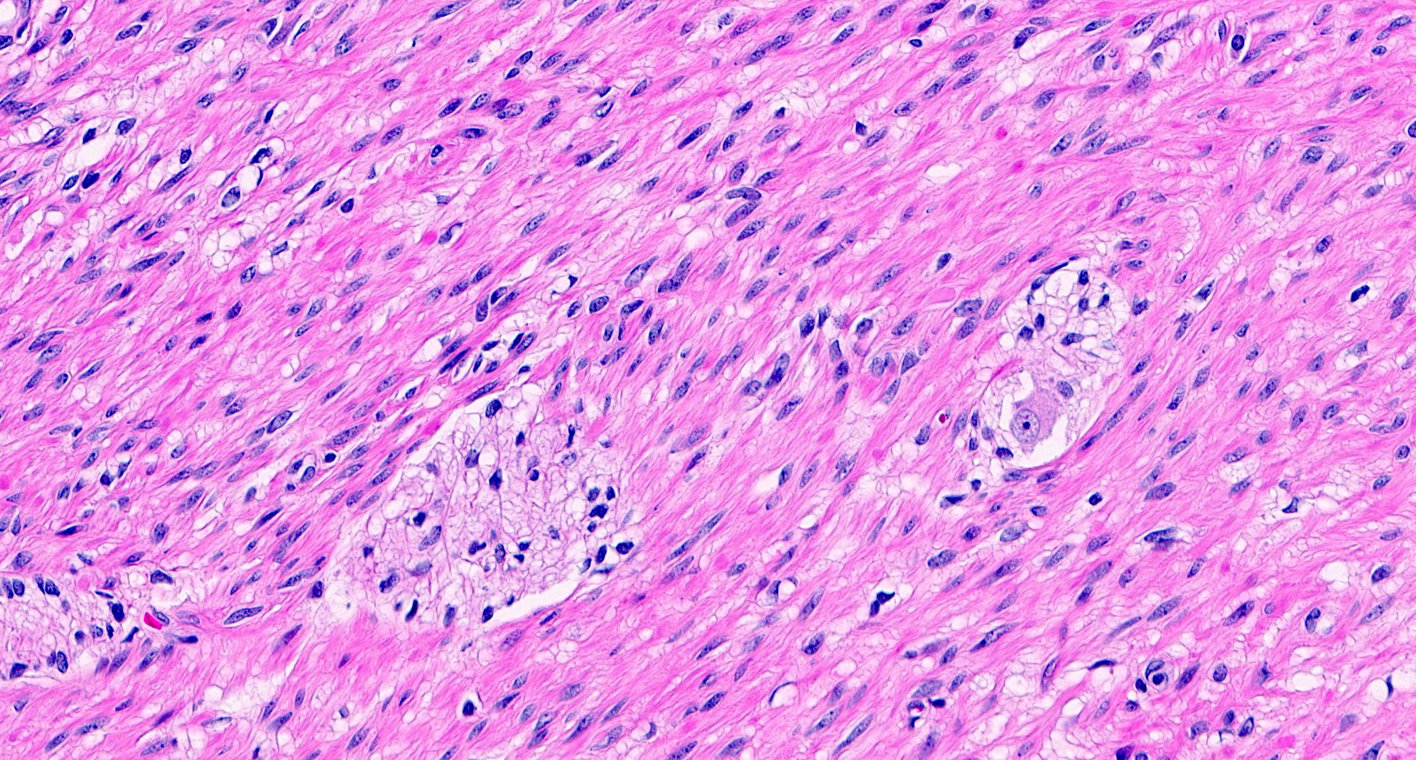
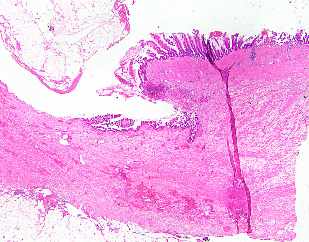
Adenocarcinoma with adjacent TVA
Infiltrative glands in stroma
Cribriforming with necrotic debris
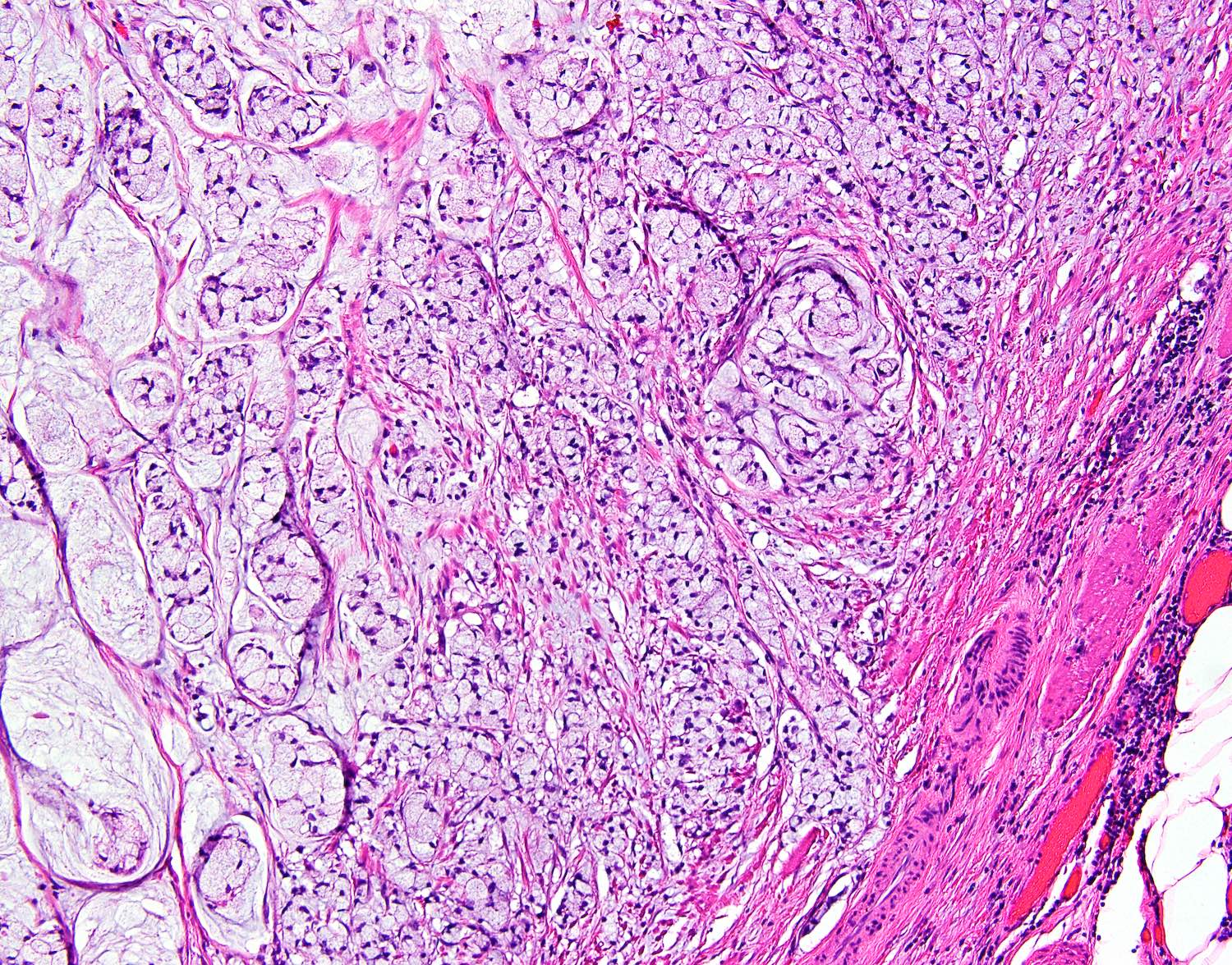
Lymphovascular invasion
Luminal obstruction
Signet ring cells
Contributed by Dr. Oleksandr Grygoruk, Boris Hospital and Medical Center, Kiev (Ukraine)


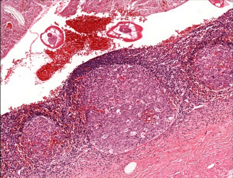
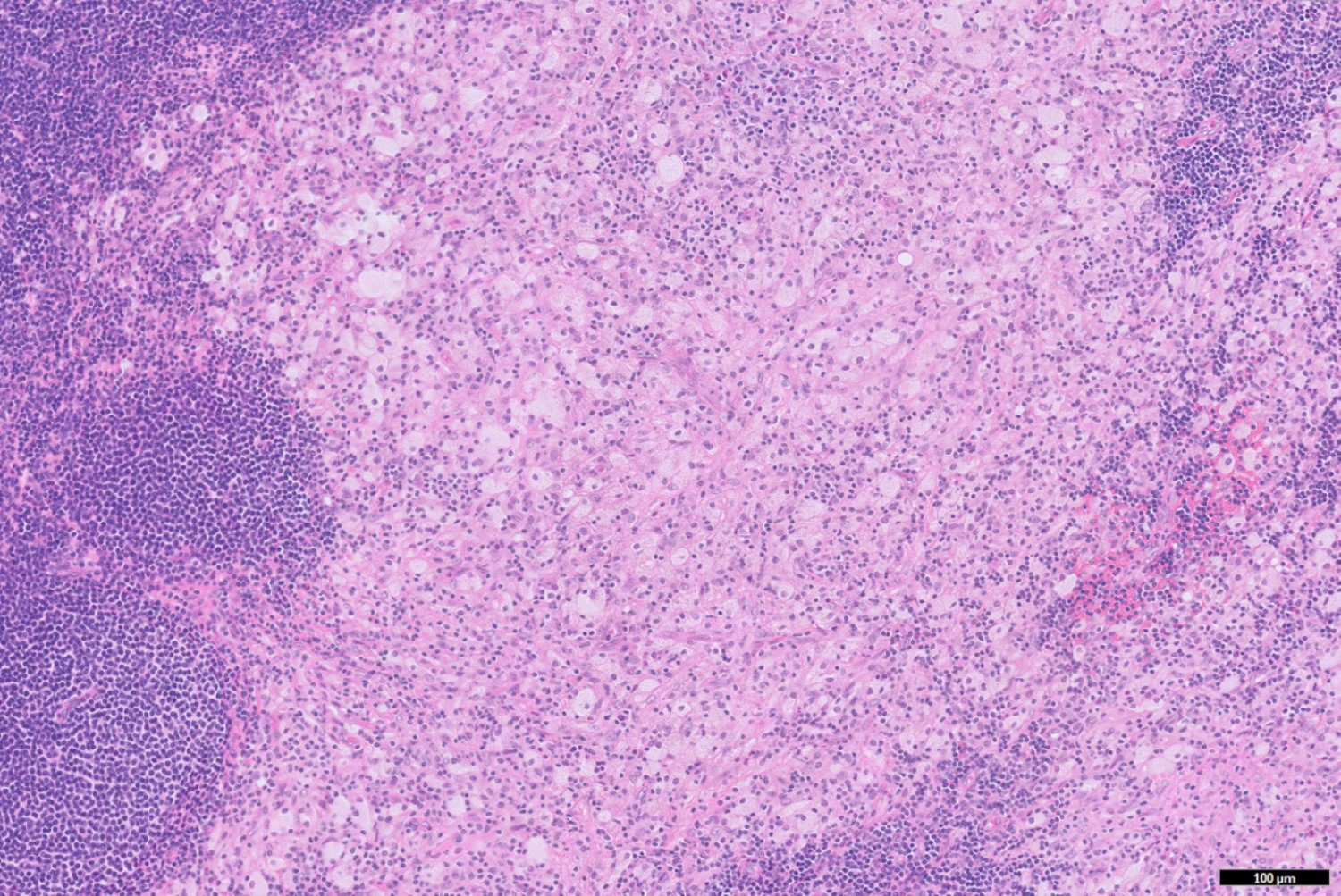
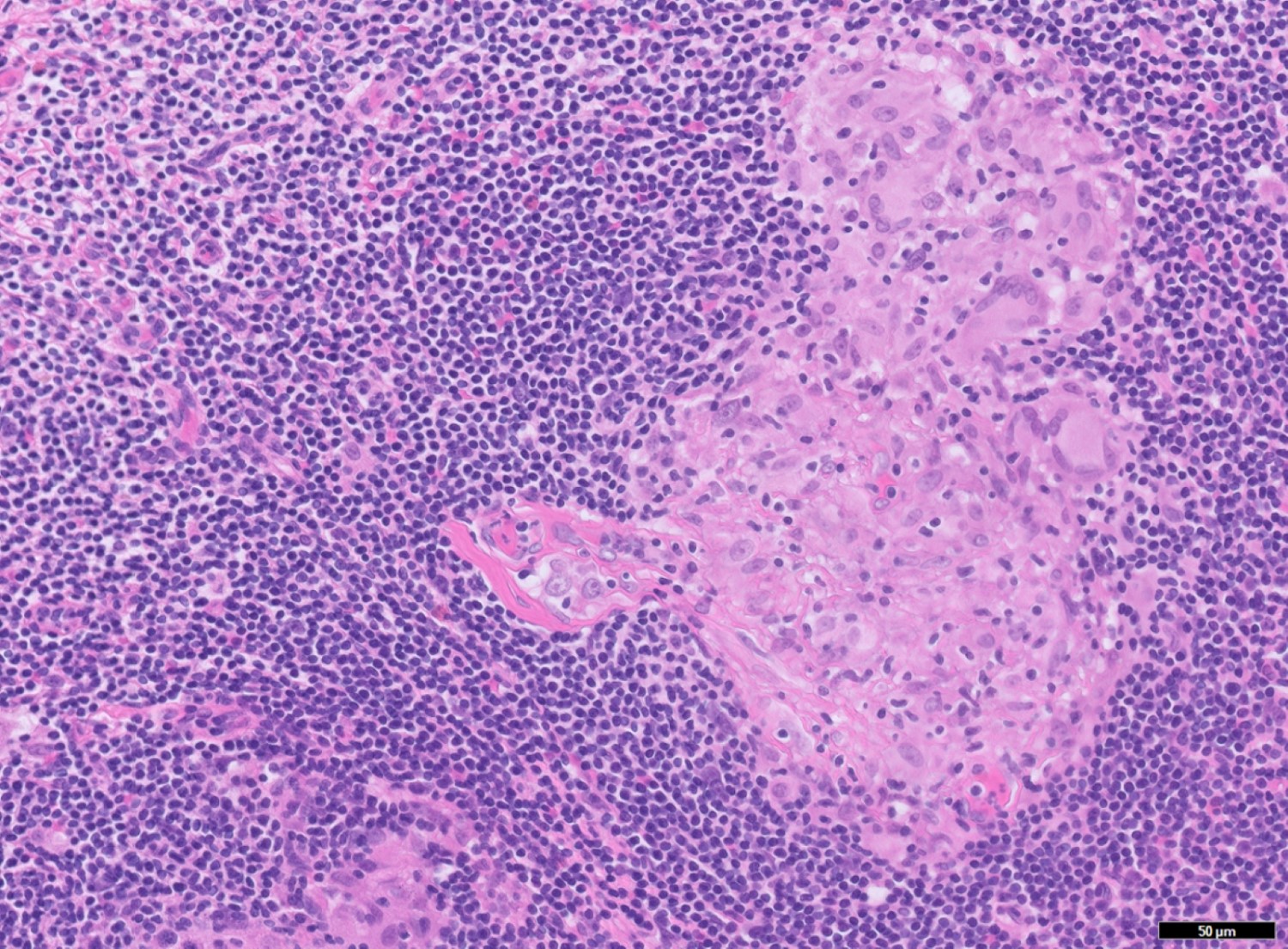
40 year old woman with prodromal measles confirmed by serology
Images hosted on other servers:

Warthin-Finkeldey cells (lymph node)
Images hosted on other servers:

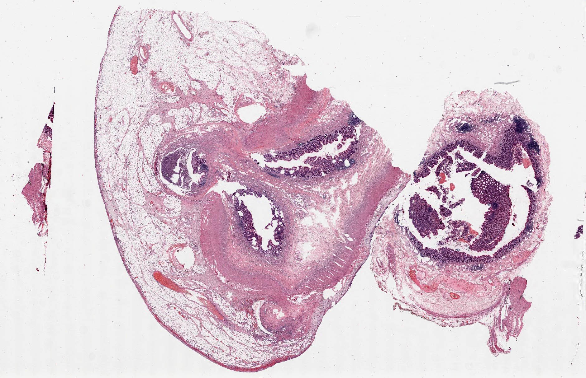
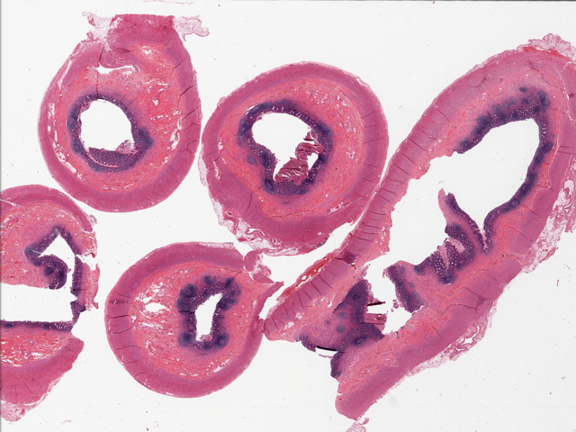
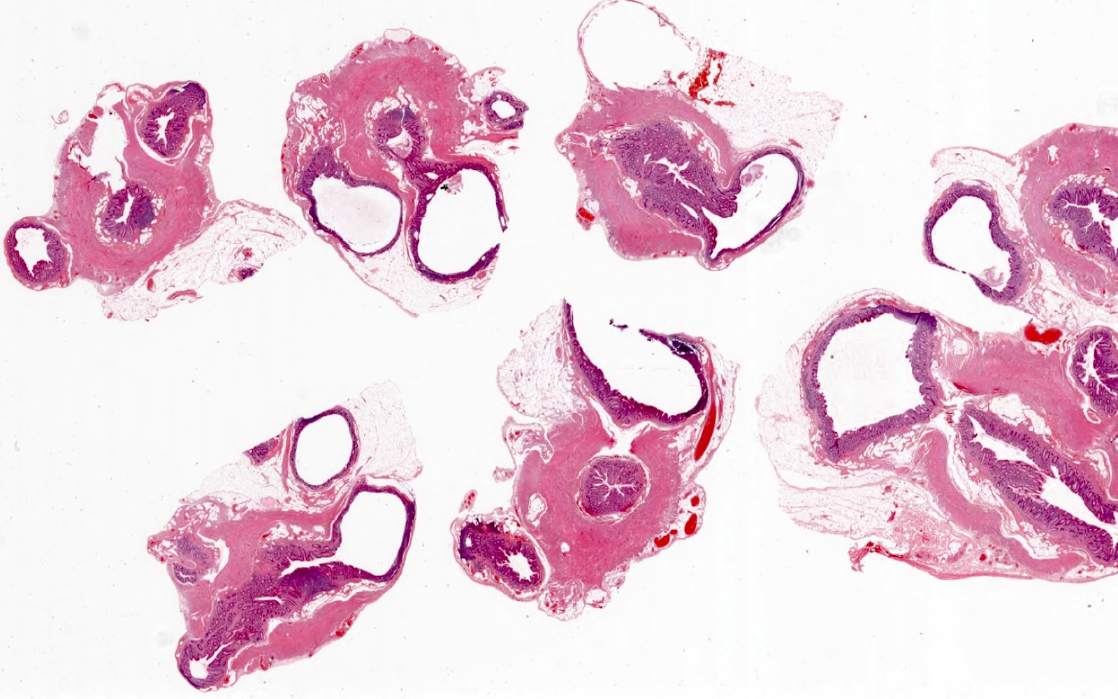
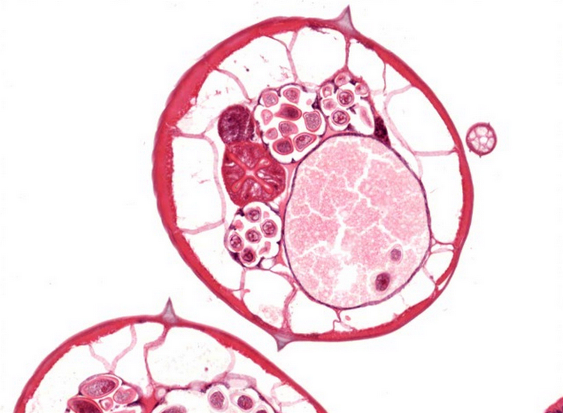
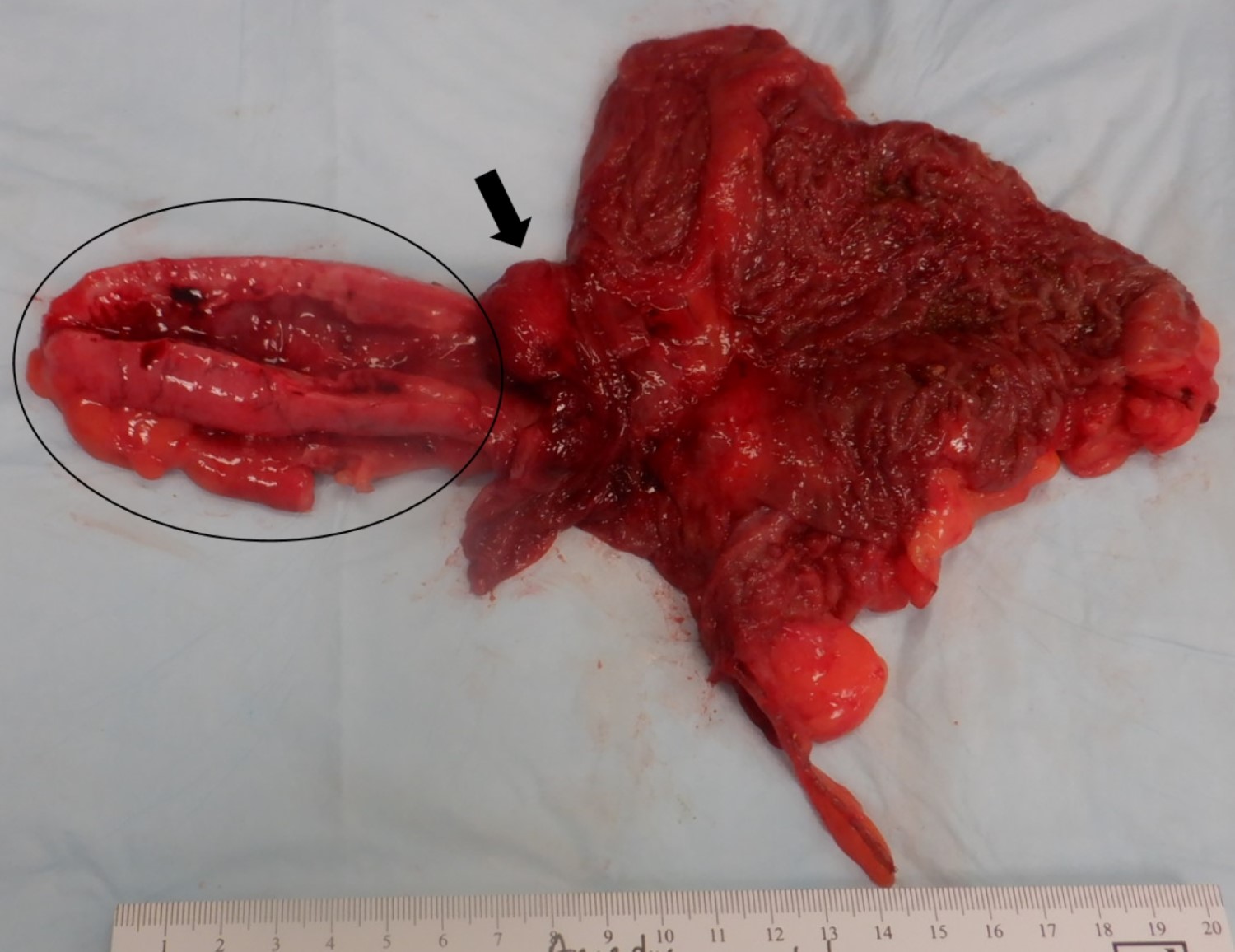
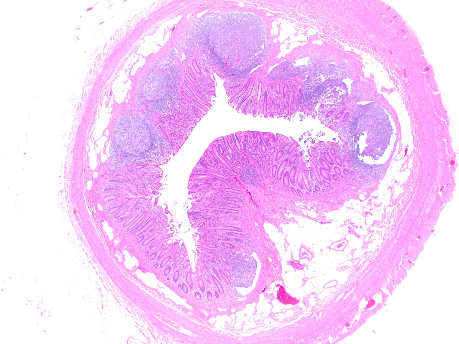
Cecum, appendix and arteries

View from cecum
Contributed by Maryam Kherad Pezhouh, M.D., M.Sc.
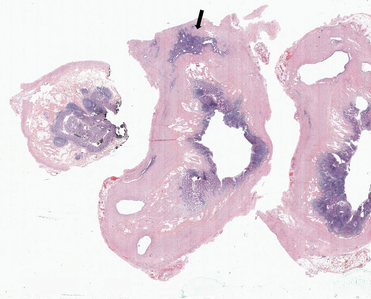
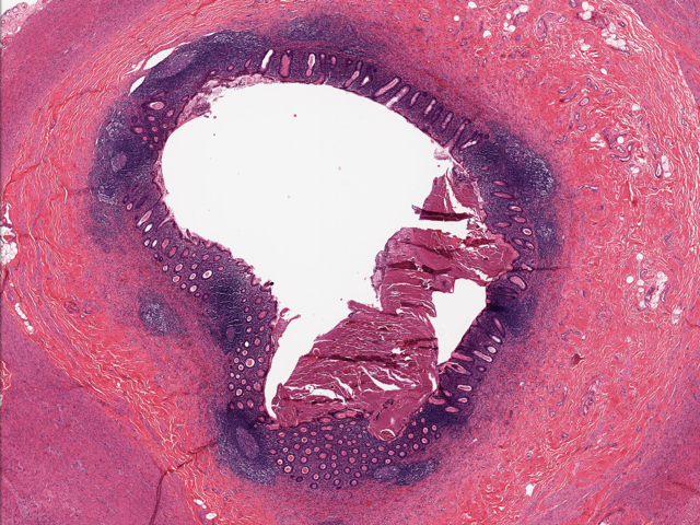
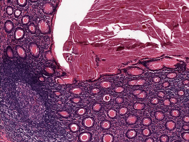
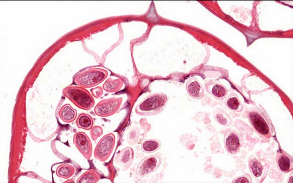
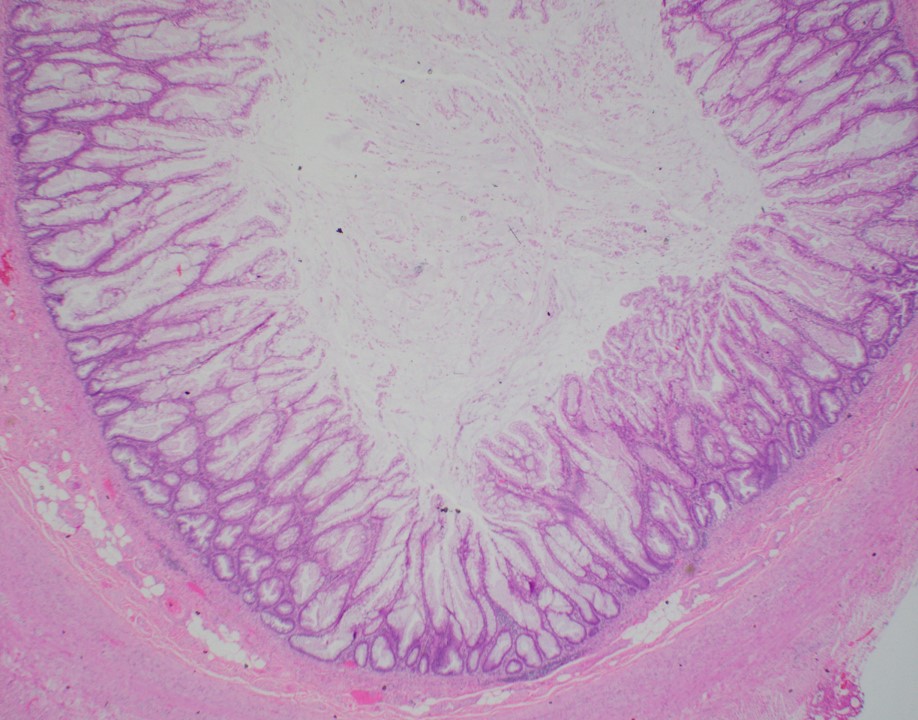
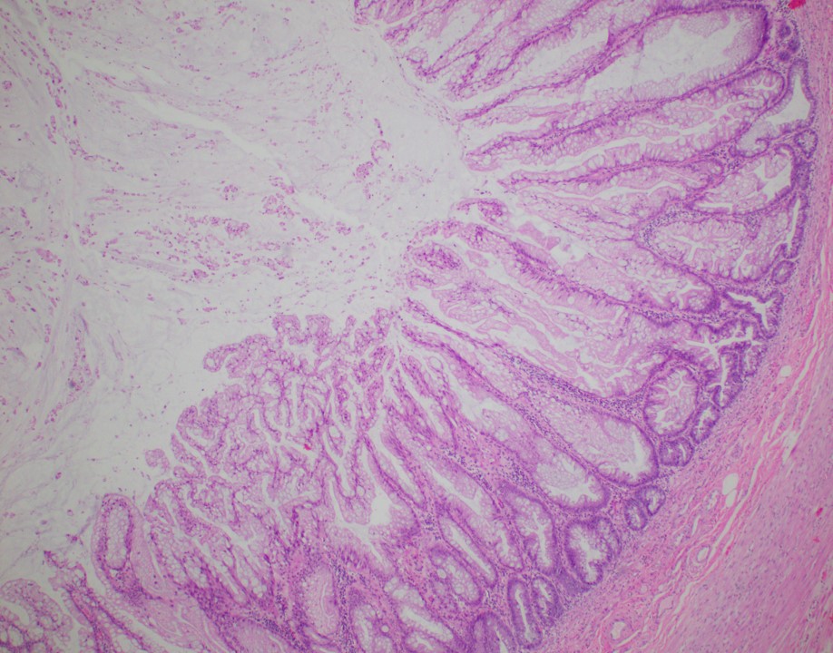
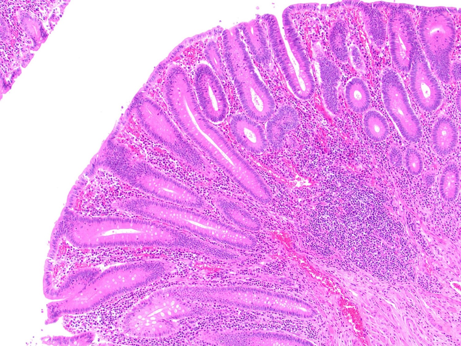
Appendix
Contributed by Maryam Kherad Pezhouh, M.D., M.Sc.
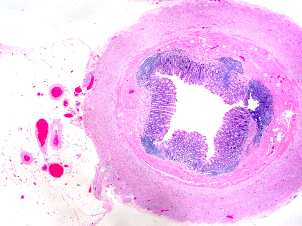
Cross section of an appendix
Images hosted on other servers:

Acute appendicitis on CT
Contributed by Danielle Hutchings, M.D.
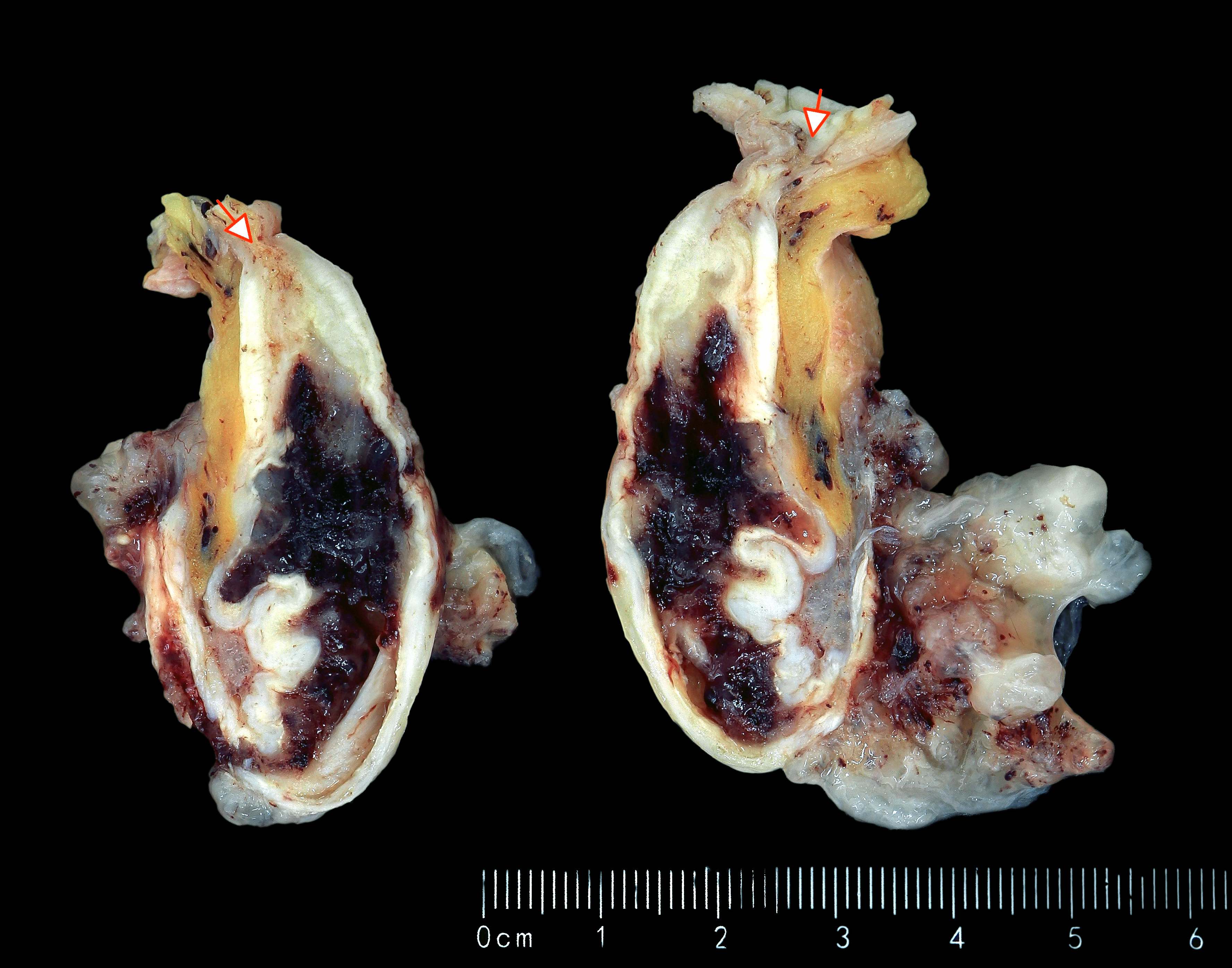
Dilatation and hyperemia
Serosal exudate
Perforation
Contributed by Danielle Hutchings, M.D.
Transmural mixed inflammatory infiltrate
Cluster of CMV infected cells
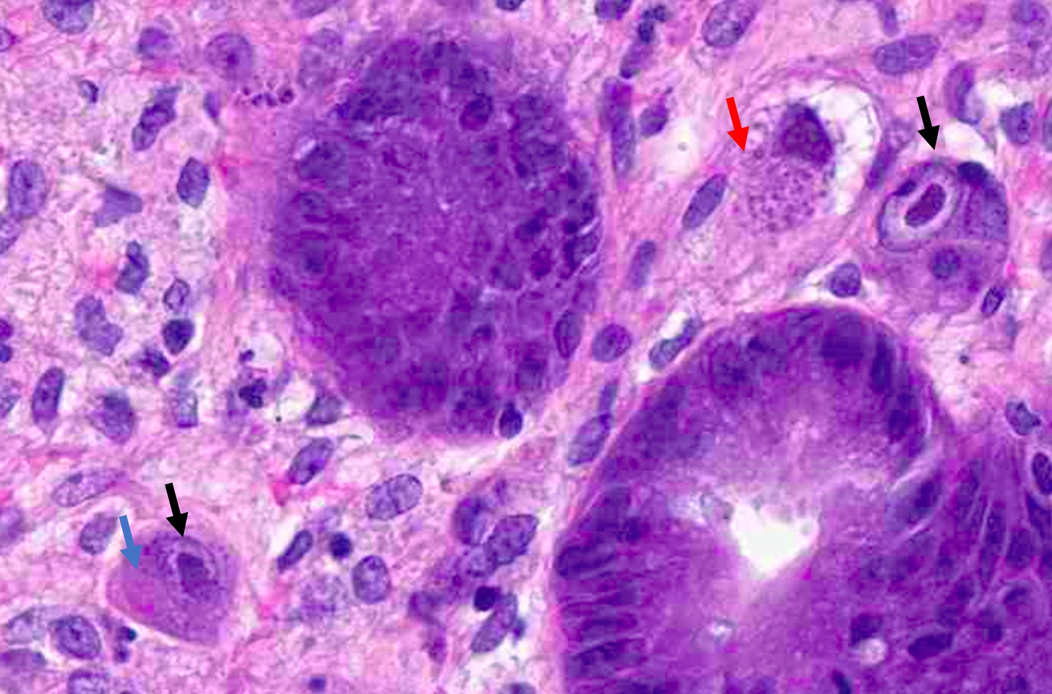
Intranuclear and intracytoplasmic inclusions
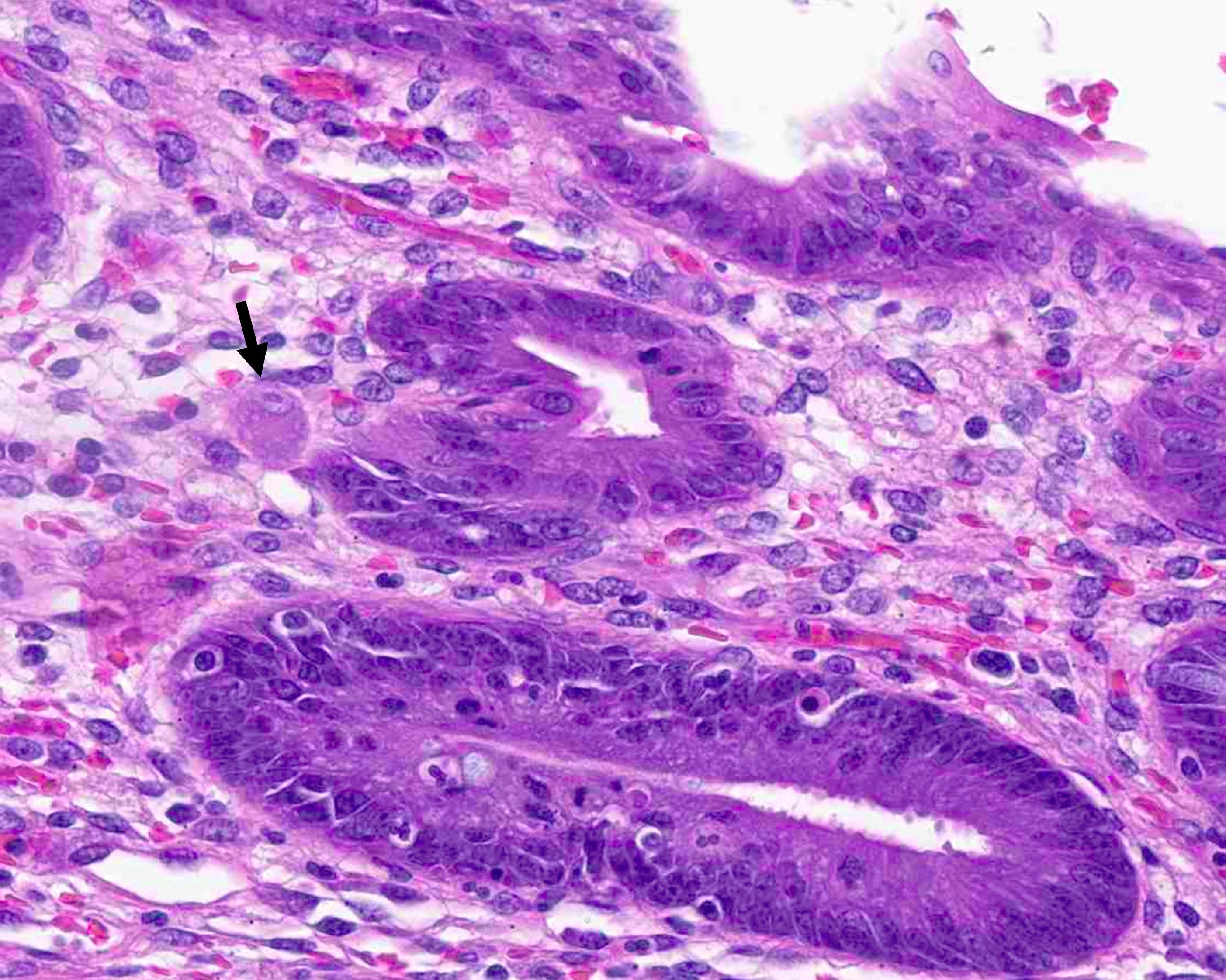
CMV infected cell
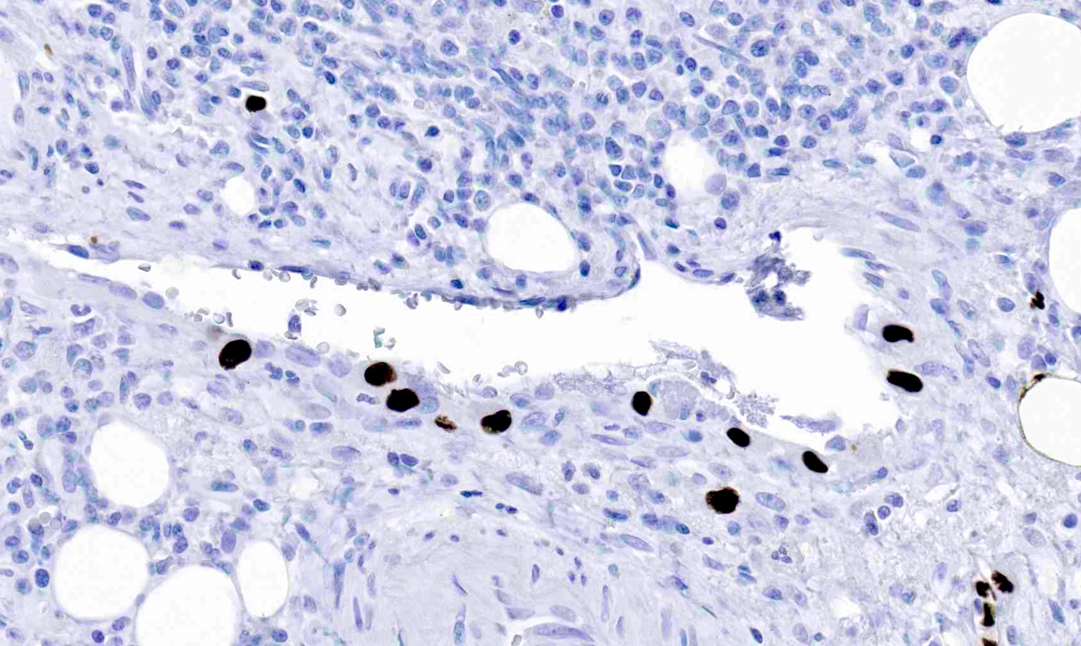
Positive CMV immunostain, endothelial cells
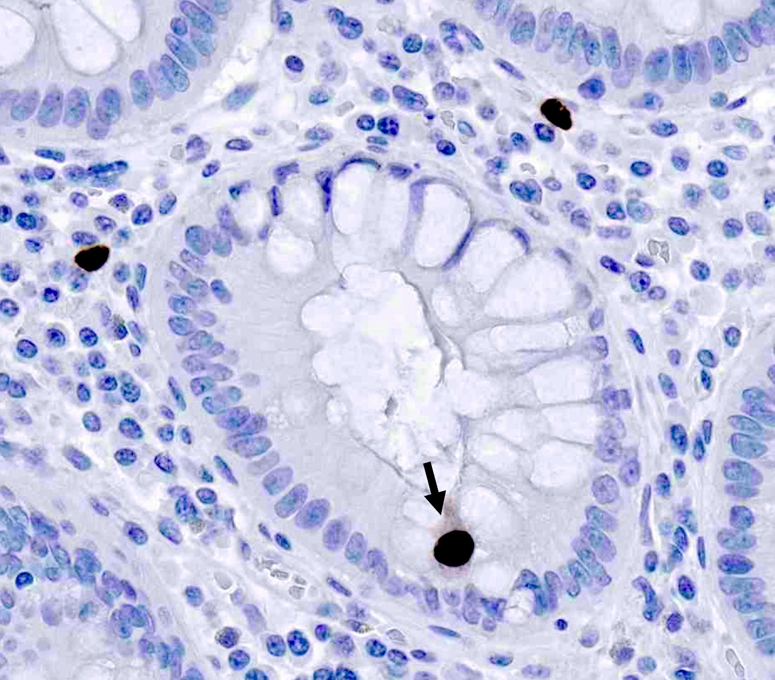
CMV immunostain, epithelial cell
Contributed by Dr. Michael Feely

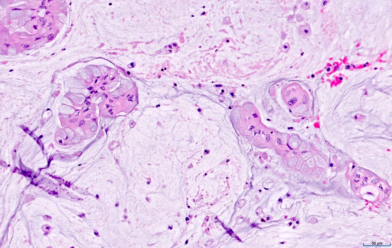
Thick eosinophillic mucus distends the lumen of the appendix as well as individual crypts
Images hosted on other servers:

Oblique coronal
reformation of
contrast enhanced
CT scan
Images hosted on other servers:

Intraoperative image
Images hosted on other servers:

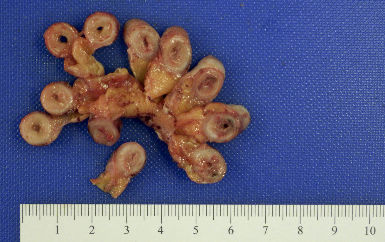
Multiple appendiceal diverticula

Congenital appendiceal diverticulum
Contributed by Qingqing Liu, M.D., Ph.D.
Multiple diverticula
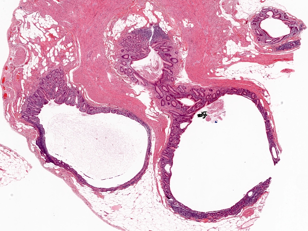
Connection between diverticulum and main lumen
Inflamed diverticulum
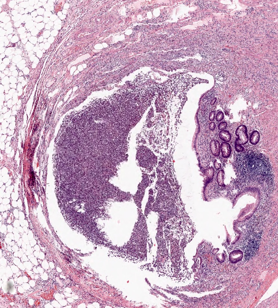
Inflammatory exudate
Acute appendicitis accompanied by appendiceal diverticulitis

Images hosted on other servers:

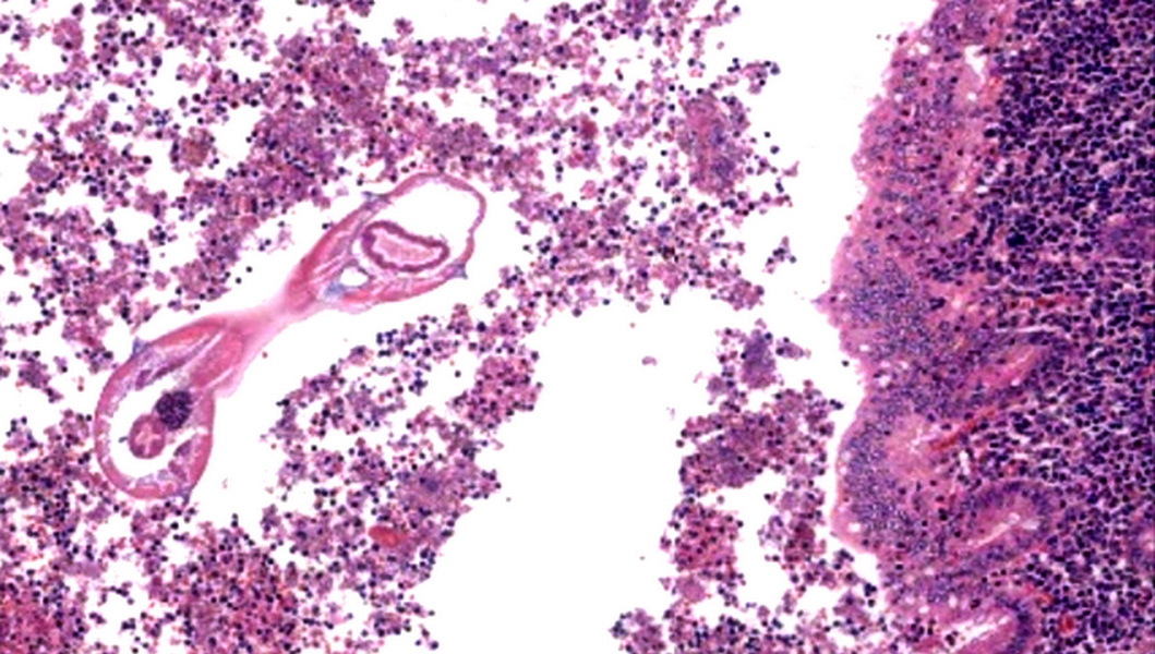
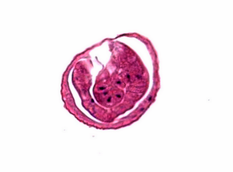
Intraoperative pinworm

Enterobius vermicularis through appendix
Images hosted on other servers:

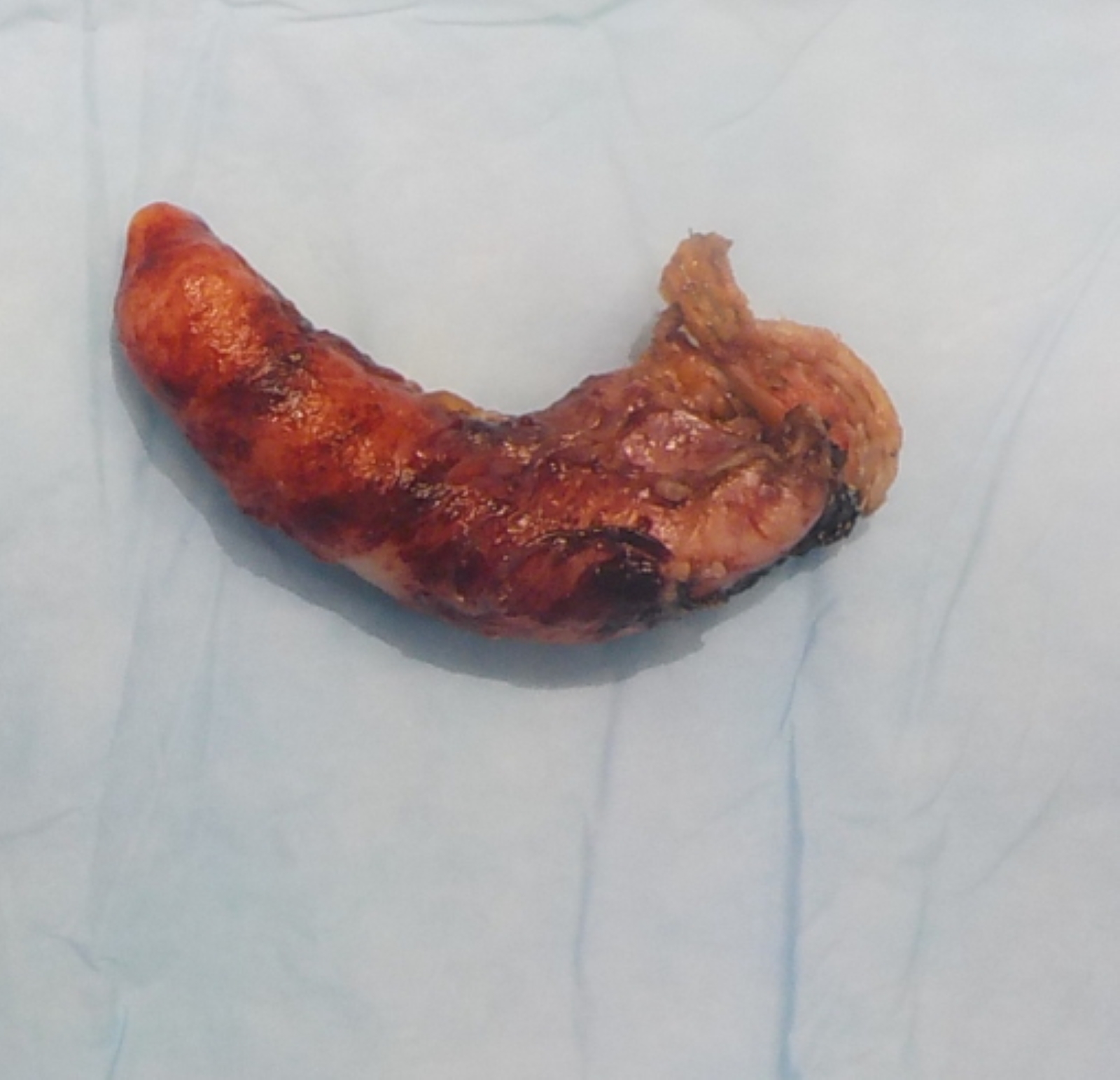
Inflamed appendix with stercolith

With retrieved worm

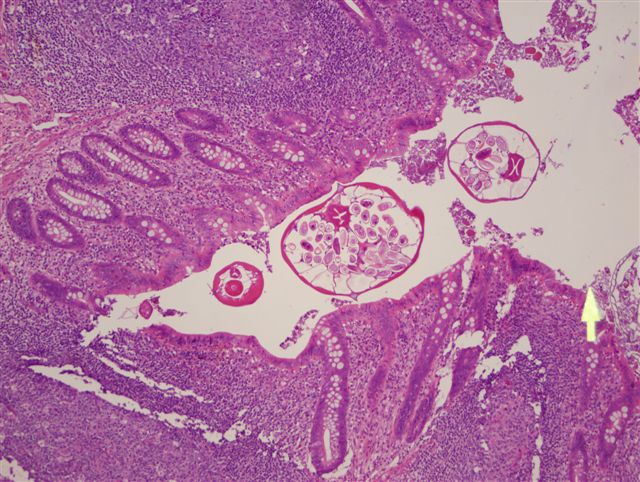
Enterobius vermicularis through appendix
Contributed by Eiman Adel Hasby, M.D. and Case #90
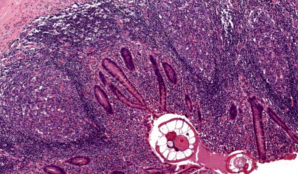
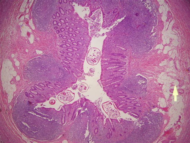
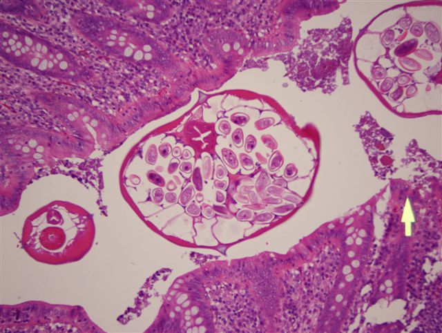
Appendix with luminal Enterobius
Enterobius in acute appenicitis
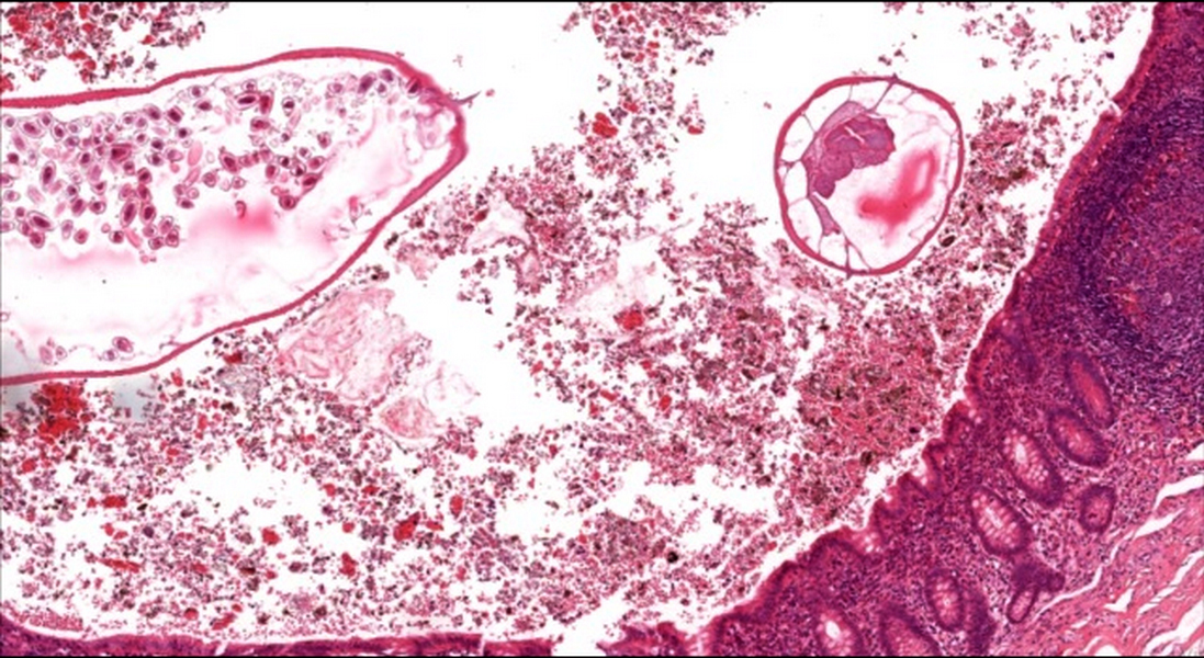
Appendicitis with luminal Enterobius
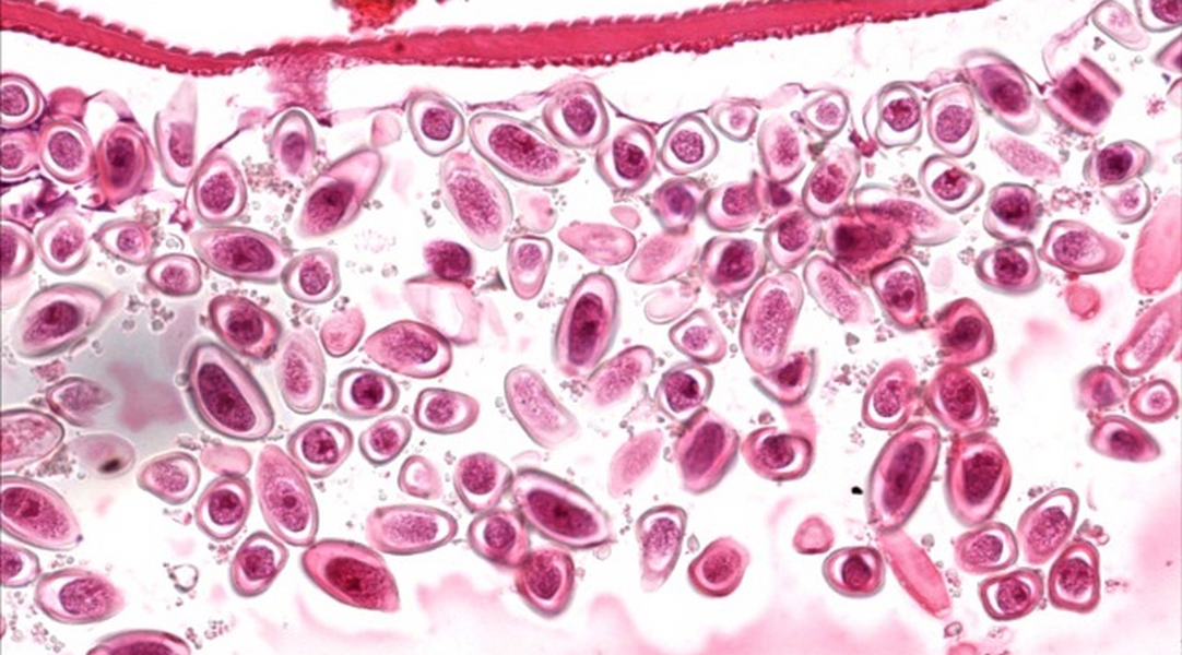
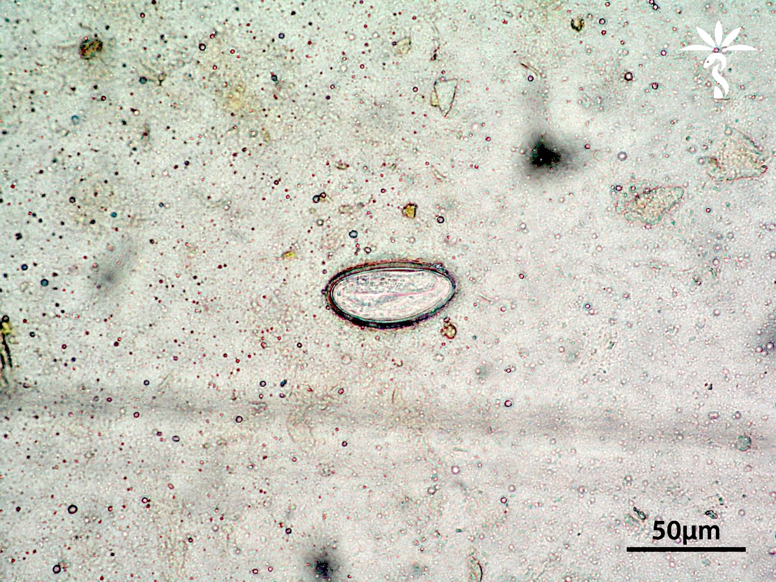
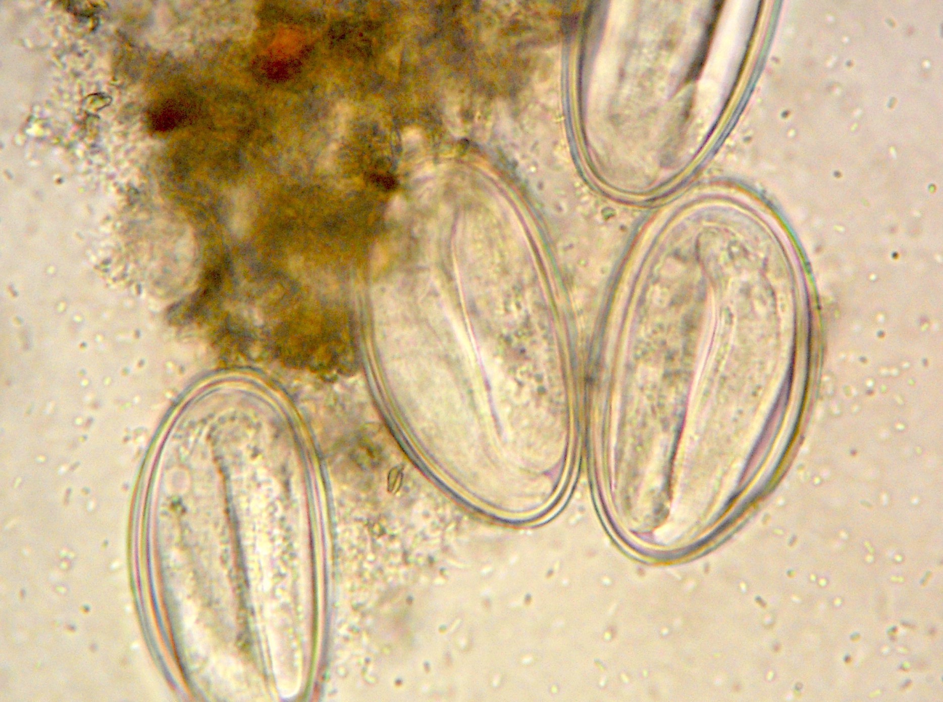
Enterobius eggs
Appendicitis with enterobiasis
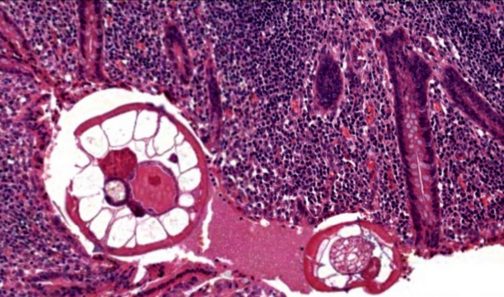
Appendicitis with enterobiasis
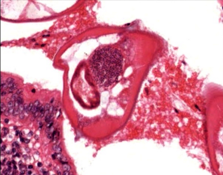
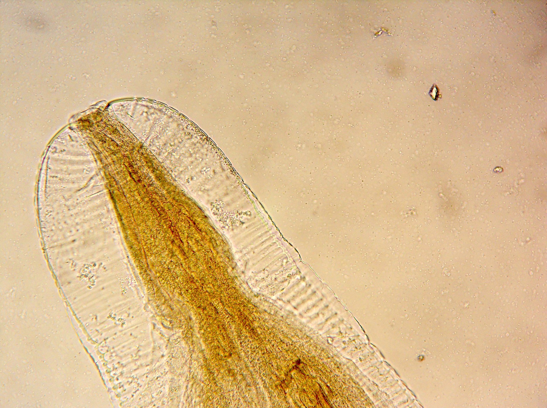
Male Enterobius adult worm

Female Enterobius adult worm
Gravid female Enterobius worm
Enterobius eggs
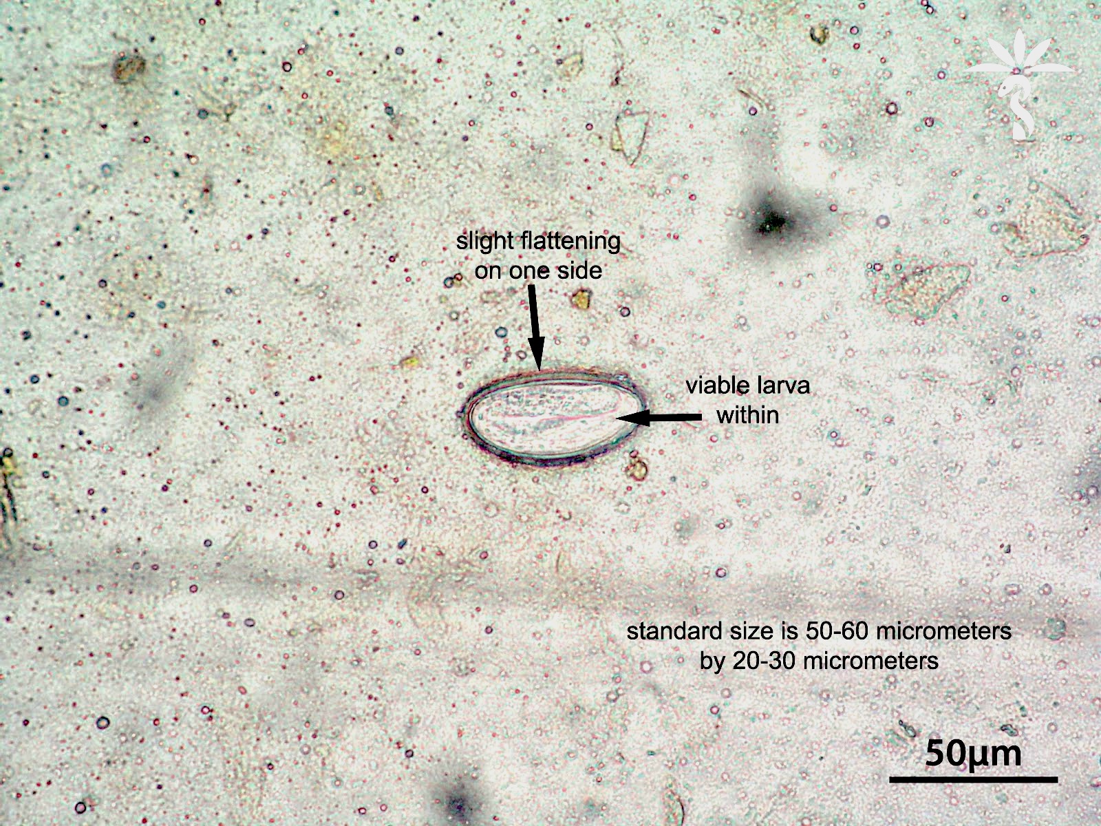
Enterobius egg with larva
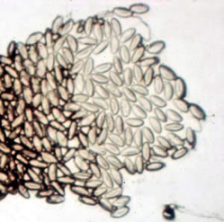
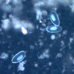
Eggs
Contributed by Bobbi Pritt, M.D. and Chamarajan Shrinivasan, M.D.
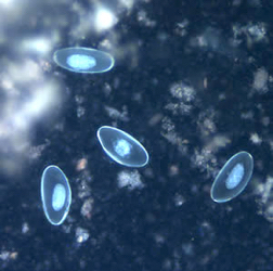
Wet mount from stool sample

Ova and larva of Enterobius vermicularis
Contributed by Centers for Disease Control and Prevention
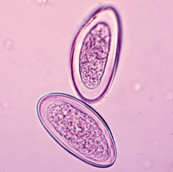
Eggs
Contributed by Raul S. Gonzalez, M.D.
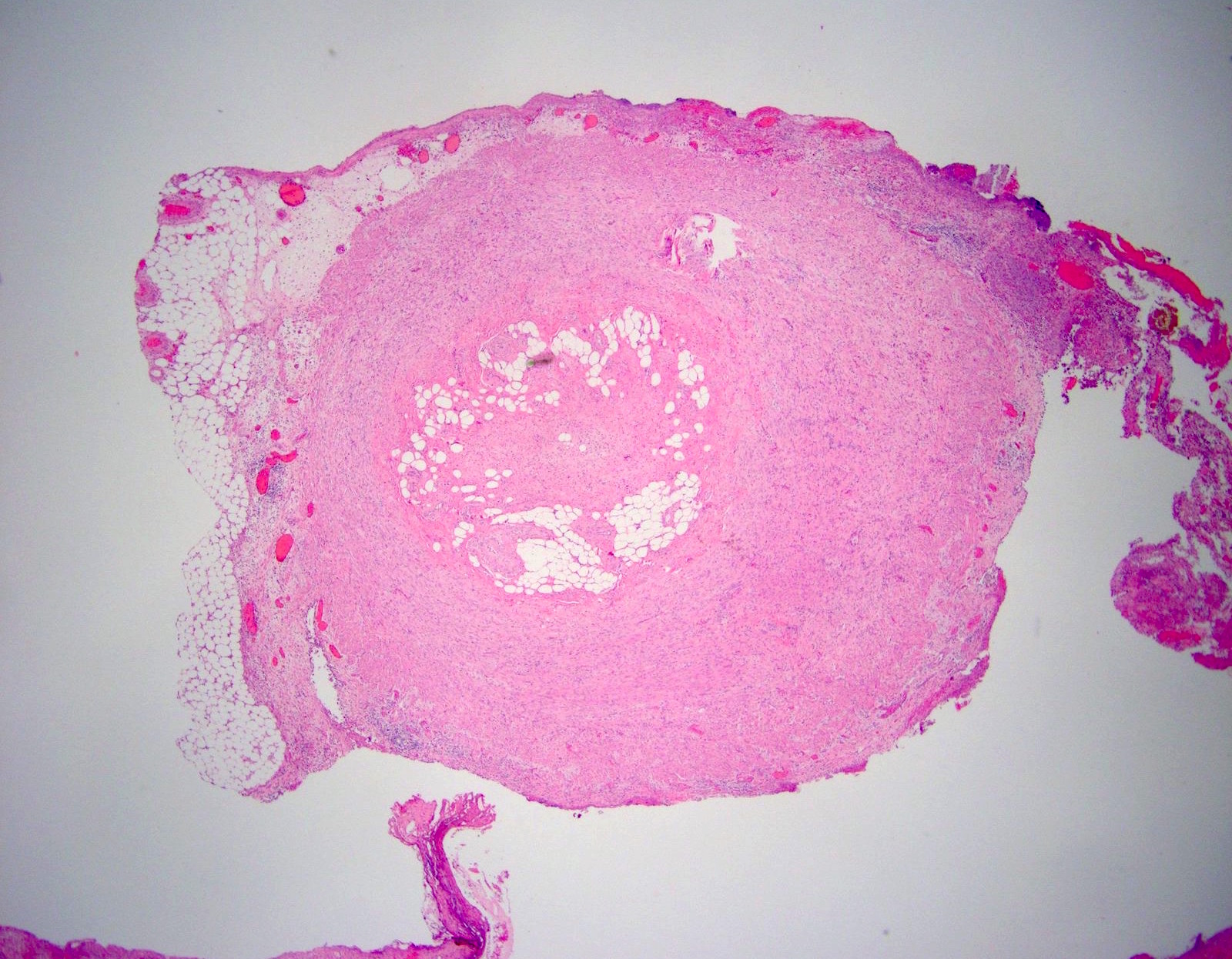
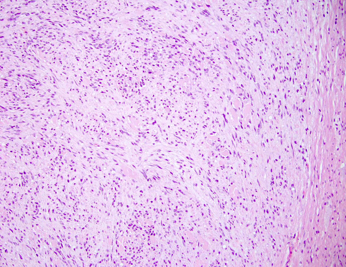
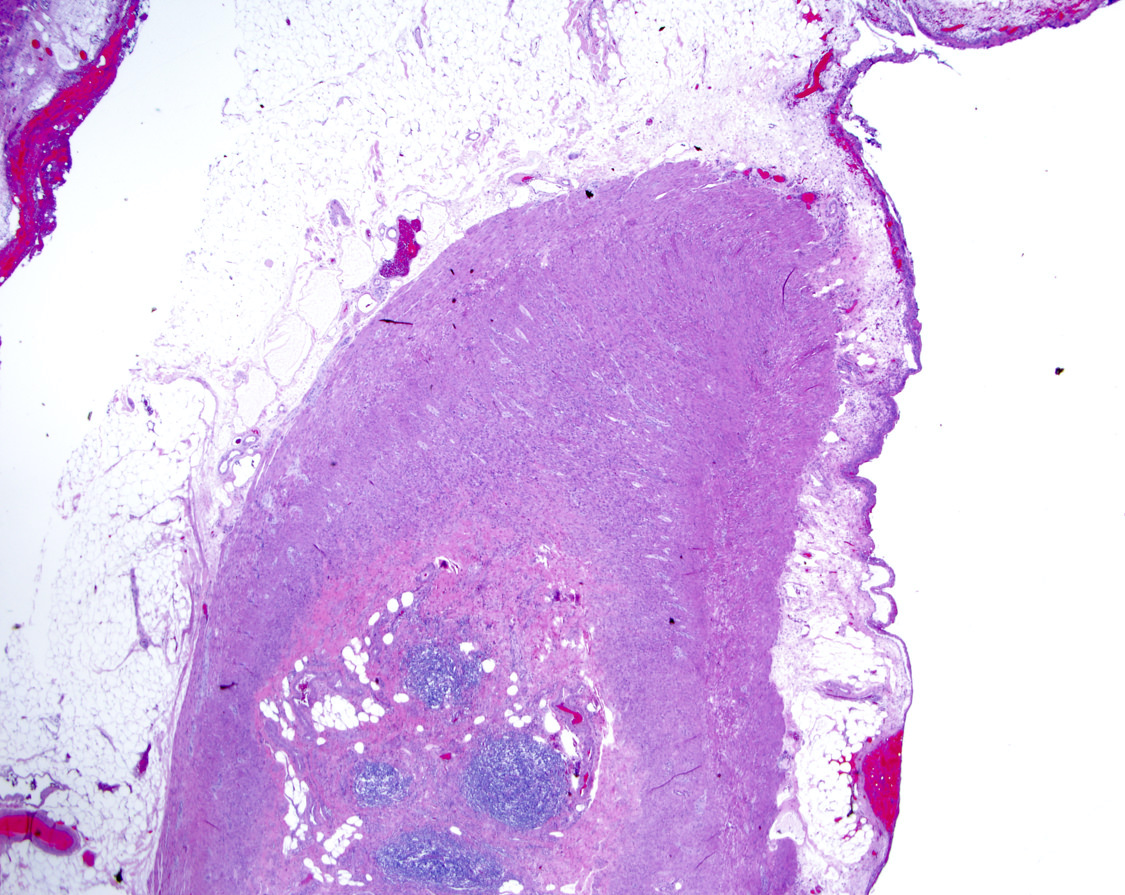
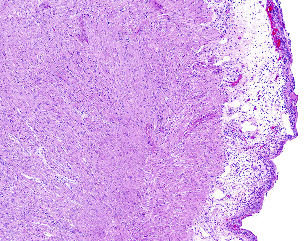
Obliterated lumen
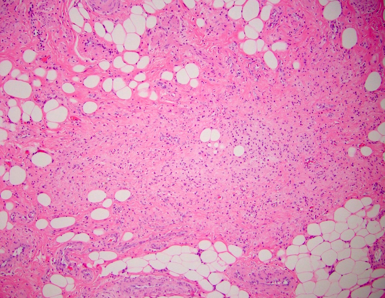
Adipose and spindle cells
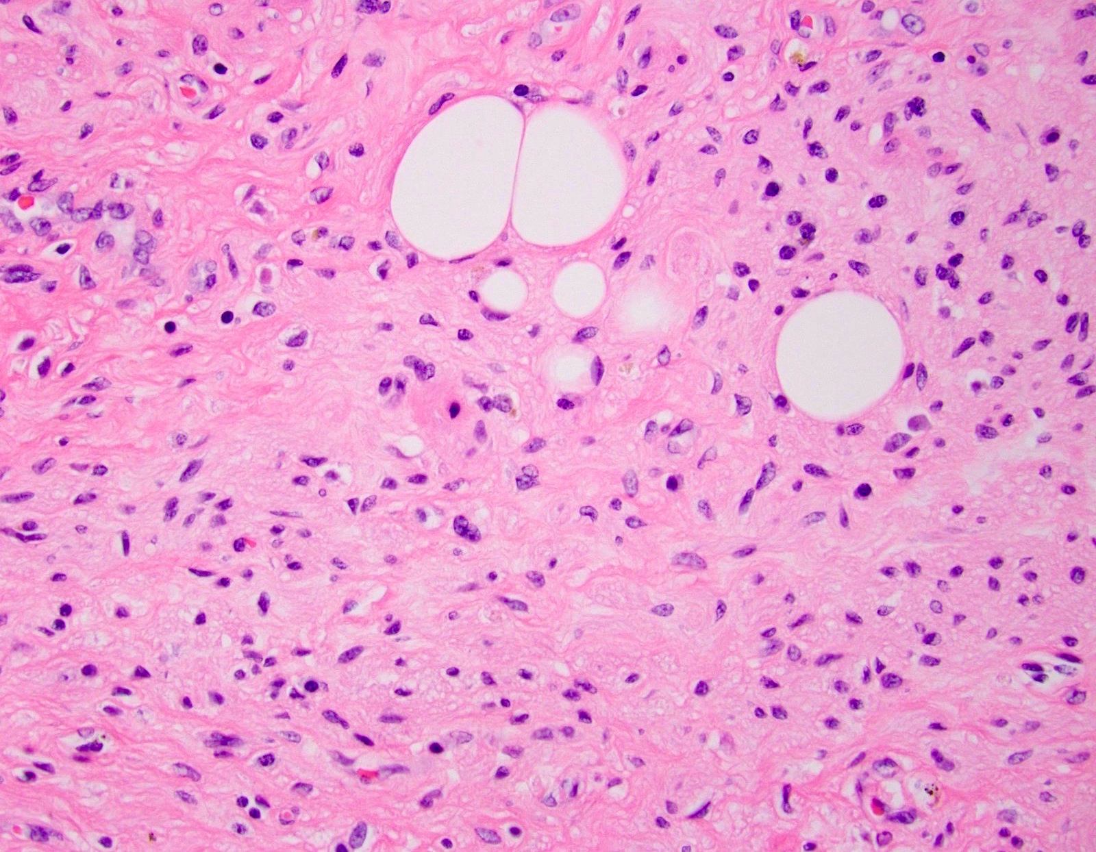
Bland spindle cells
Superimposed acute appendicitis
Images hosted on other servers:

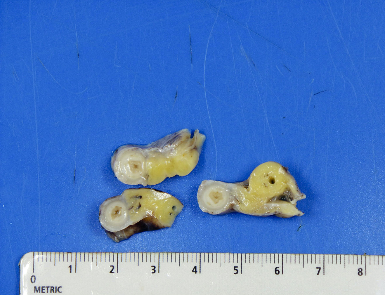
Small GIST of appendix

Pedunculated cystic GIST

Pedunculated GIST after imatinib
Contributed by Raul S. Gonzalez, M.D.
Well circumscribed tumor
Bland spindle cells
Images hosted on other servers:

Small appendiceal GIST

Bland appendix GIST
Images hosted on other servers:

CT abdomen: dilated and inflamed appendix

CT / MRI: ovary and appendix
Images hosted on other servers:

Laparoscopic view of an inflamed appendix
Contributed by Trang Lollie, M.D.
Concentric growth
Low grade growth pattern

High grade growth pattern
High grade growth pattern
Perineural invasion
Lymphovascular invasion
Images hosted on other servers:

Pleomorphic electron dense granules

Electron dense granules within mucin
Contributed by Maryam Kherad Pezhouh, M.D., M.Sc. and Masoumeh Peykan Heyraty, M.D.
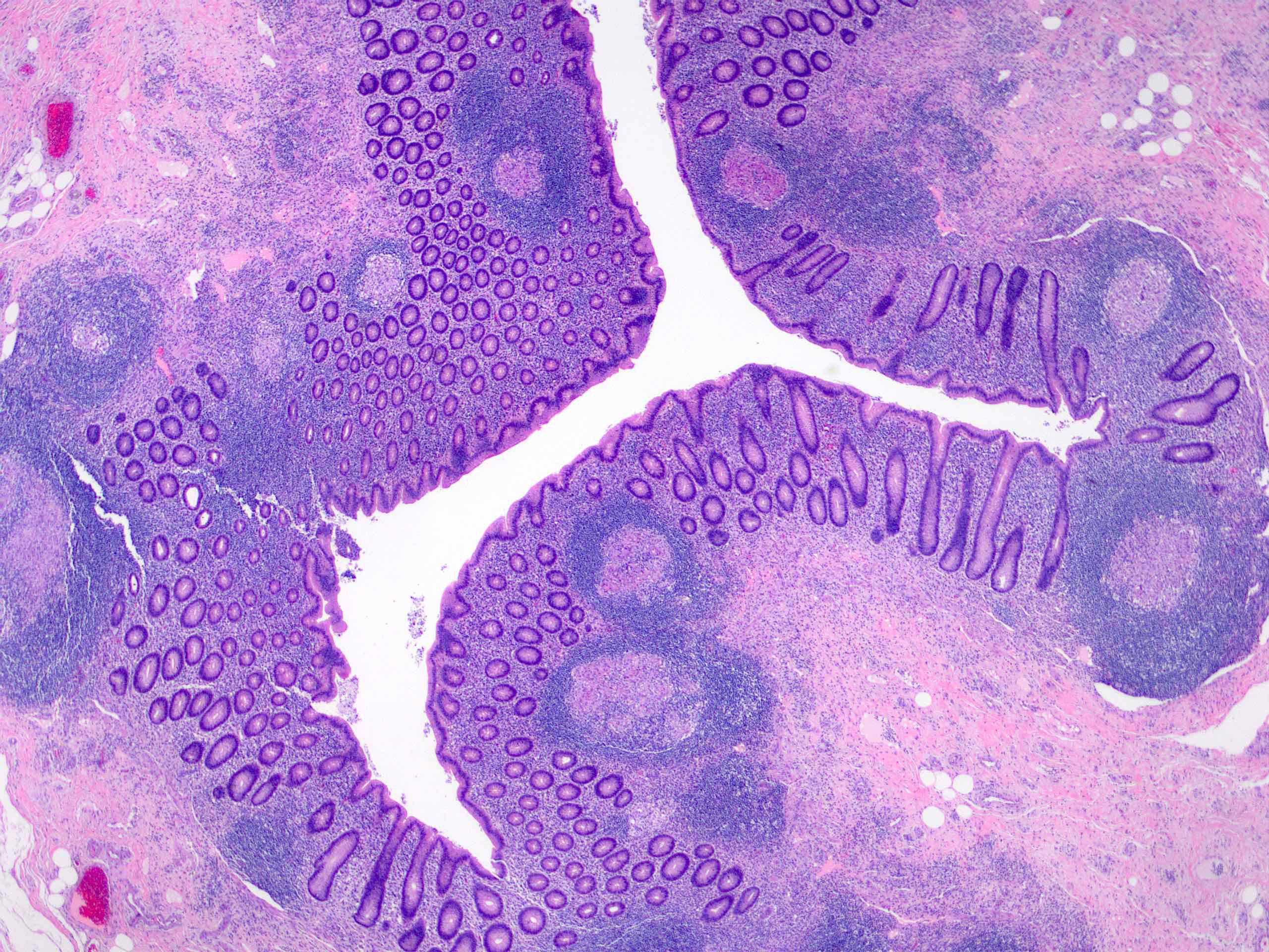
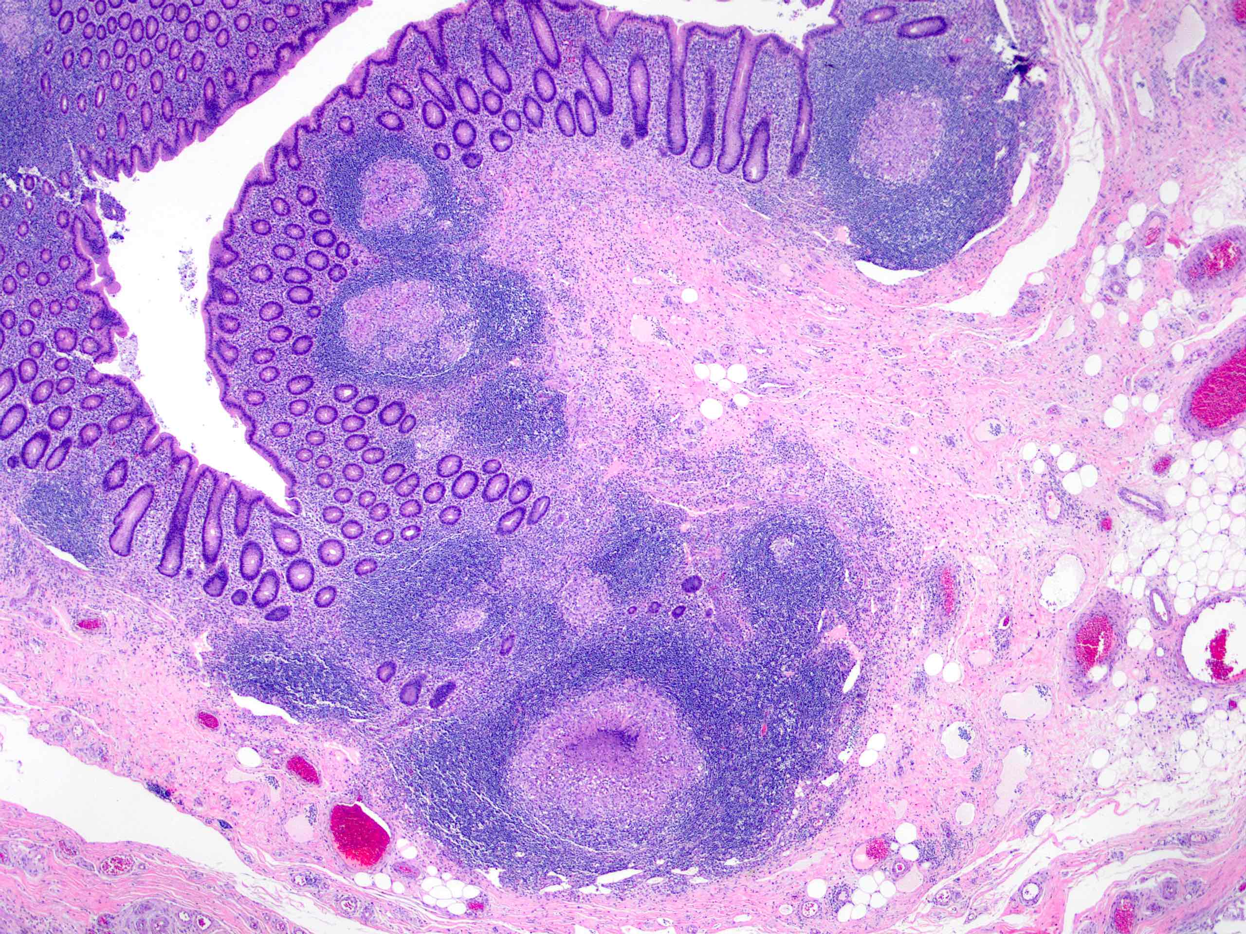
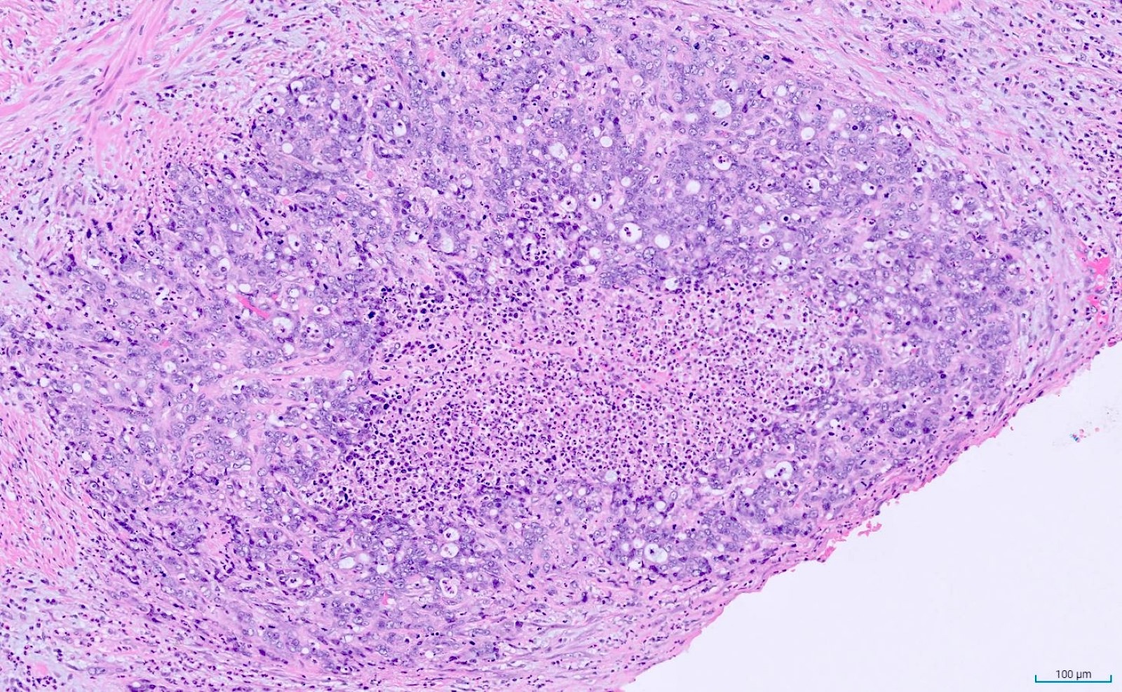
Multiple granulomas
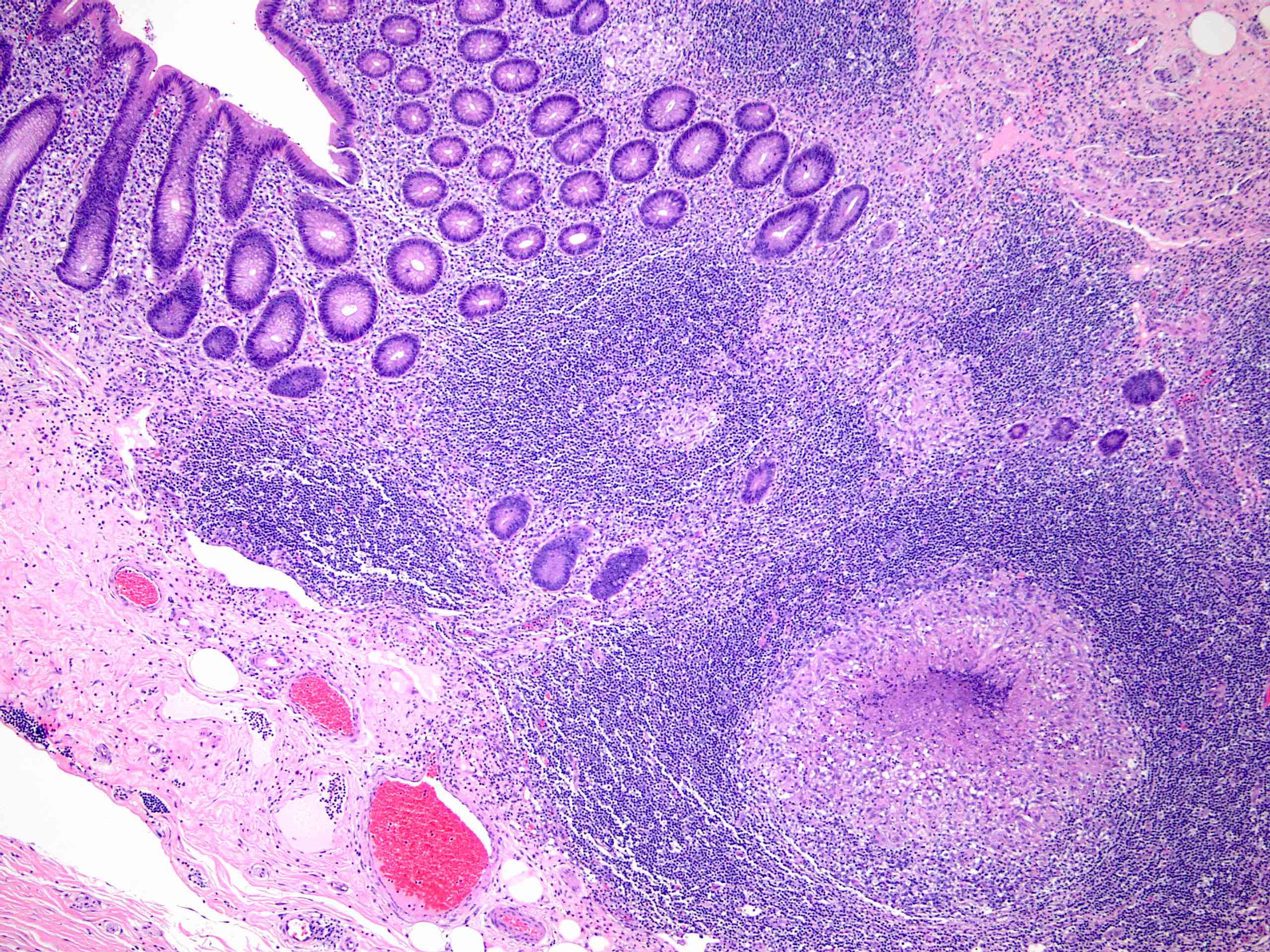
Focal necrotizing granuloma
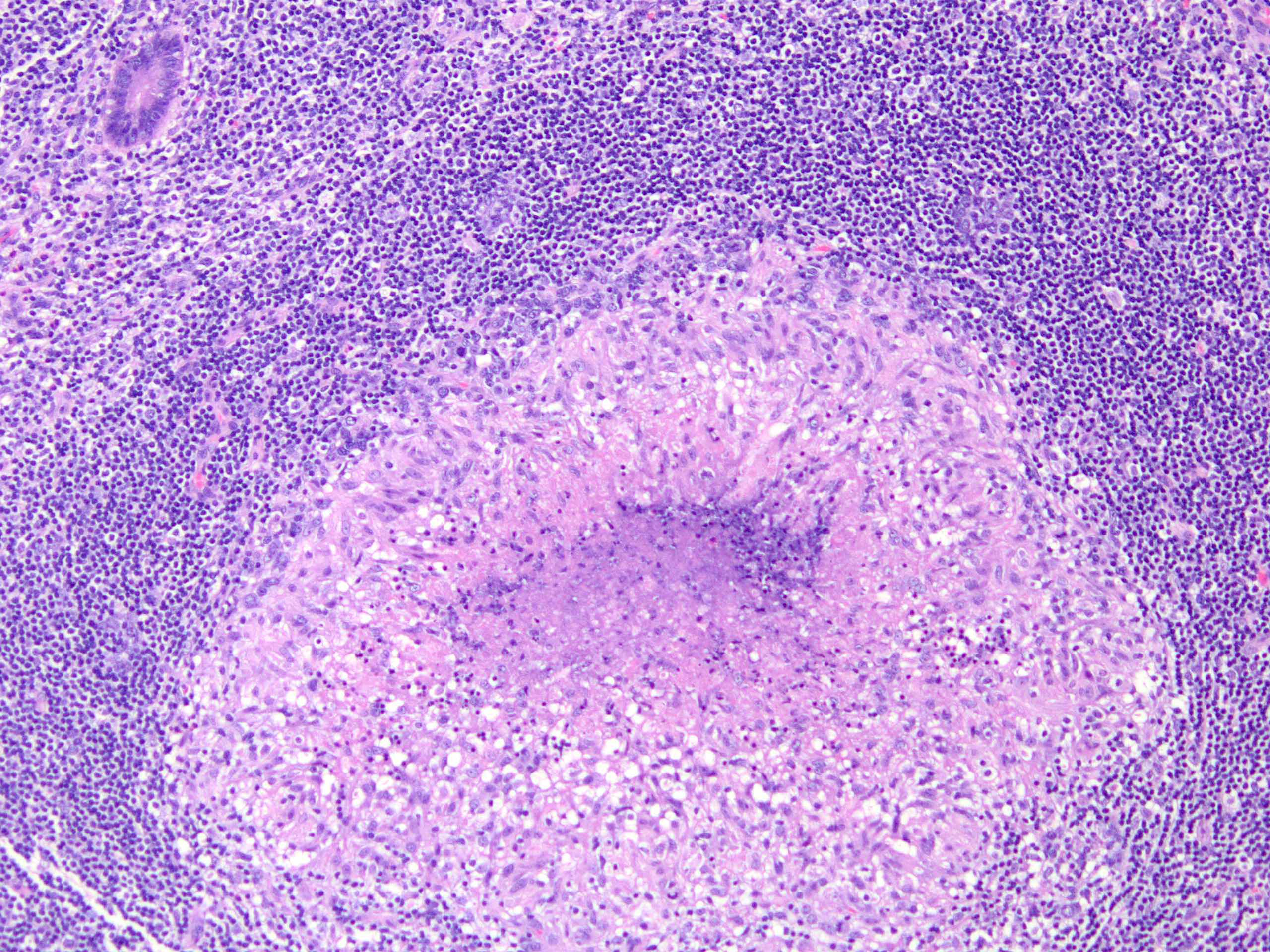
Necrotizing granuloma

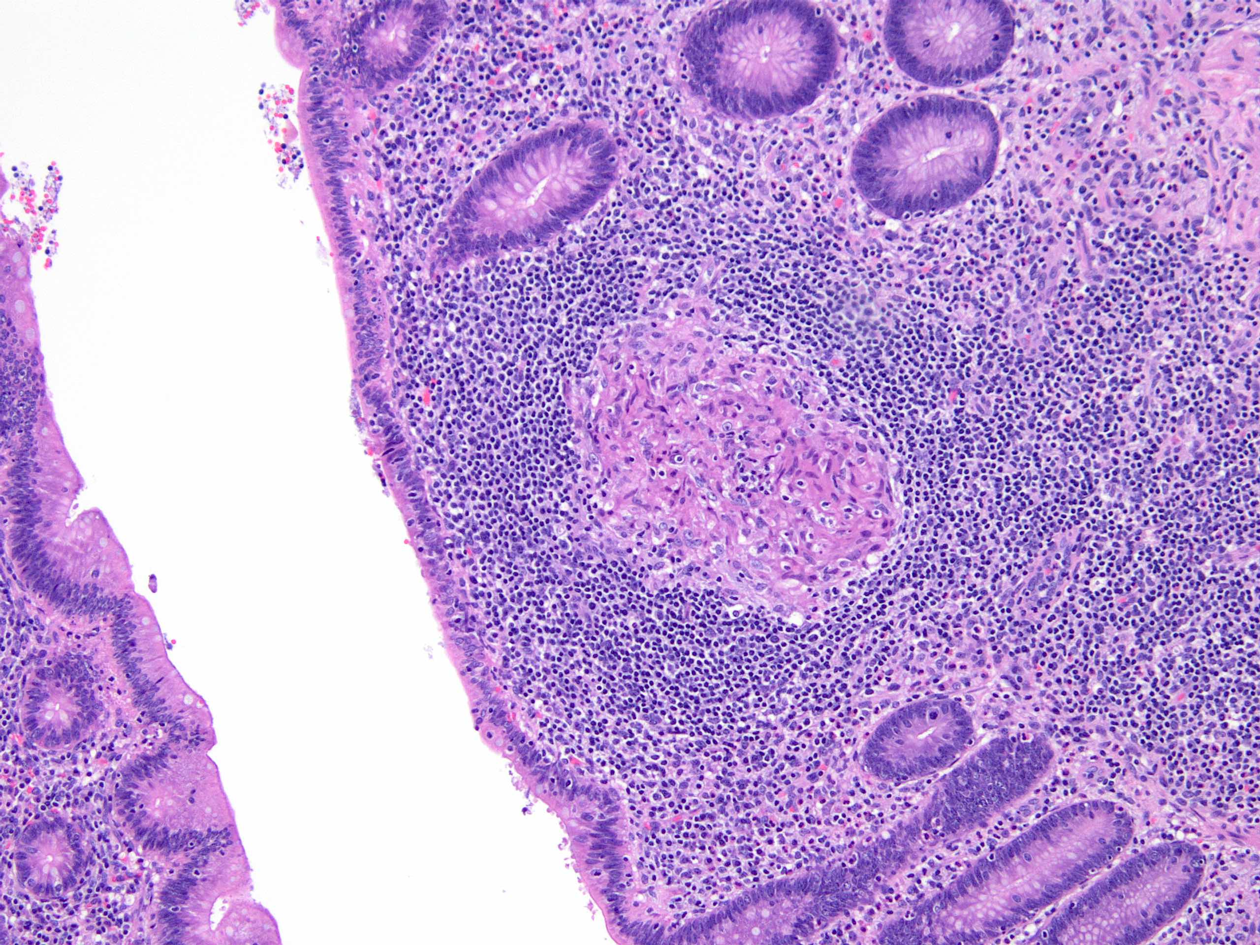
Well formed granulomas
Contributed by Aaron R. Huber, D.O.
Associated acute appendicitis
Serrated epithelium
Images hosted on other servers:

Abscess resolved after treatment
Images hosted on other servers:

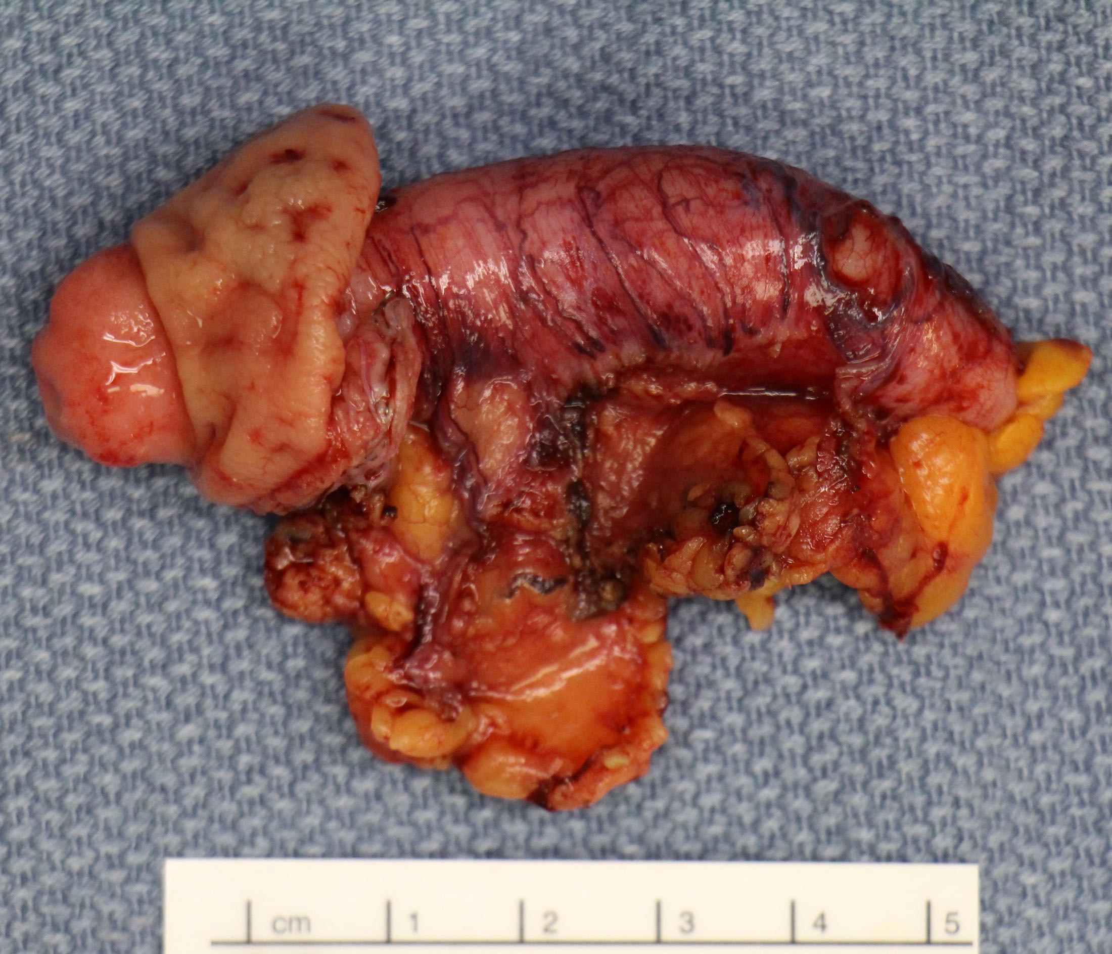
Interval appendectomy
Contributed by Pu Ni, M.D. and Qingqing Liu, M.D., Ph.D.
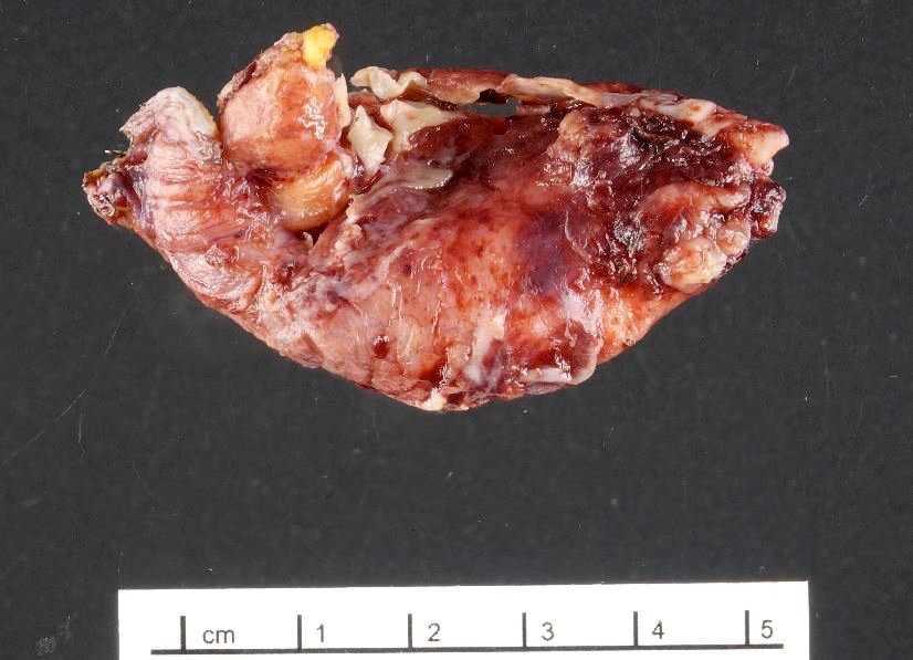
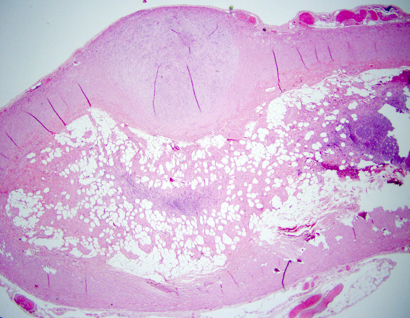
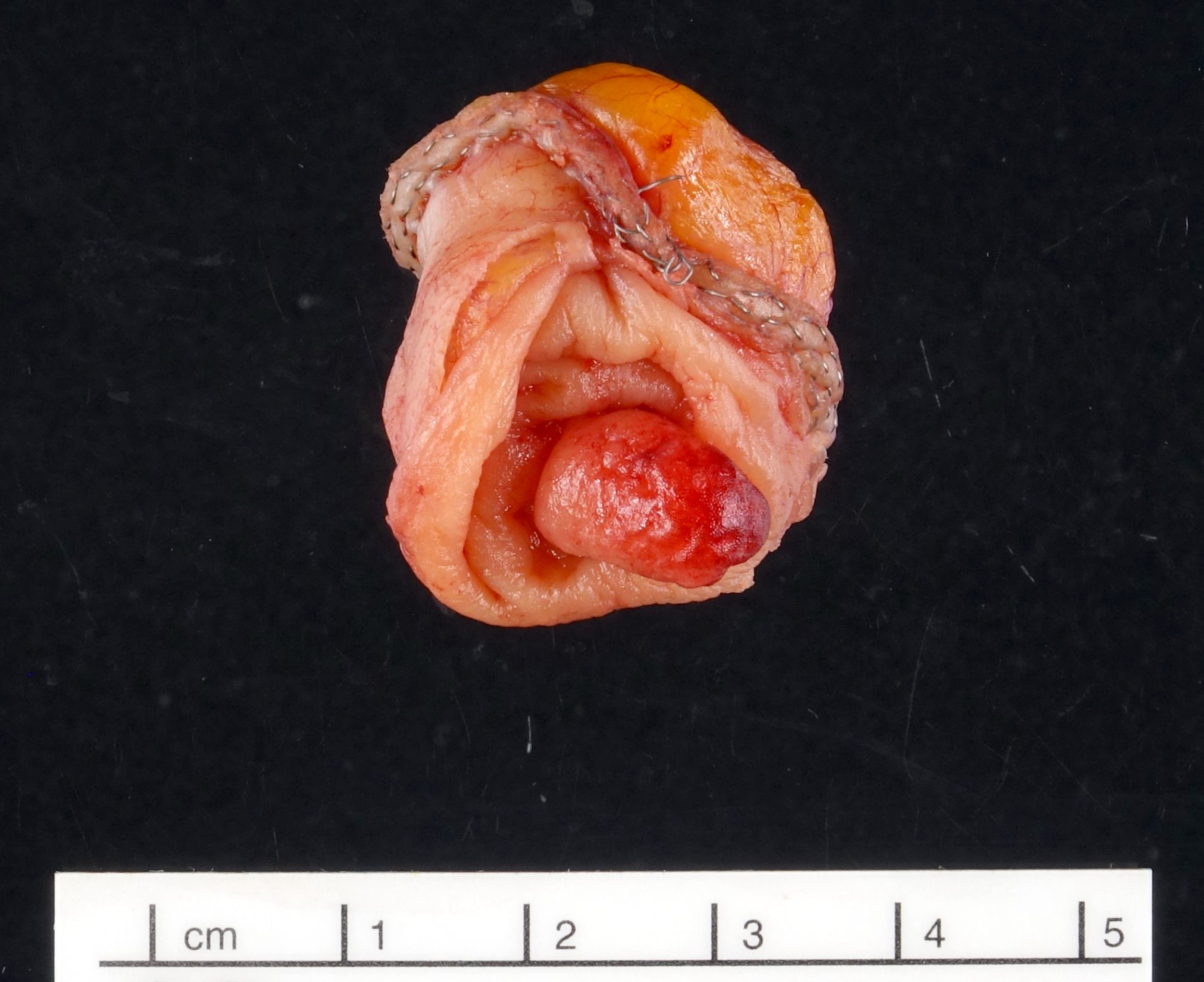
Mildly enlarged appendix
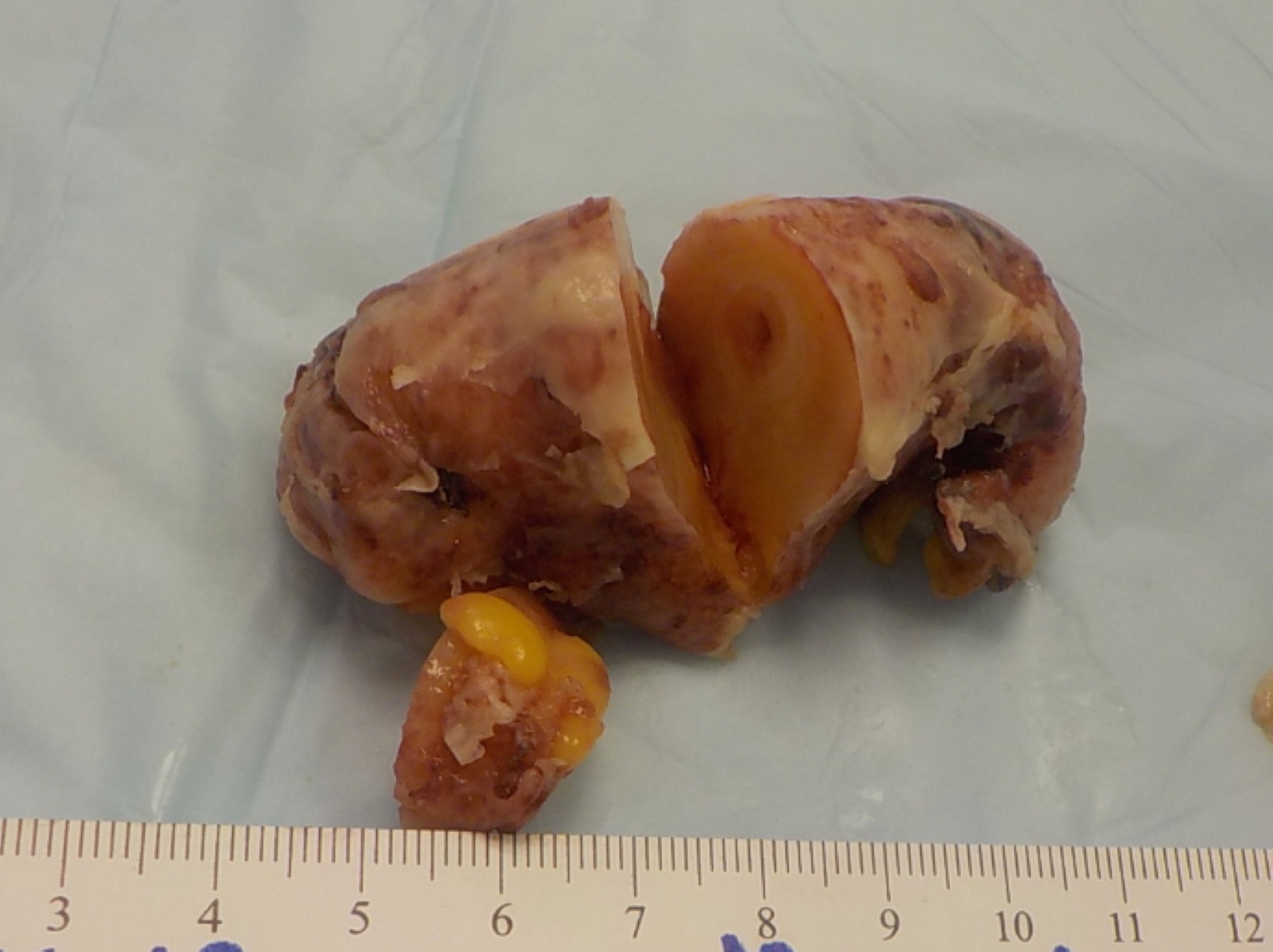
Significantly enlarged appendix
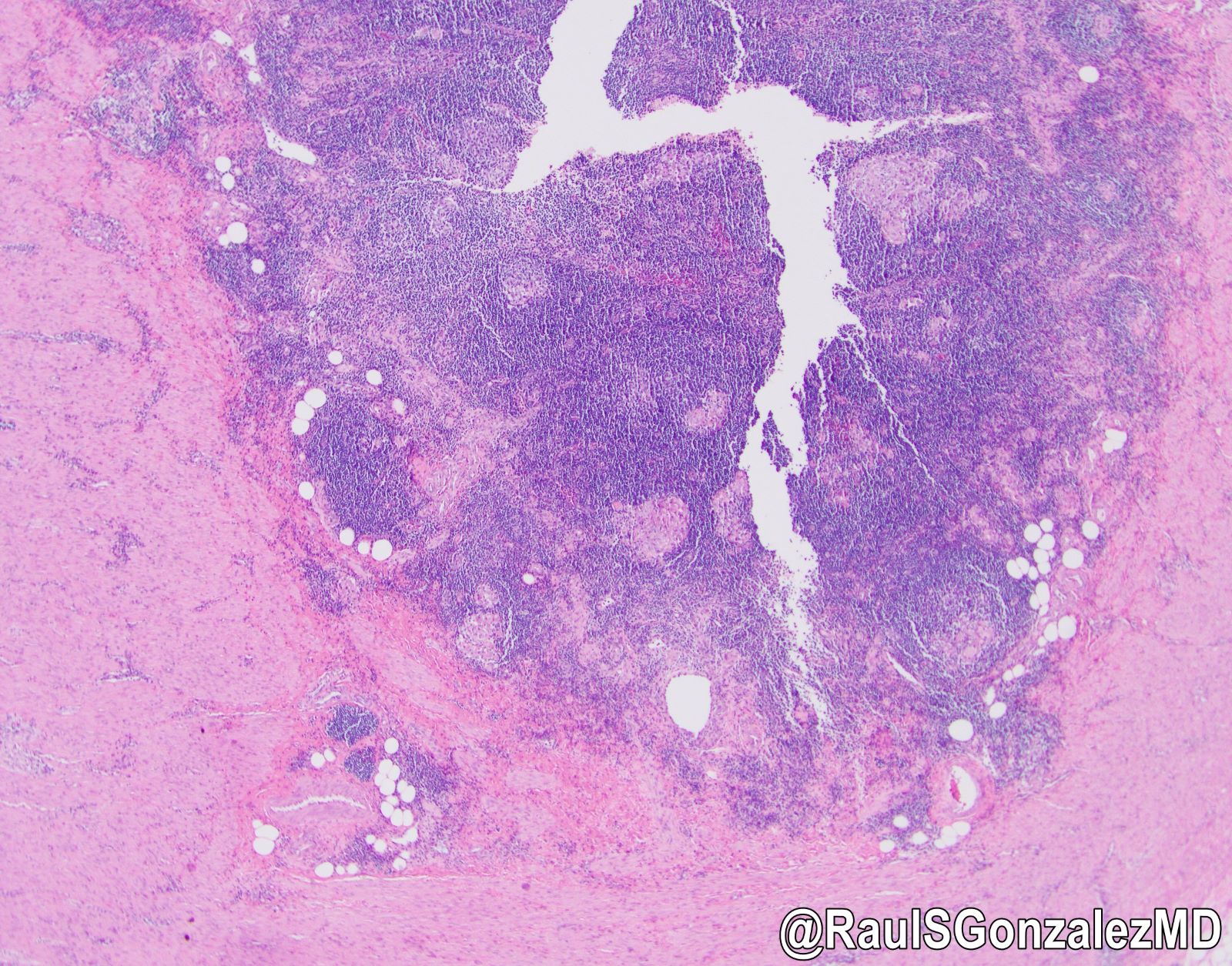
Inflammatory polyp mimicking neoplasm
Contributed by Pu Ni, M.D., Qingqing Liu, M.D., Ph.D. and @RaulSGonzalezMD on Twitter
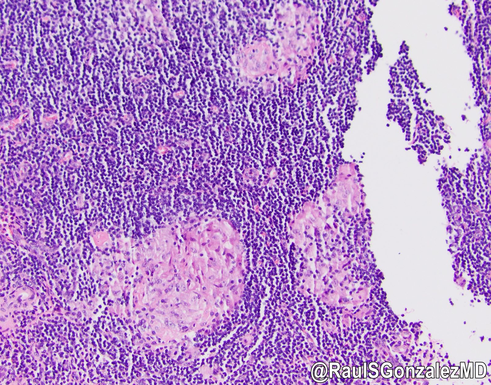
Granulomas and
xanthogranulomatous
inflammation
Xanthogranulomatous
inflammation
Granulomas
Mural fibrosis
Transmural chronic inflammation
Contributed by @RaulSGonzalezMD on Twitter (see original post here)">
 tinyurl.com/2ndx69rz / #pathology #PathTwitter #gipath #PathOutPic"
Contributed by @RaulSGonzalezMD on Twitter (see original post here)">
Interval appendicitis
Images hosted on other servers:

CT demonstrating an intraluminal tubular structure

Intraluminal protruding mass with central fat component

Cecal thickening without visualization of appendix
Images hosted on other servers:

Elongated lesion at appendiceal orifice

Tubular structure in cecum

Endoscopy
Contributed by Mahzad Azimpouran, M.D. and Danielle Hutchings, M.D.
Elongated tubular lesion
Inversion of appendiceal base
Associated with mucinous neoplasm
Serosal surface
Contributed by Mahzad Azimpouran, M.D. and Danielle Hutchings, M.D.
Dome shaped configuration
Polypoid configuration
Ganglion cell aggregates
Ganglion cells and nerves
Lymphoid aggregates
Appendiceal mucinous neoplasm
Contributed by @Andrew_Fltv on Twitter

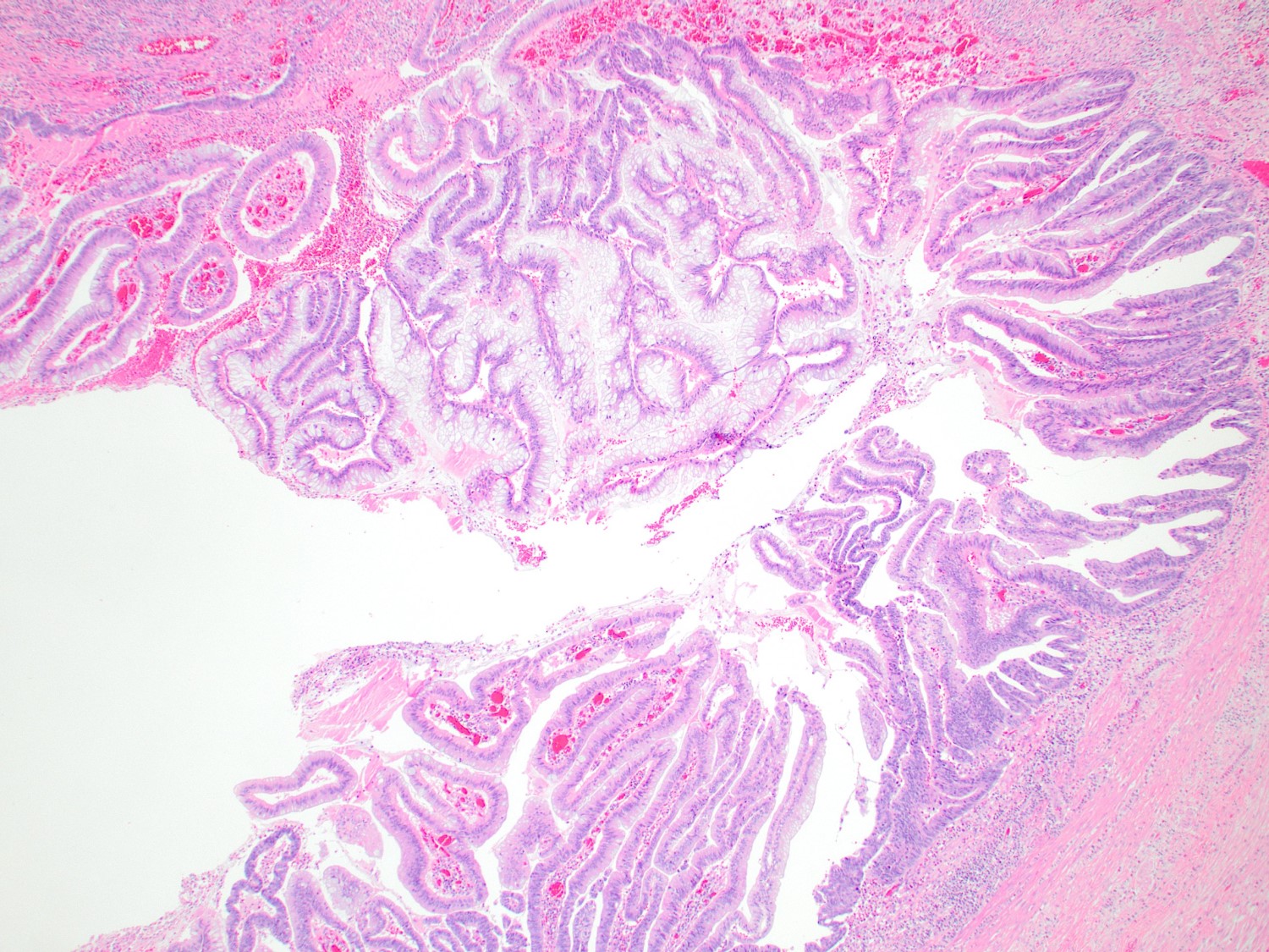
LAMN and HAMN
Images hosted on other servers:

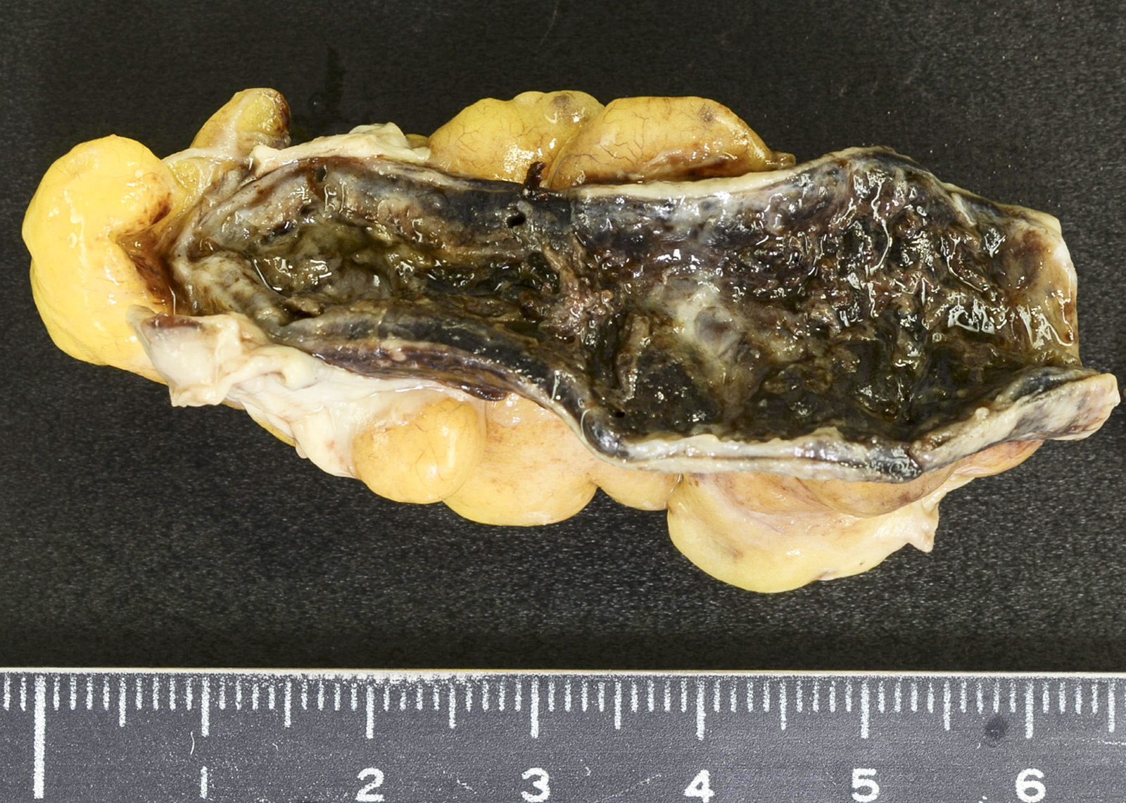
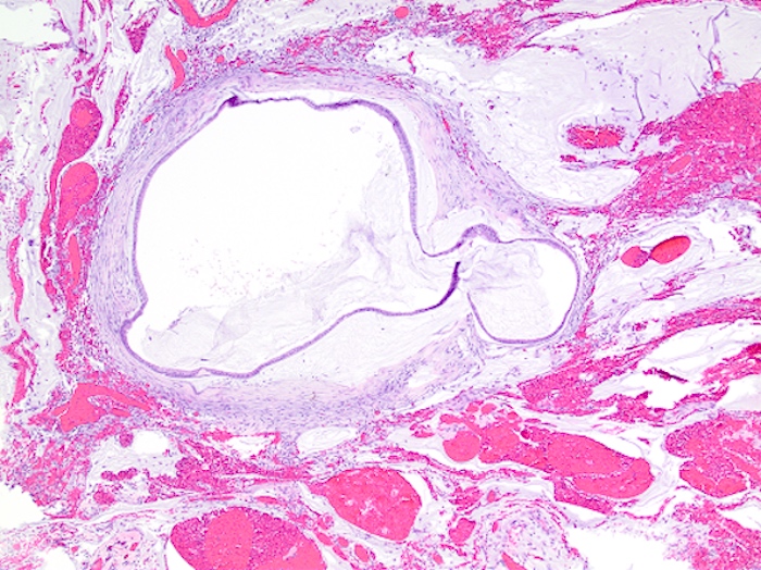
Mucocele: dilated appendix
Contributed by Raul S. Gonzalez, M.D. and Michael Feely, D.O.
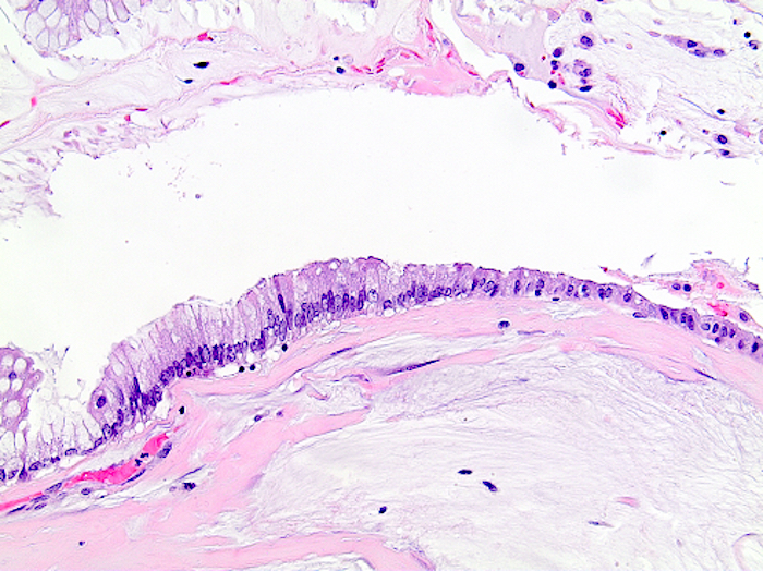
Denuded LAMN
LAMN with loss of muscularis mucosae
LAMN with pushing invasion
HAMN

LAMN in cross section

Villous architecture in LAMN
Low grade epithelium in LAMN

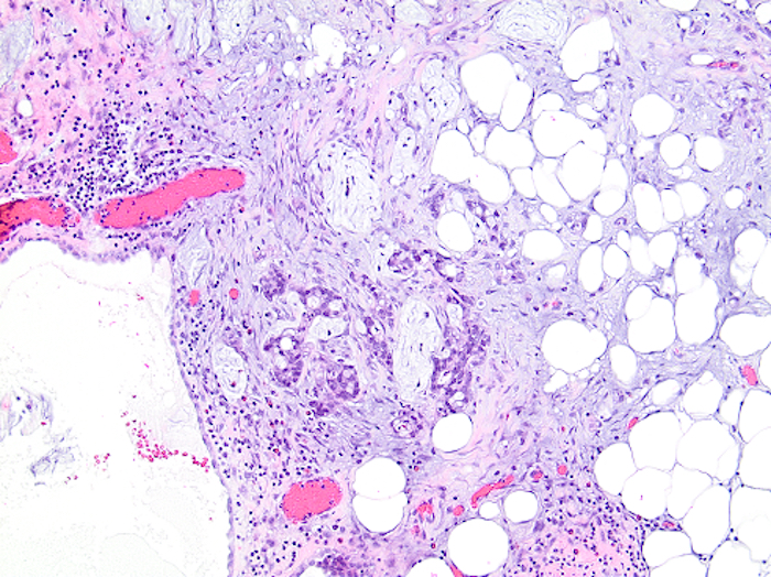
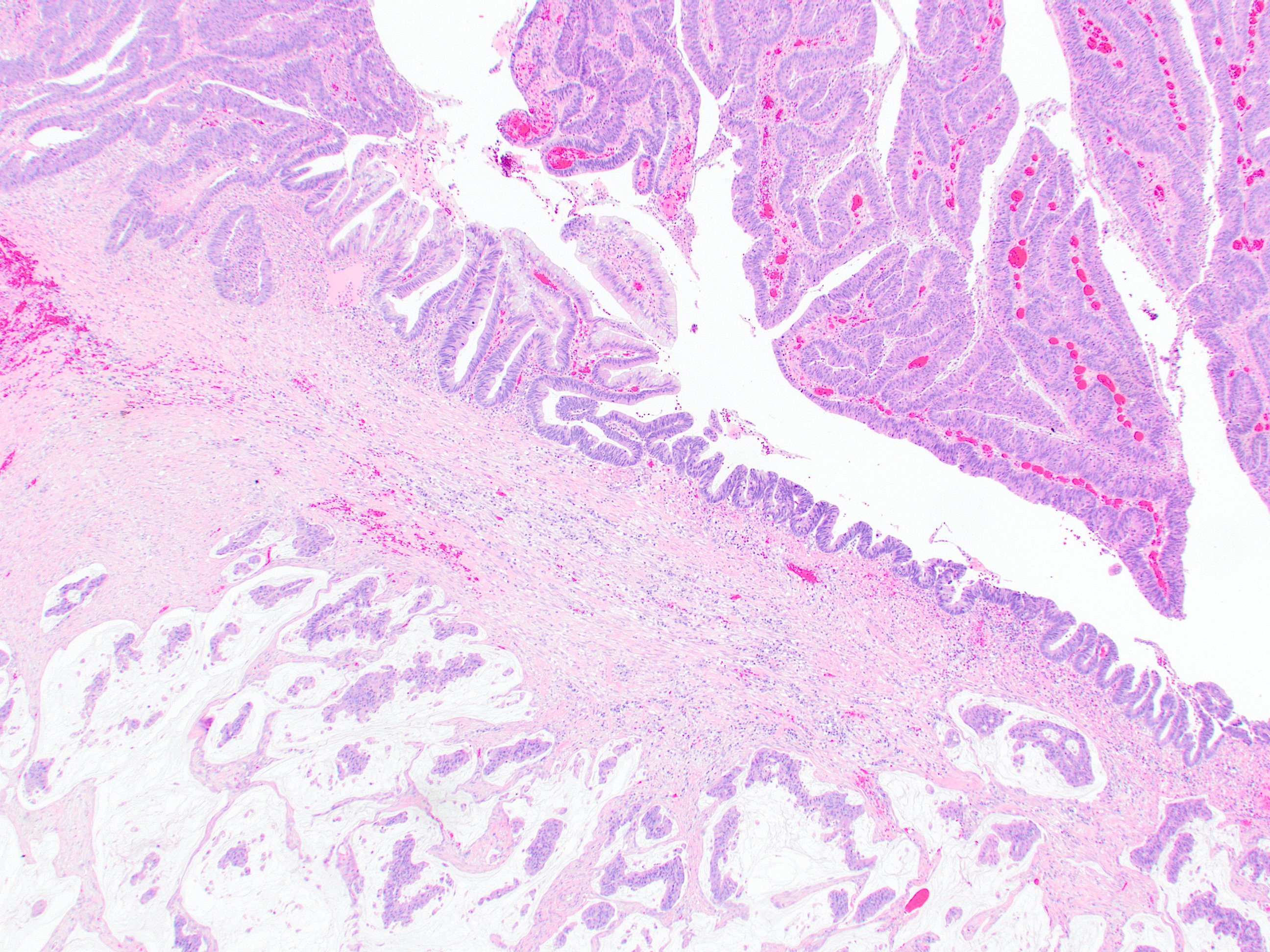
LAMN with extra-appendiceal mucin

Extra-appendiceal mucin with serosal reaction

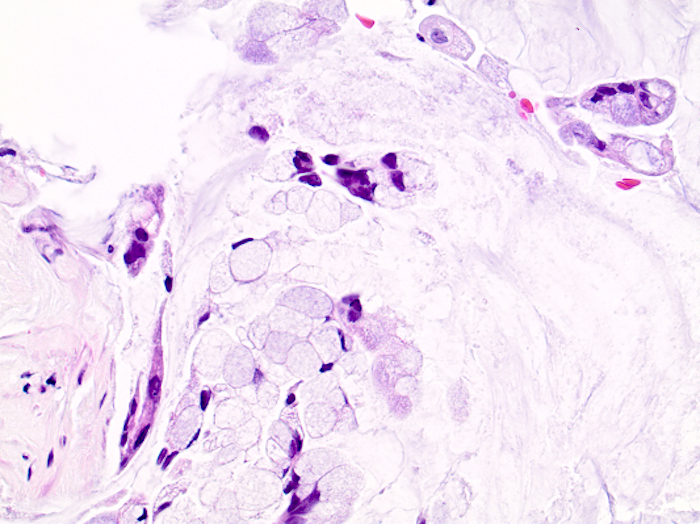
Cellular extra-appendiceal mucin
Grading appendiceal mucinous neoplasms
| Tumor grade | In the appendiceal primary tumor | In the peritoneal metastasis |
| 1 | Low grade cytology with a pushing margin (low grade appendiceal mucinous neoplasm) | Hypocellular mucinous deposits Neoplastic epithelial elements have low grade cytology No infiltrative type invasion |
| 2 | High grade cytology with a pushing margin (high grade appendiceal mucinous neoplasm) Invasive mucinous adenocarcinoma without a signet ring cell component | Hypercellular mucin deposits as judged at 20x magnification
High grade cytological features Infiltrative type invasion characterized by jagged or angulated glands in a desmoplastic stroma or a small mucin pool pattern with numerous mucin pools containing clusters of tumor cells |
| 3 | Signet ring cell adenocarcinoma with numerous signet ring cells in mucin pools or infiltrating tissue | Mucinous tumor deposits with signet ring cells |
- Note: several different neoplasms are covered by this table
- LAMN should be grade 1 (primary)
- HAMN should be grade 2 (primary)
- Mucinous adenocarcinoma should be grade 2 (primary)
- Signet ring cell carcinoma should be grade 3 (primary)
- Pseudomyxoma from LAMN or HAMN can be any grade (peritoneal)
- Pseudomyxoma from adenocarcinoma can be any grade (peritoneal) but is almost always grade 2 or 3
Images hosted on other servers:

Hypodense ovoid structure
Images hosted on other servers:

Distended appendix with intact wall
Images hosted on other servers:

Mucin and globules
Images hosted on other servers:

Alcian blue staining of globules
Images hosted on other servers:


Ultrasound and CT scan
Contributed by Robert Grefka, P.A. (ASCP)CM

Appendiceal NET involving mesentery
Contributed by Kwun Wah Wen, M.D., Ph.D. and @jgwBMS on Twitter
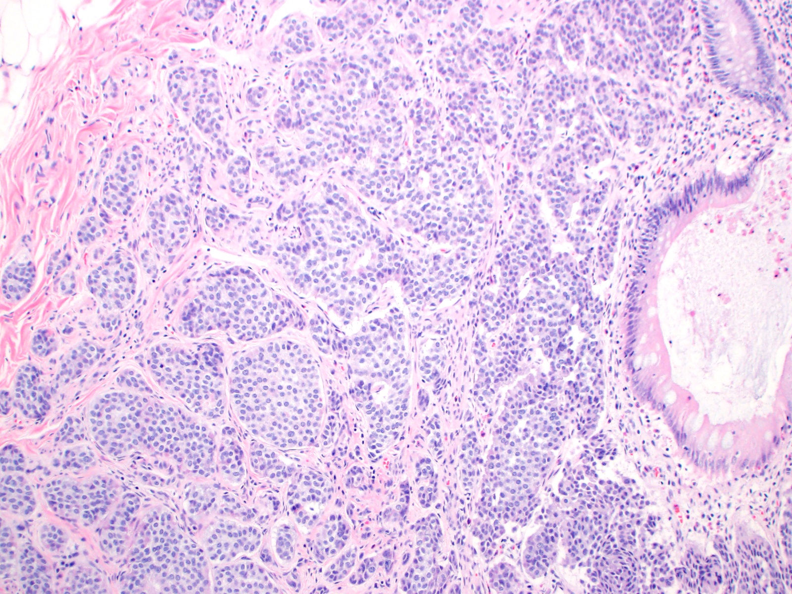
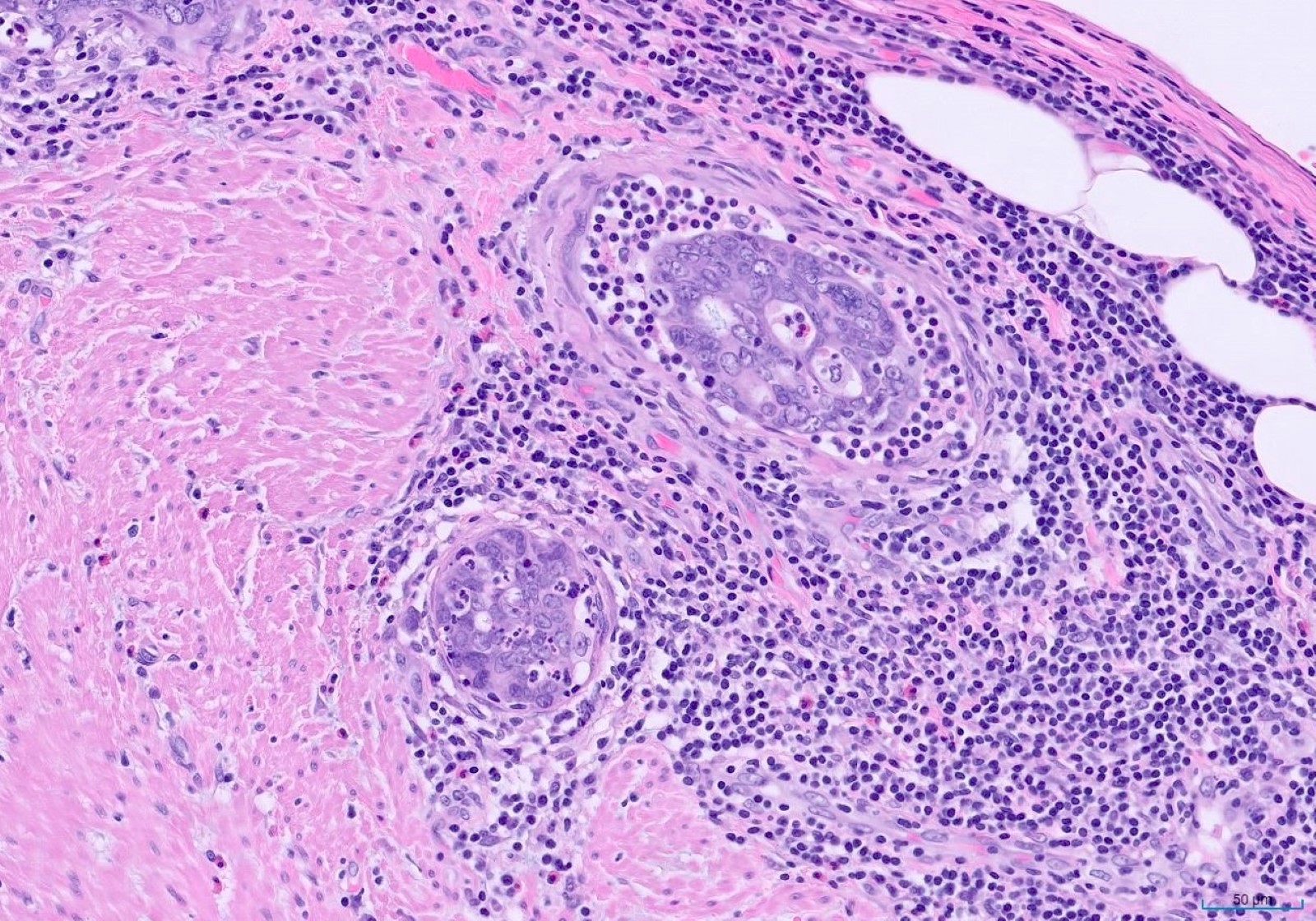
NET in nests

Nests and trabeculae
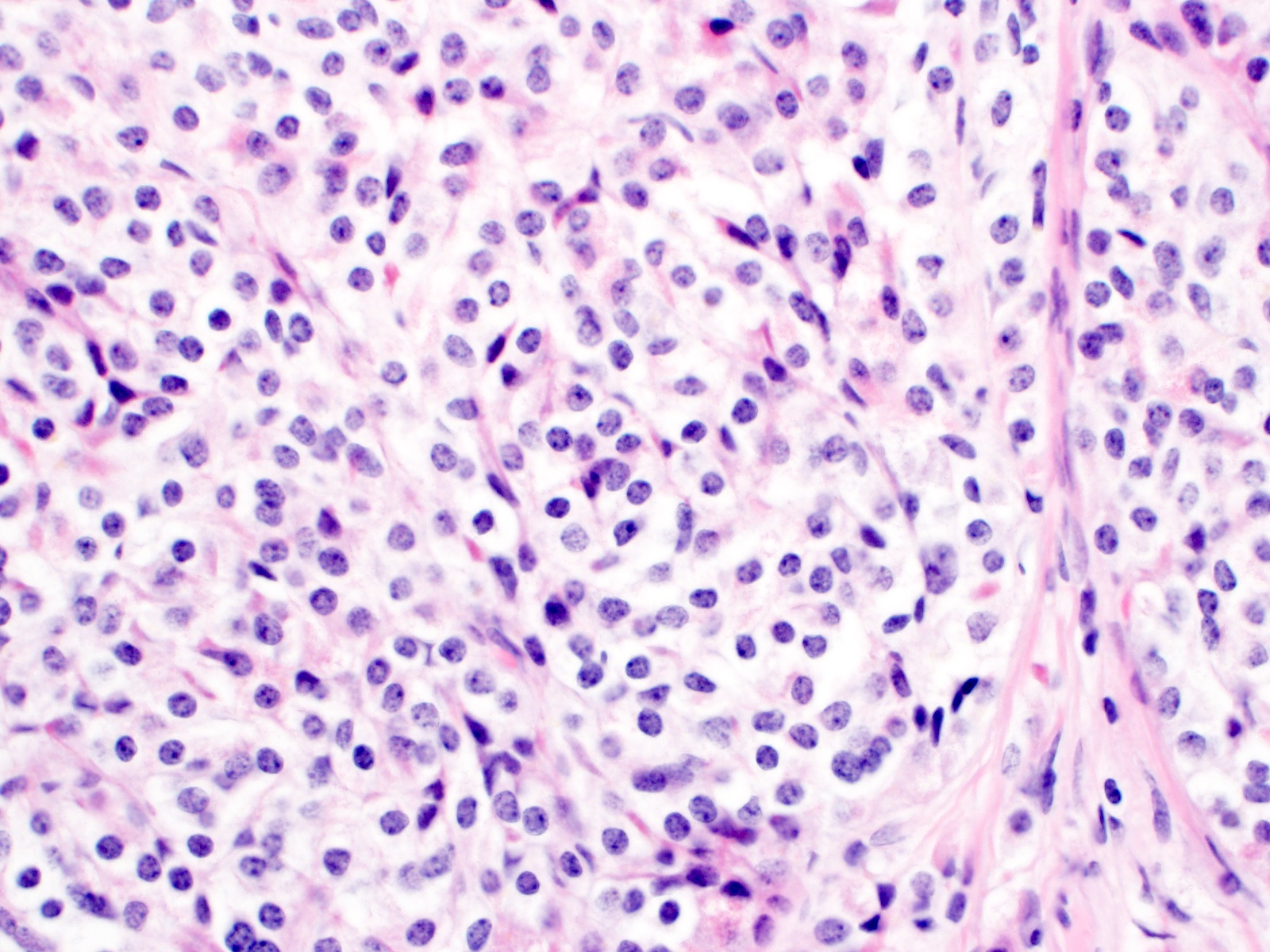
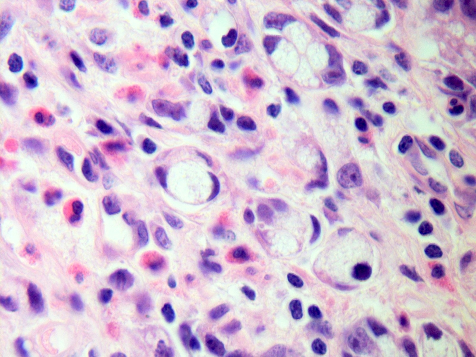
Monomorphic, low grade
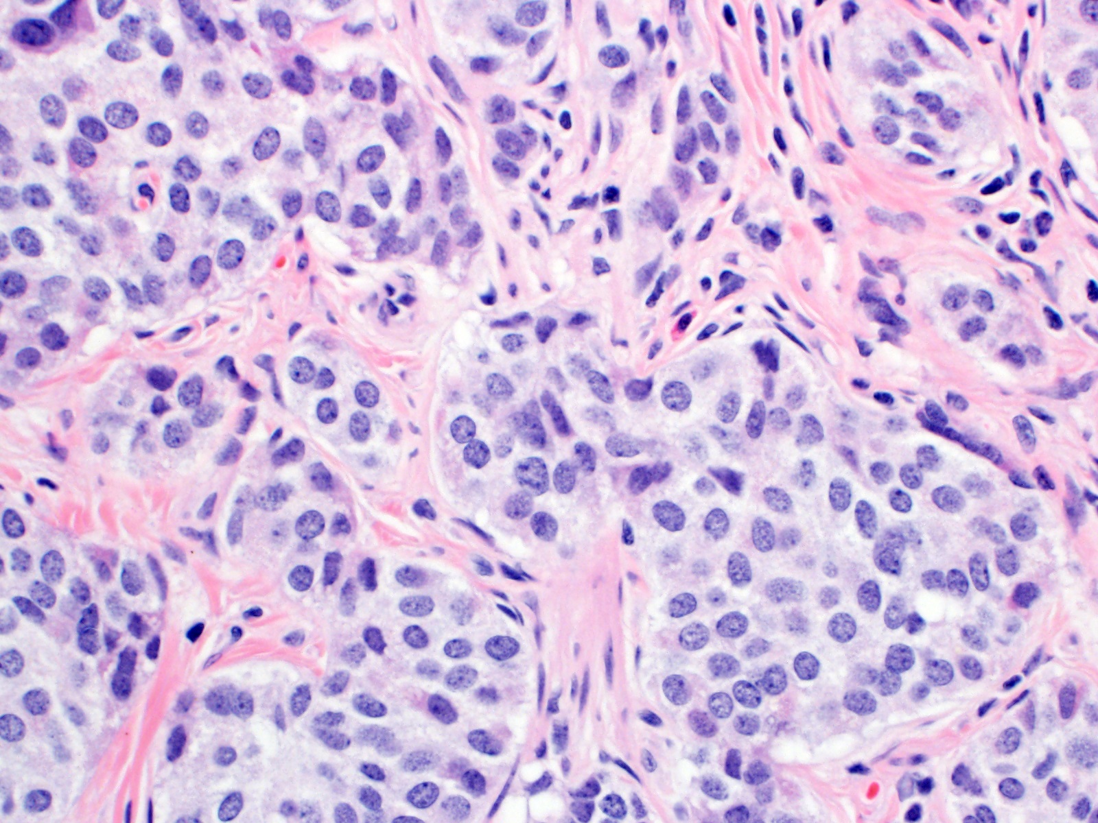
Salt and pepper chromatin

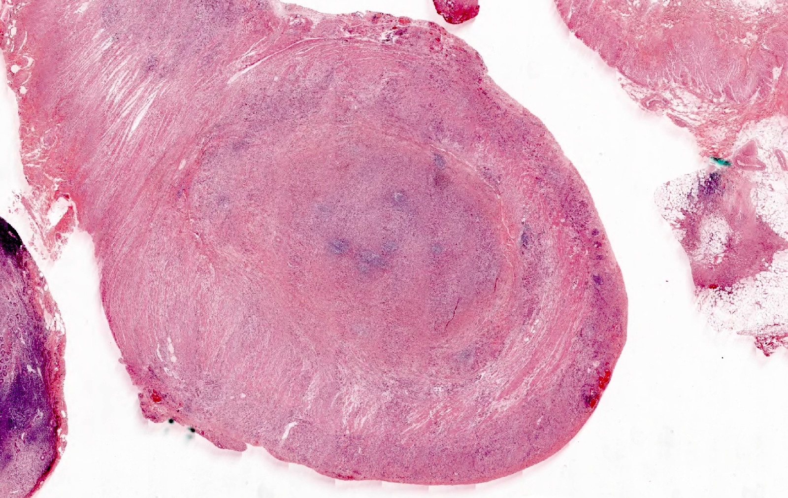
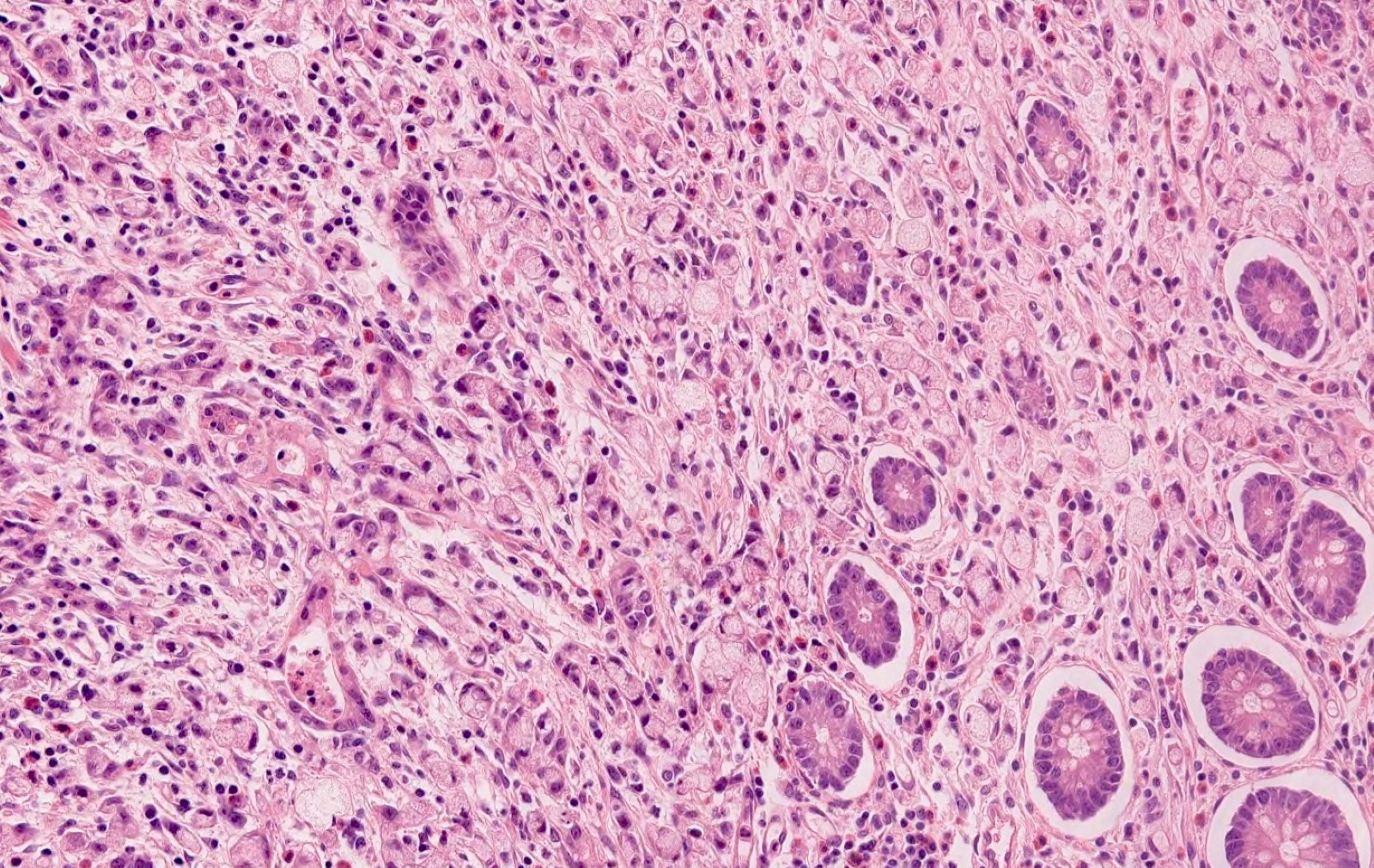
Well differentiated neuroendocrine tumor
Negative mucicarmine
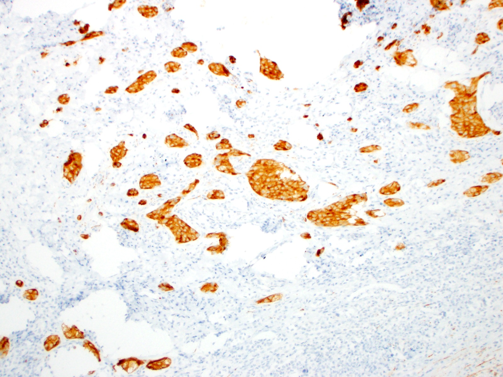
Diffusely strong synaptophysin
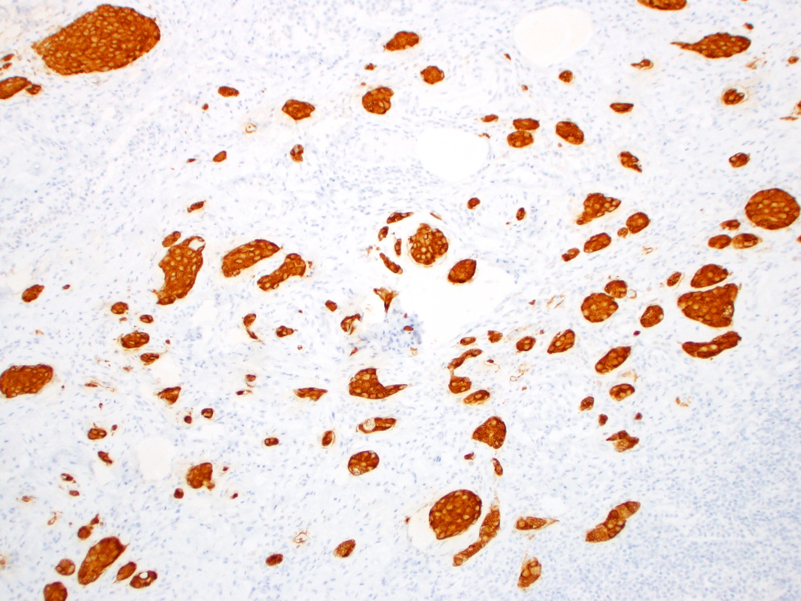
Diffusely strong chromogranin A
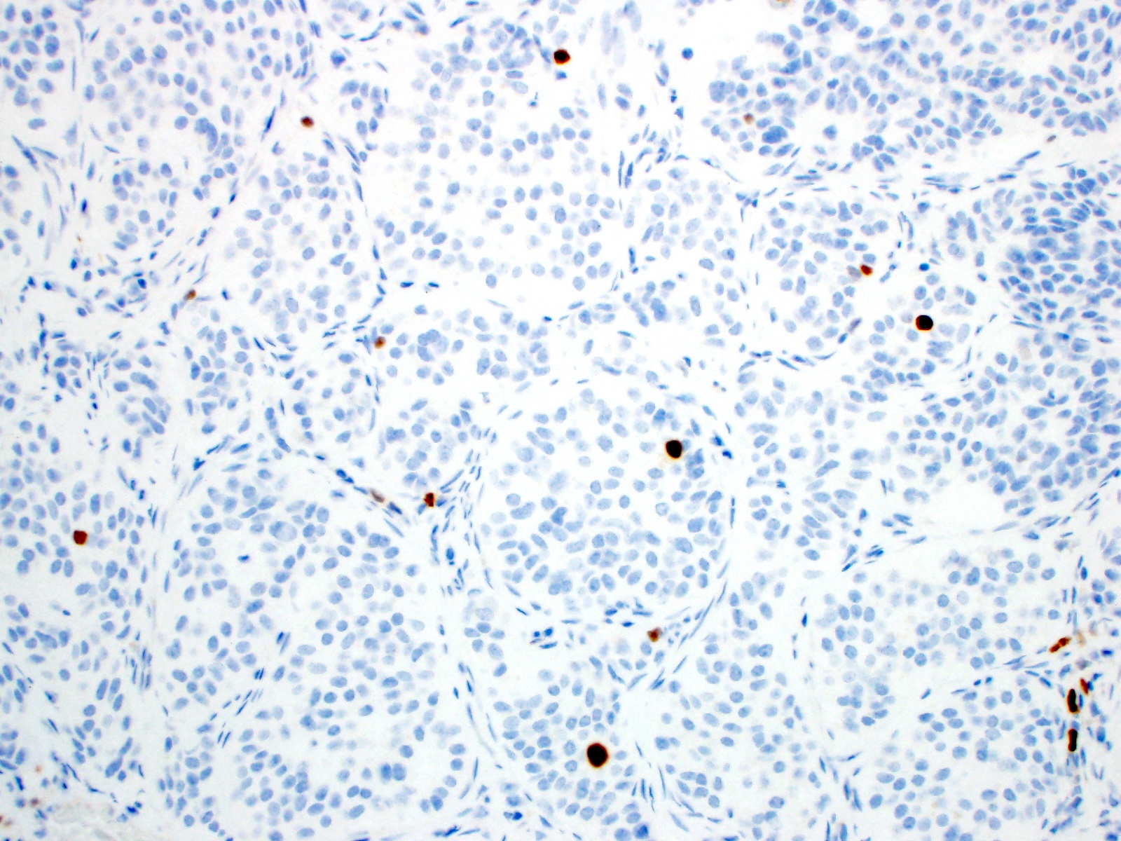
Low Ki67 index
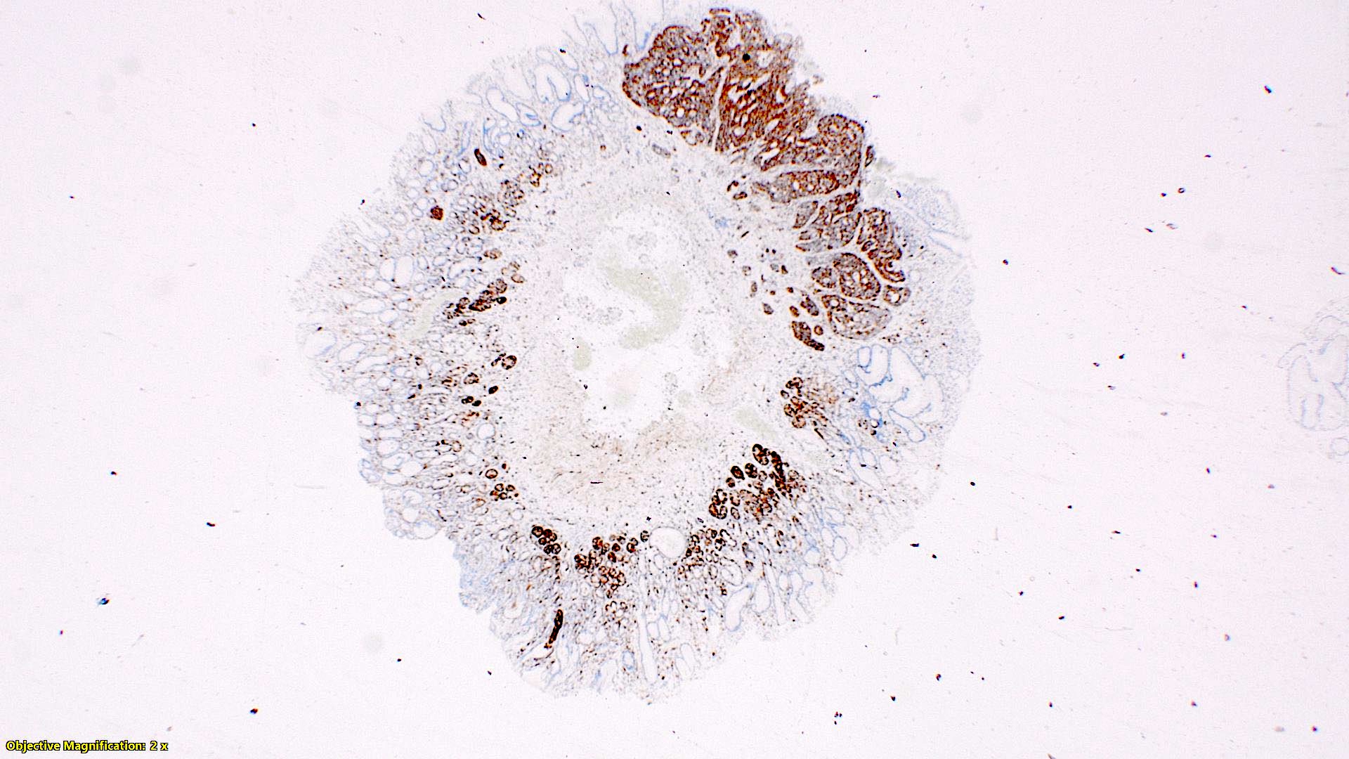
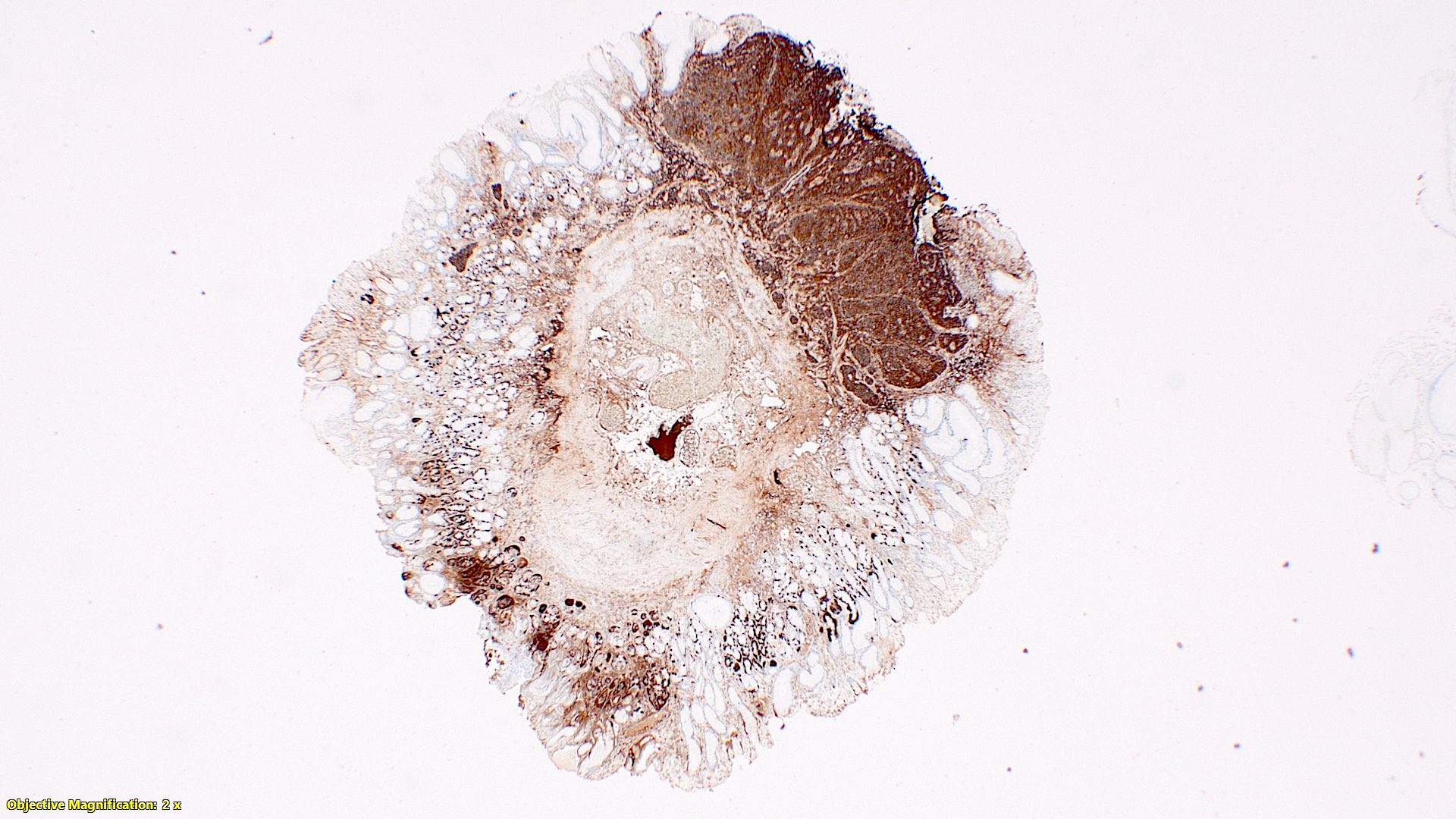
Well differentiated neuroendocrine tumor
Neuroendocrine tumor, appendix - histopathology
Contributed by Lukas Streich, M.D.
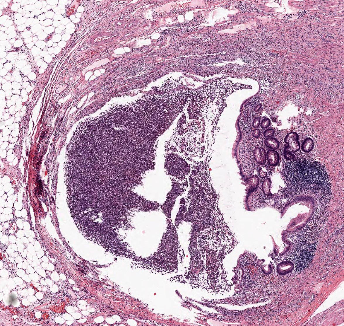
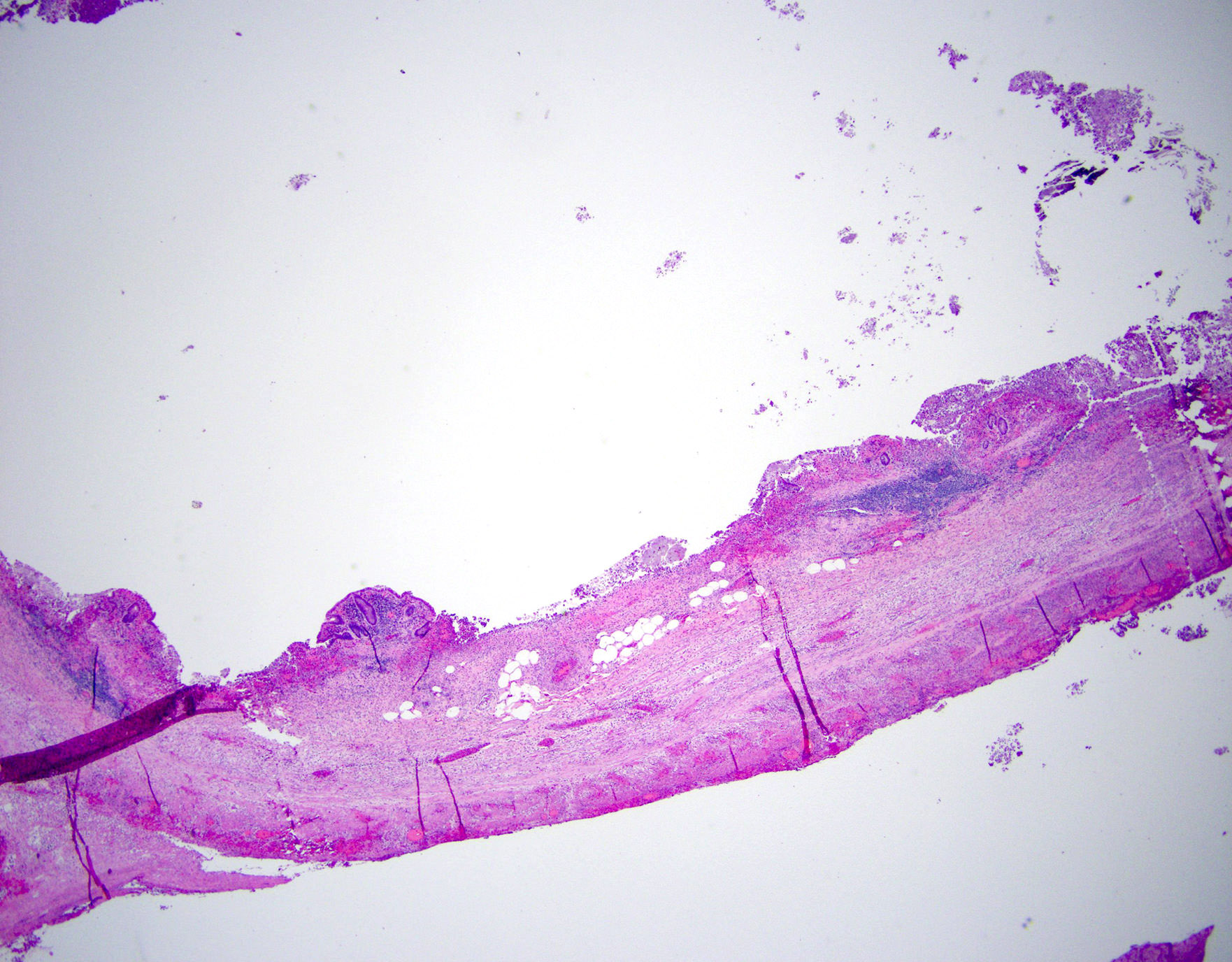
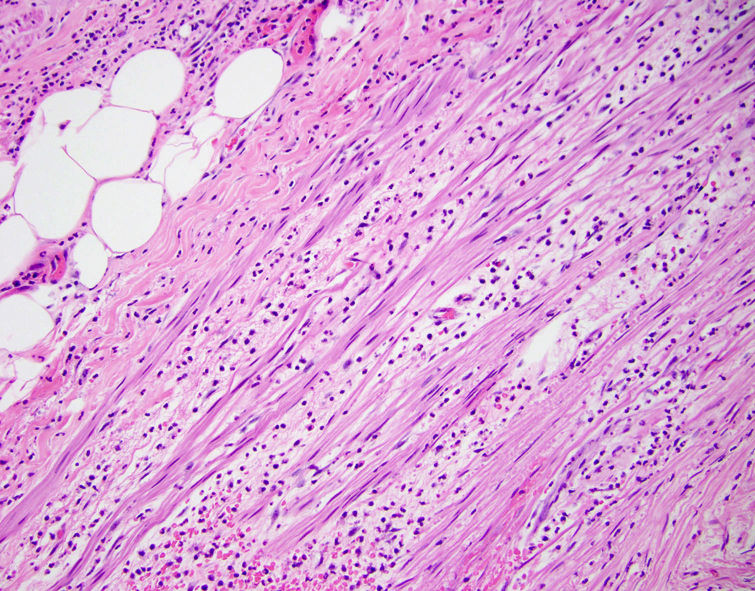
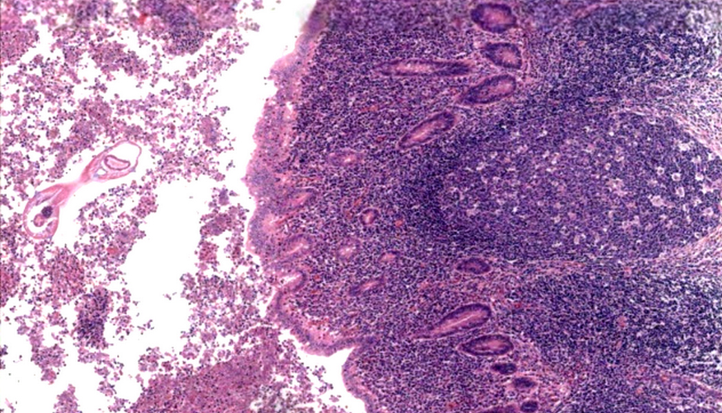
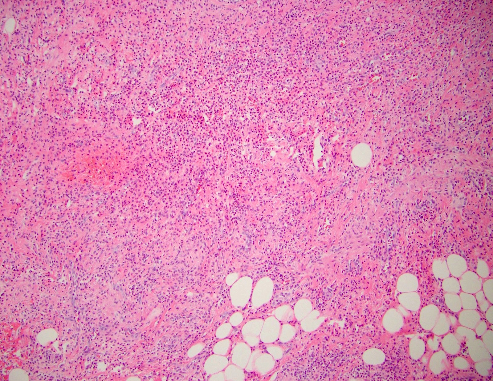
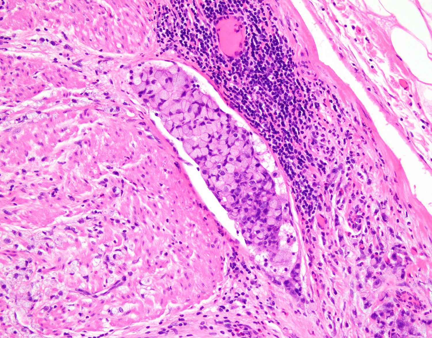
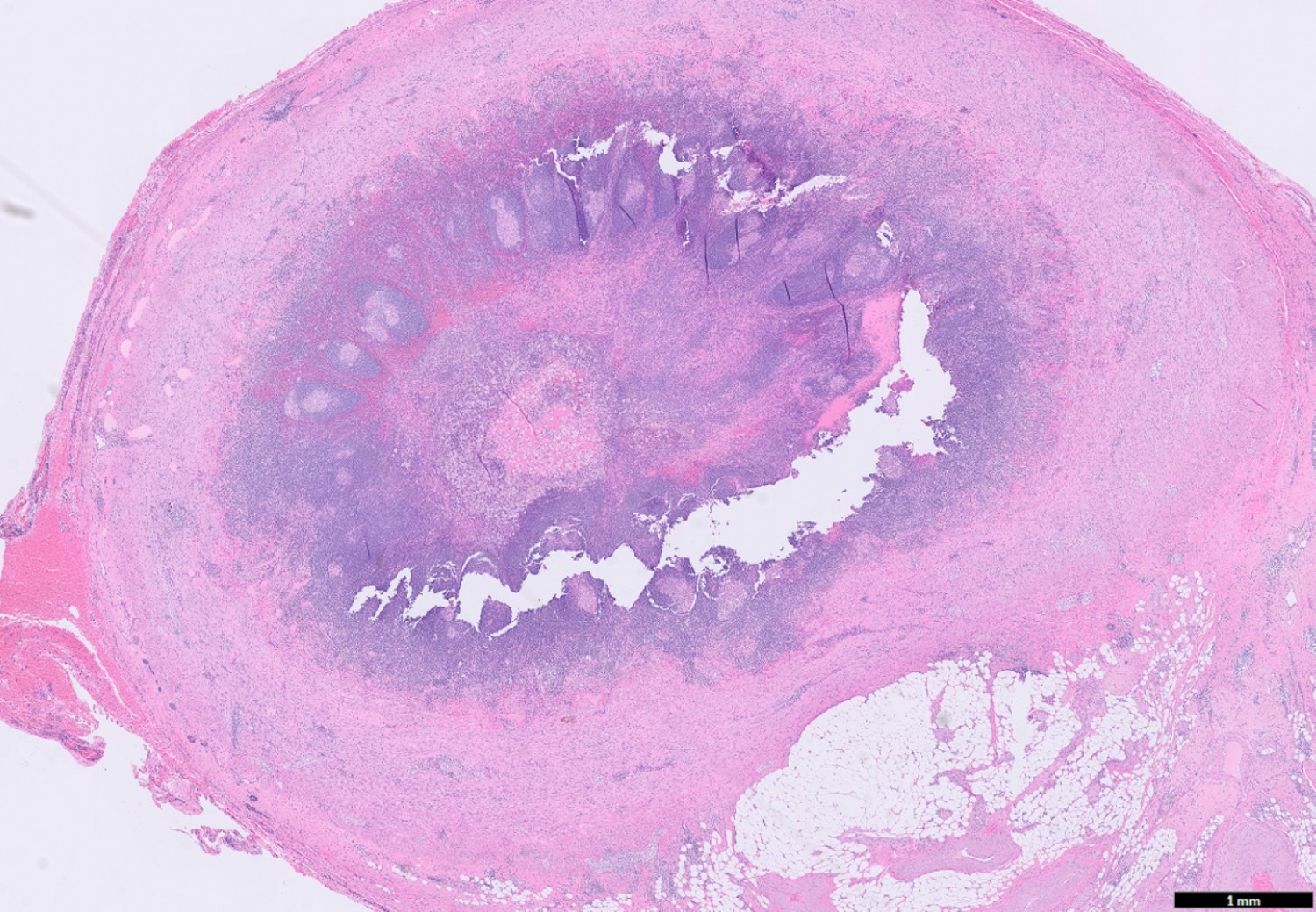
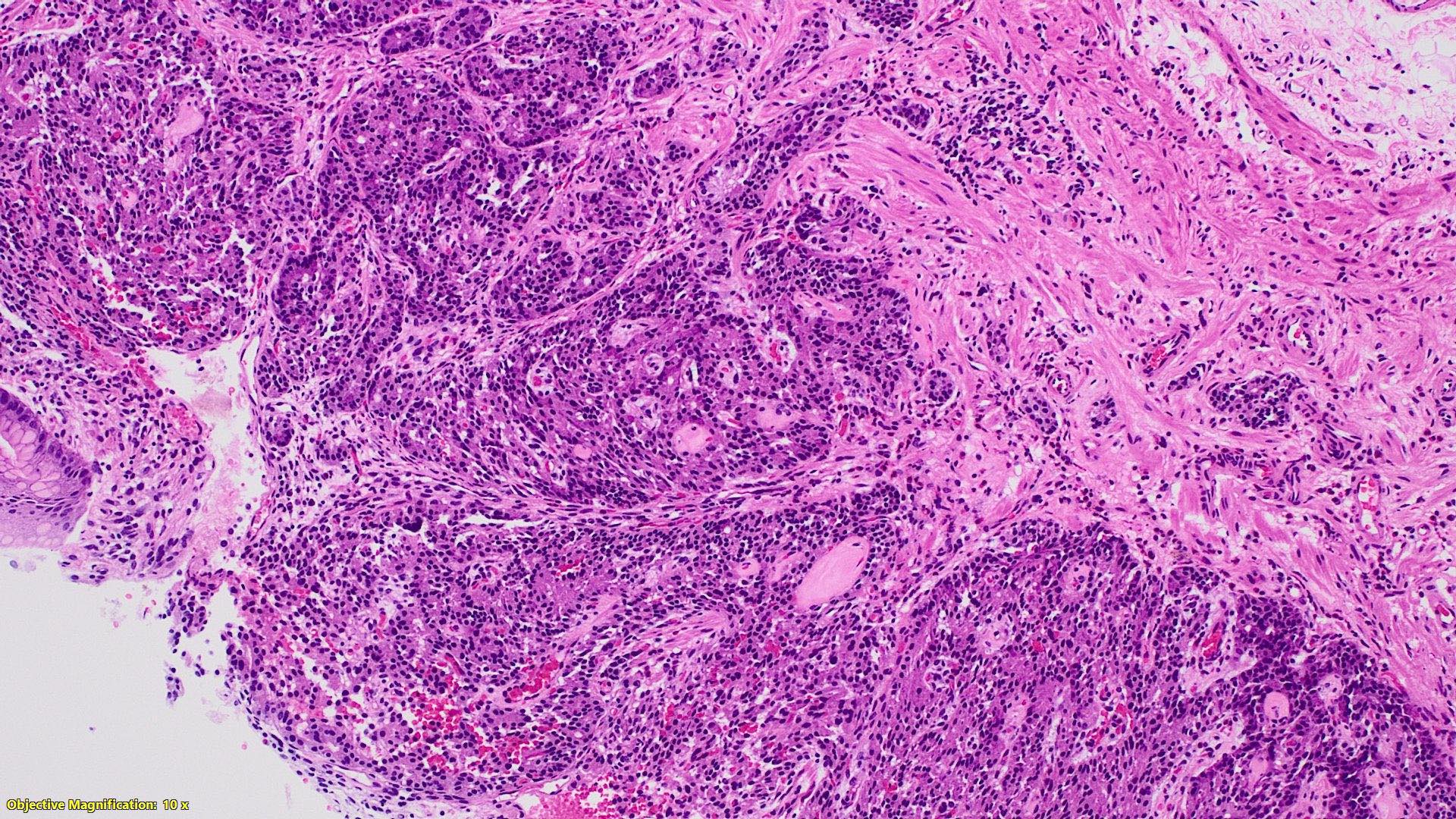
Periappendicitis
Contributed by Maryam Kherad Pezhouh, M.D., M.Sc.
Absent luminal inflammation
Serosal inflammation
Inflammation and edema
Neutrophils at serosa
Sparing of muscularis
Images hosted on other servers:

Morphological patterns of PMP
Images hosted on other servers:

Ascitic fluid

Mucinous ascites

Cytoreductive surgery

Cytoreductive surgery with HIPEC
Contributed by Alex Placek, M.D.
Omental nodules
Contributed by Alex Placek, M.D.
Low cellularity
Low grade cytology

Increased cellularity
Invasion with desmoplasia

Signet ring cells
Degenerated mucinous cells
Contributed by Michael Feely, D.O.

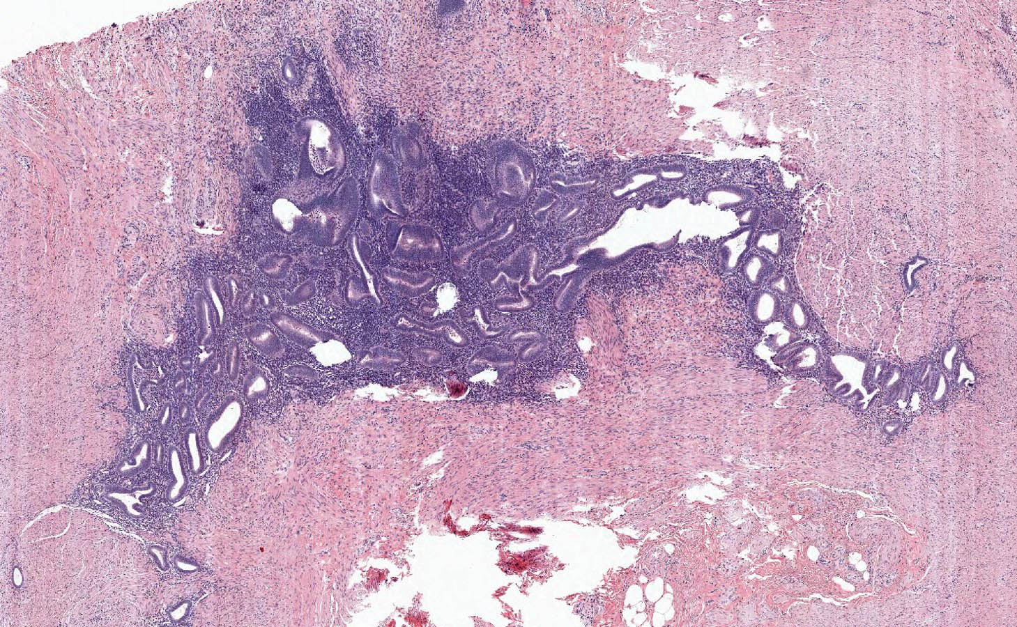
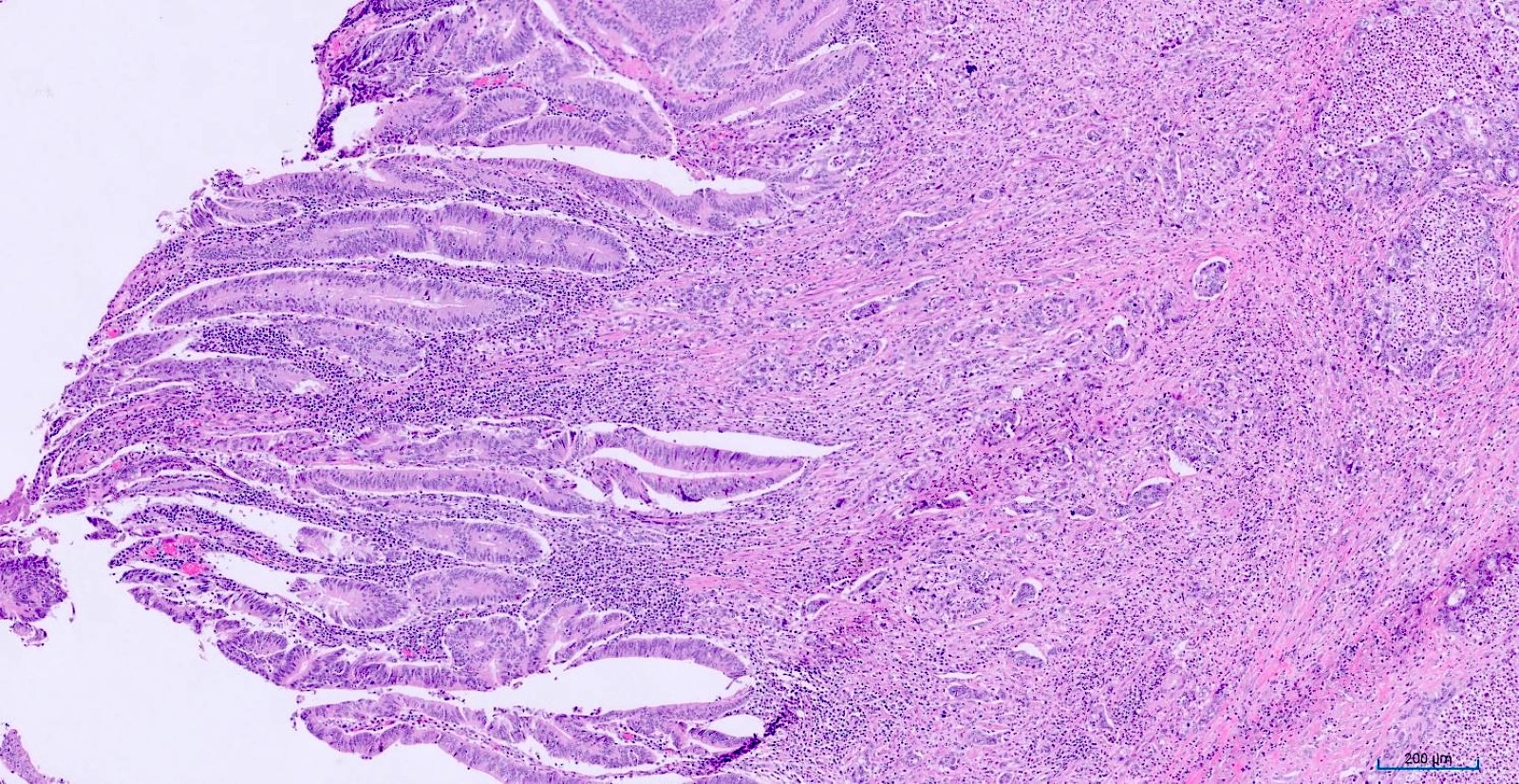
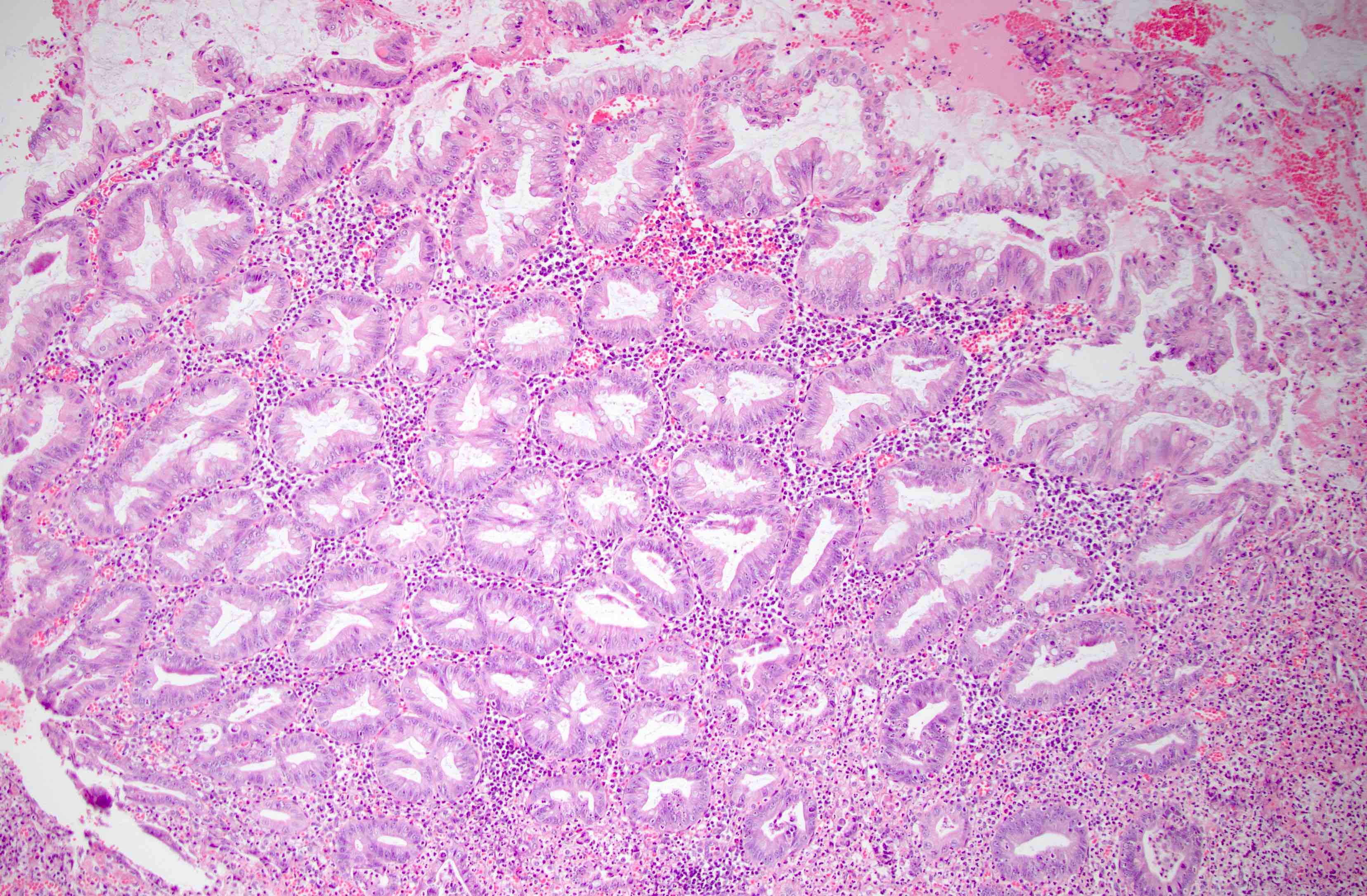
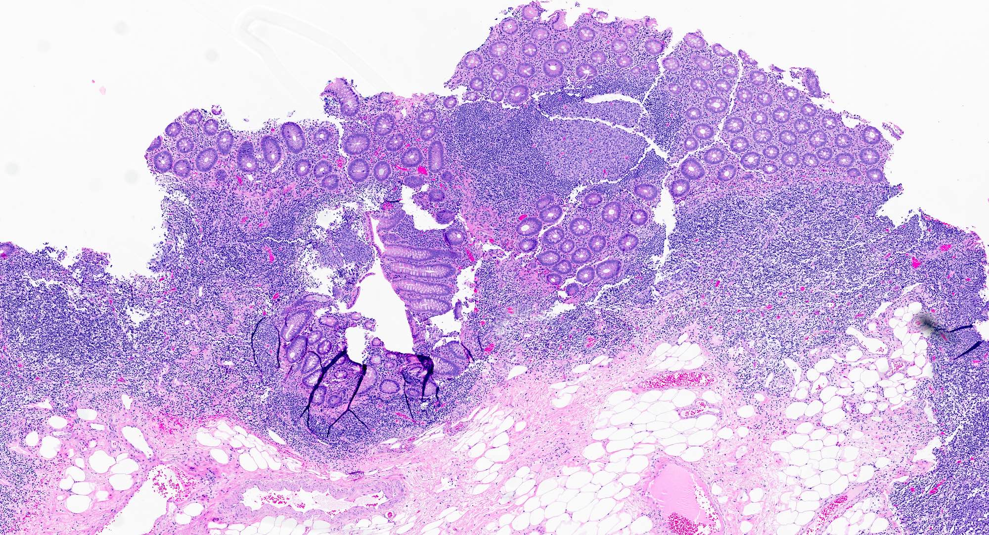
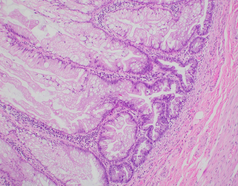
Serrated polyp of appendix
Images hosted on other servers:

Double contrast barium enema

Abdominal CT scan
Images hosted on other servers:

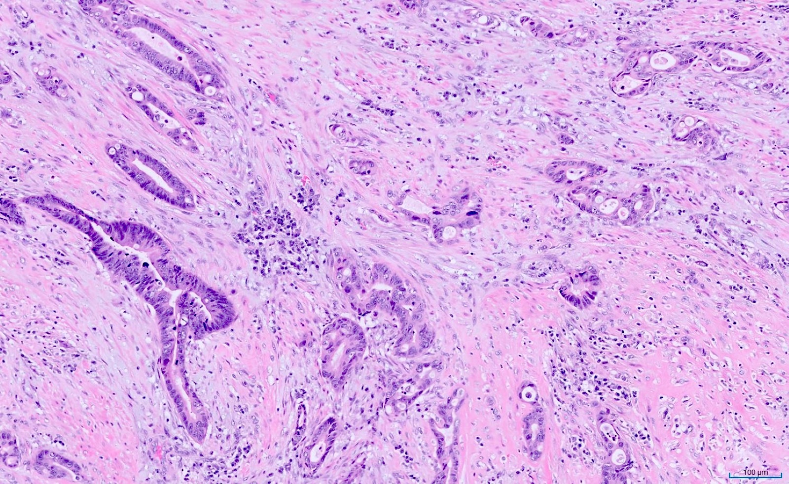
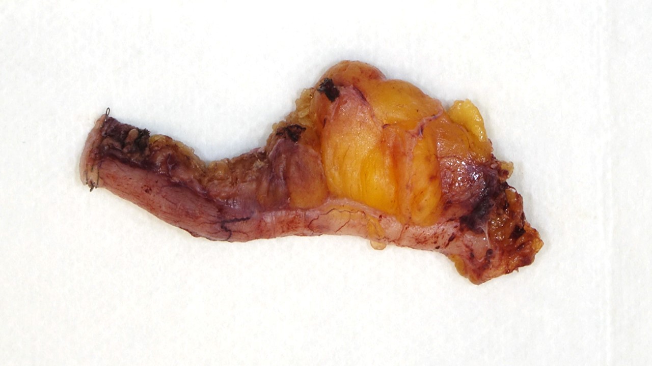
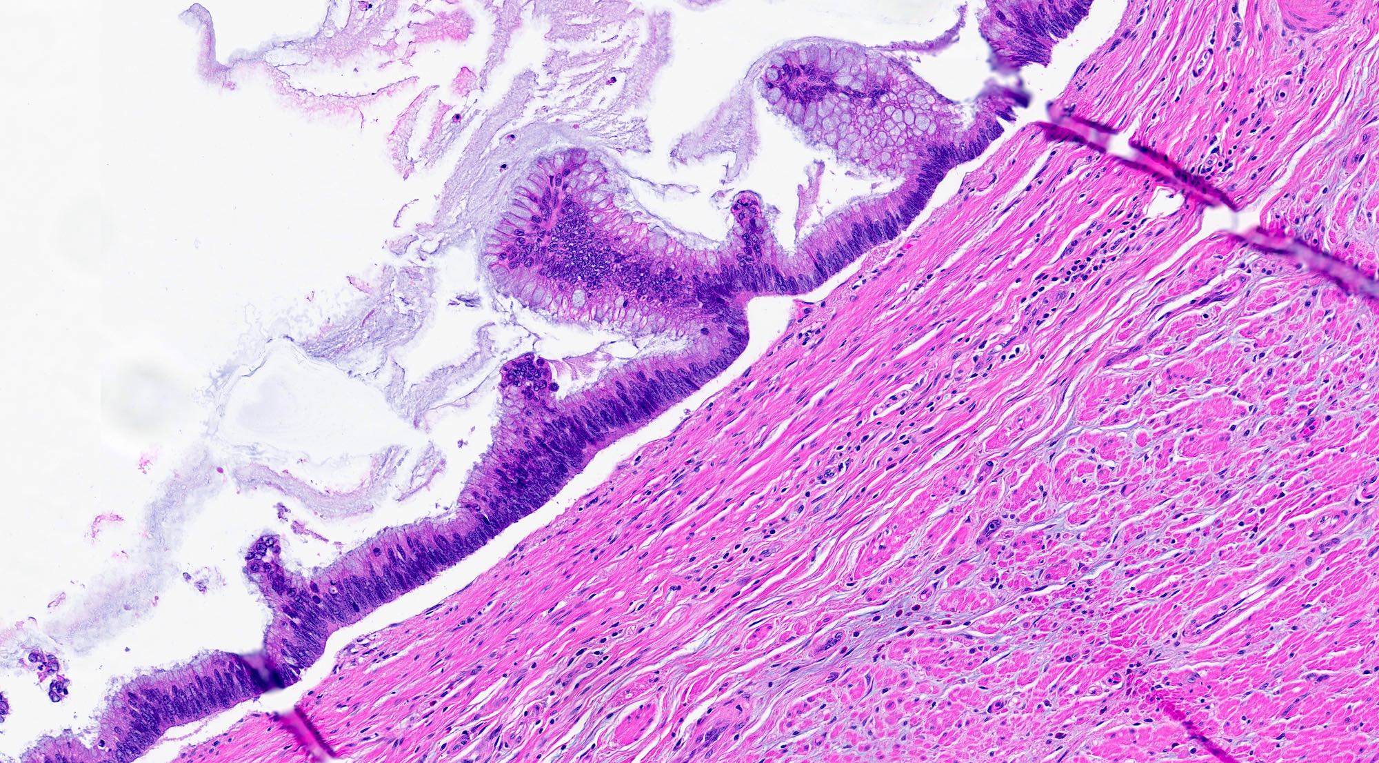
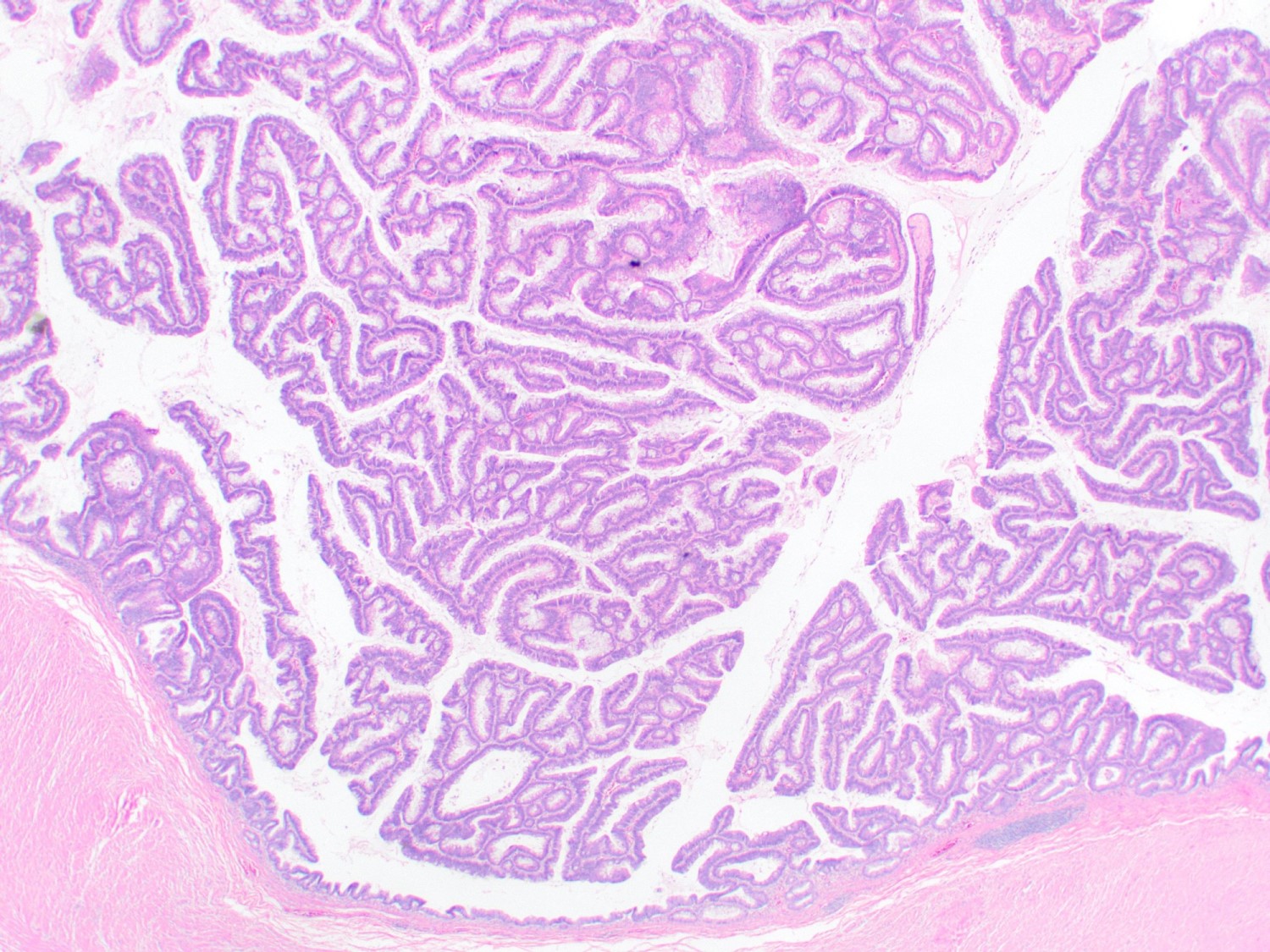
Grossly visible appendiceal adenoma
Contributed by Raul S. Gonzalez, M.D.
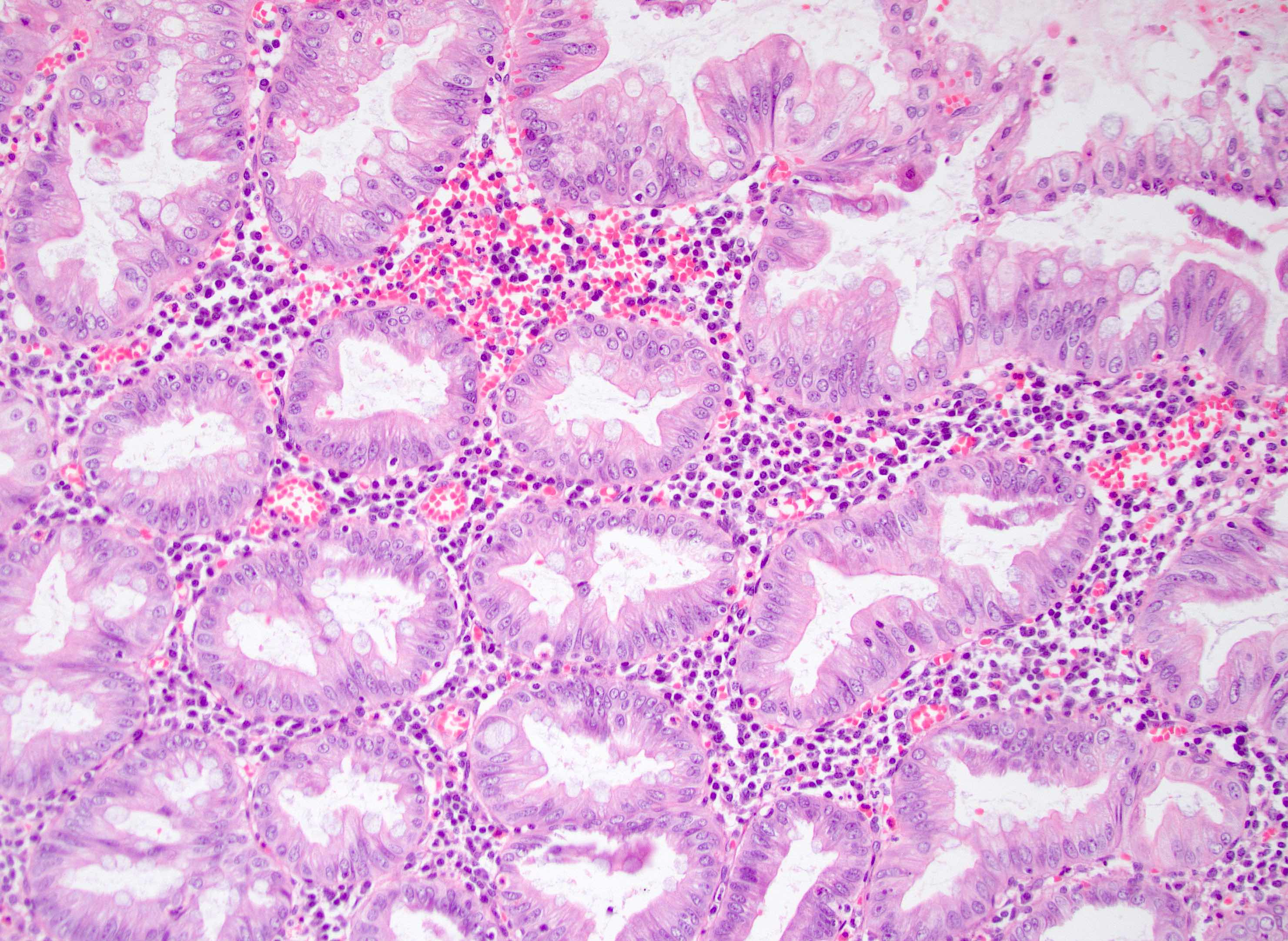
Tubular glands with elongated, hyperchromatic nuclei and crowded glands
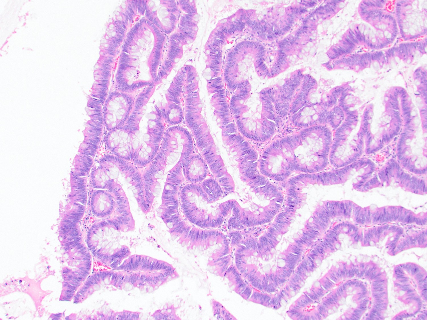
Crowded tubular and villous glands with elongated hyperchromatic nuclei
Mucinous appearance
Adenocarcinoma arising in adenoma
Greenson: 2019

IARC: 2019
Montgomery: 2017
Montgomery: 2017
Odze: 2022
Srivastava: 2023
Yantiss: 2021
Find related Pathology books: GI, liver
