


Contributed by Toral Patel, M.D.
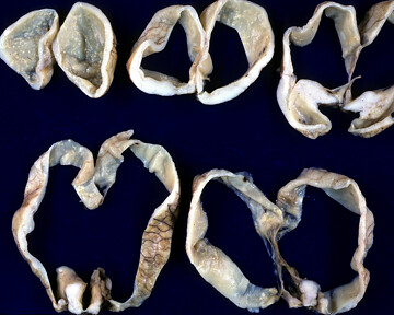
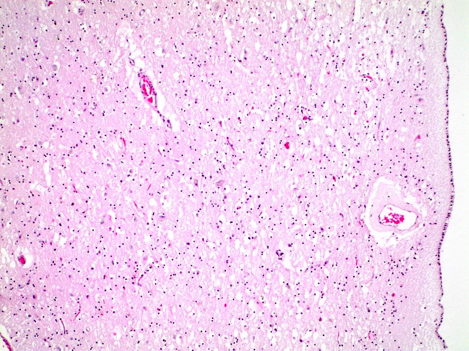
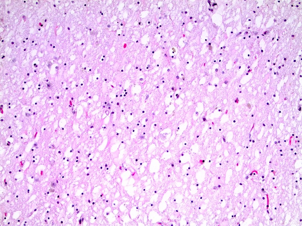
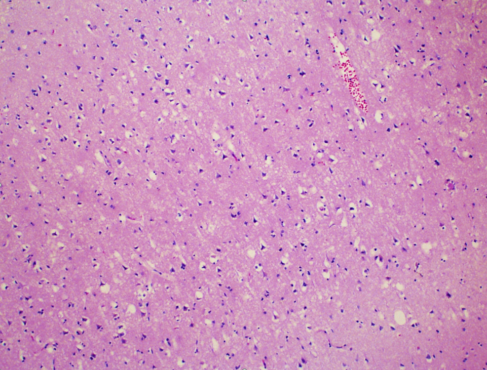
Left parietal white matter
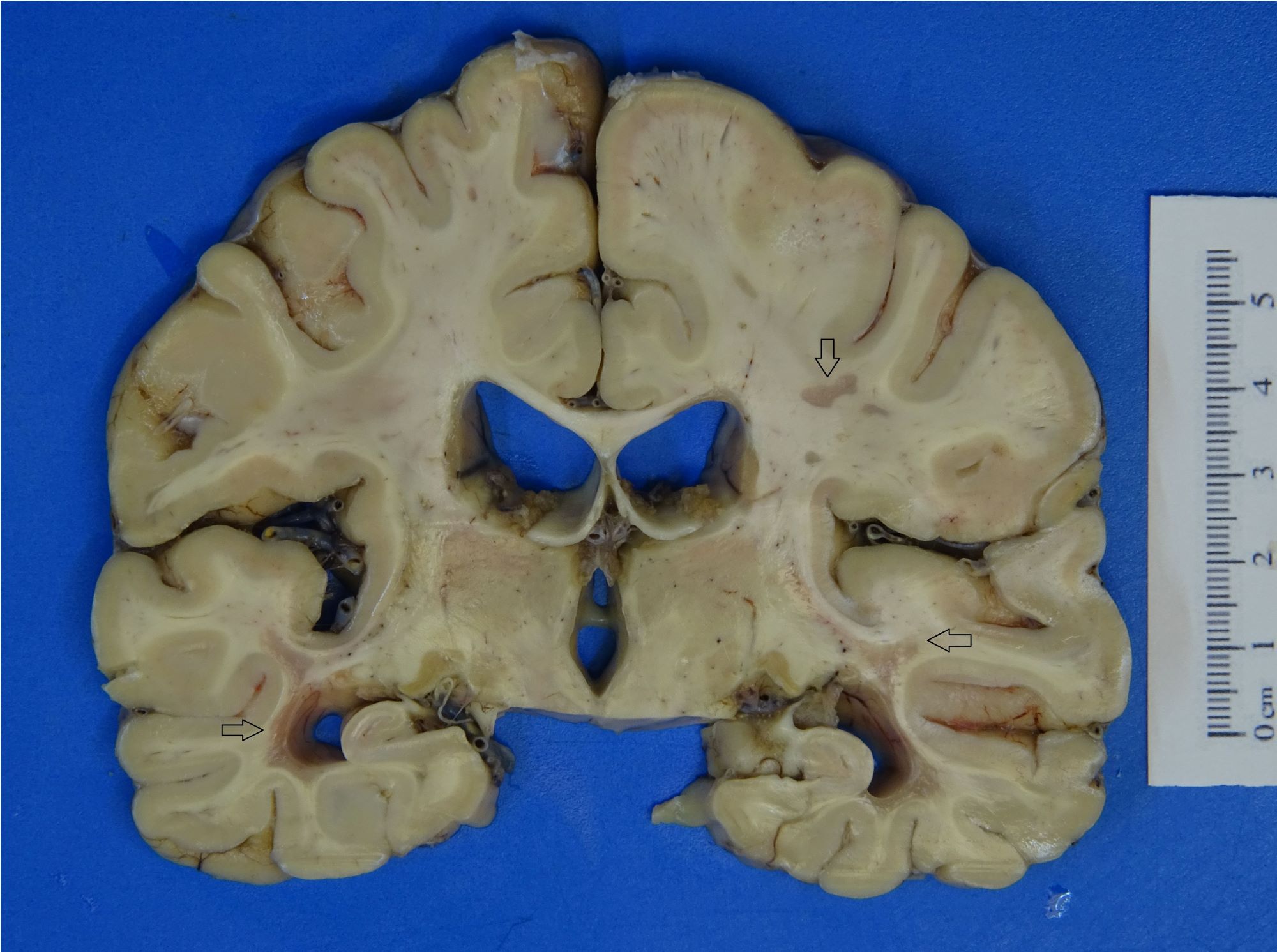
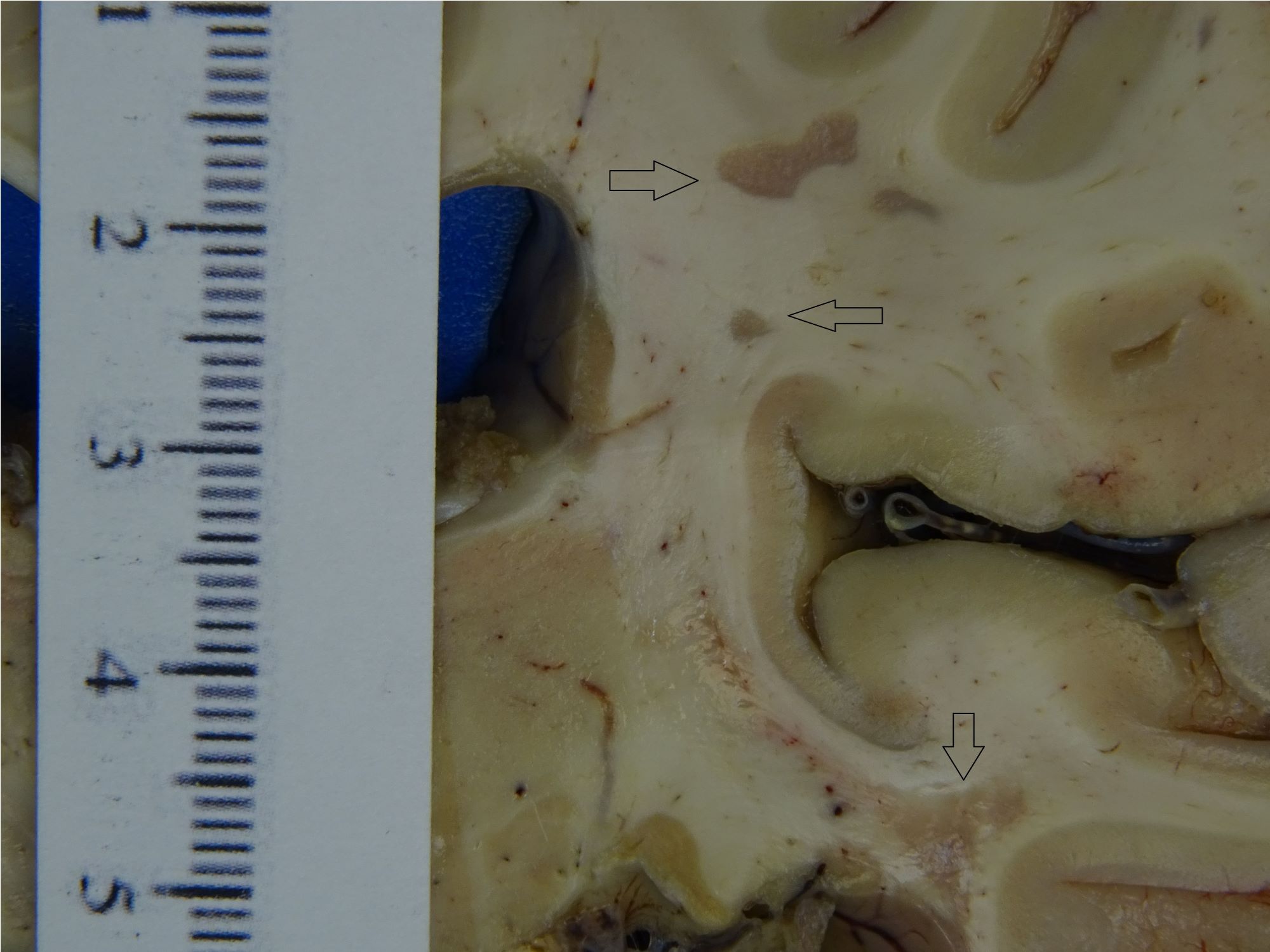
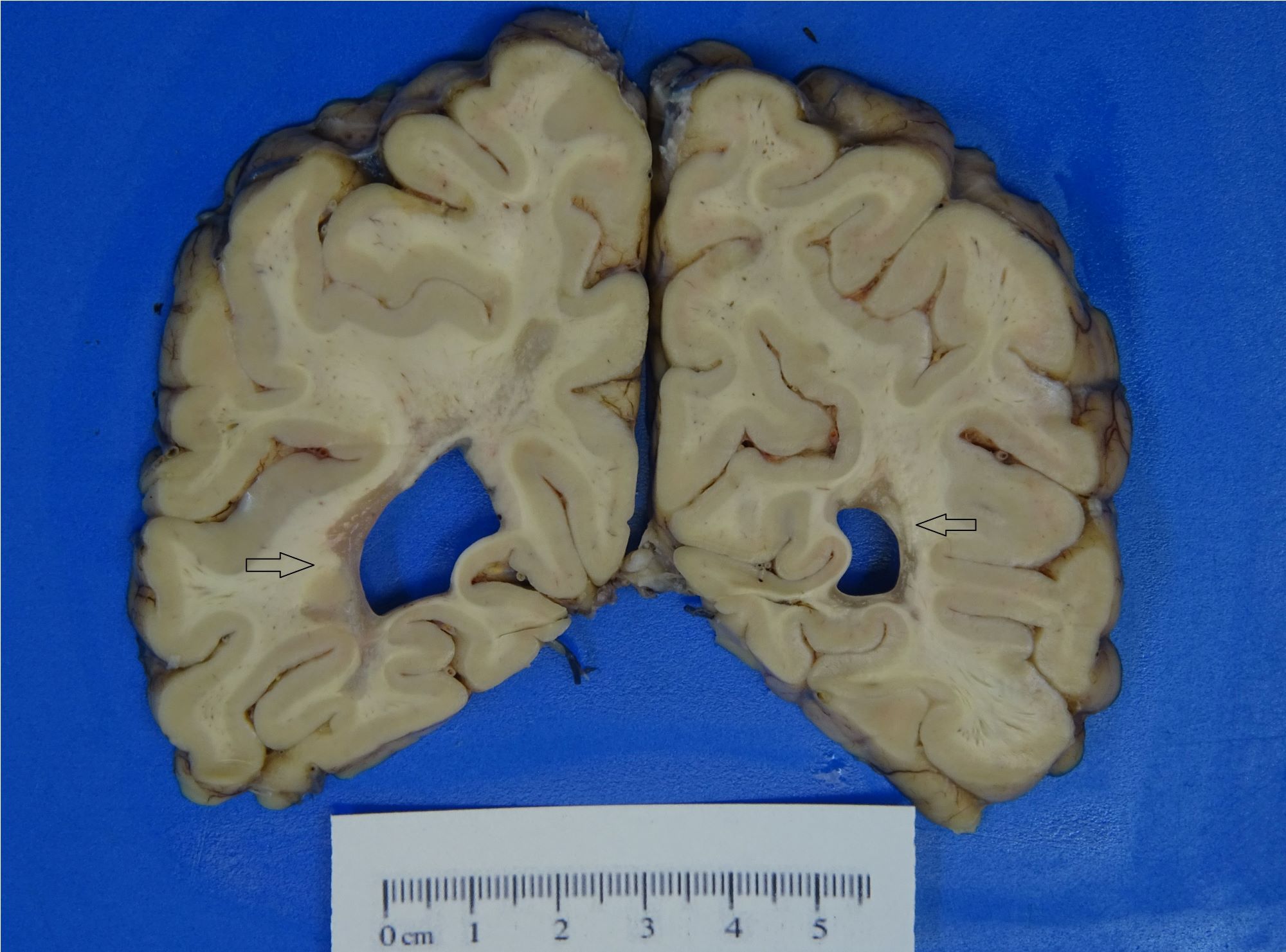
Contributed by Dennis K. Burns, M.D.
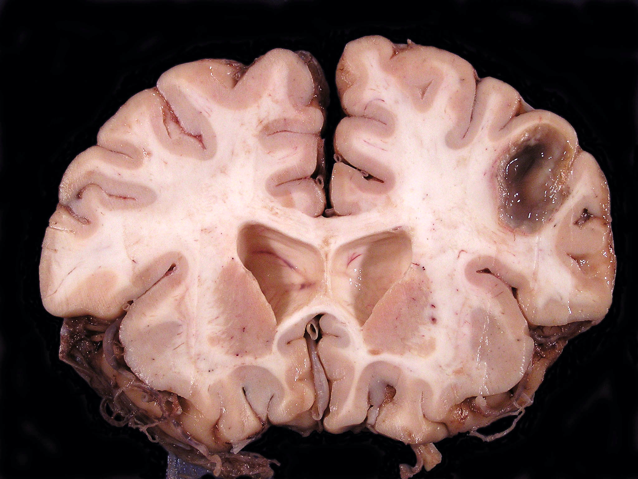
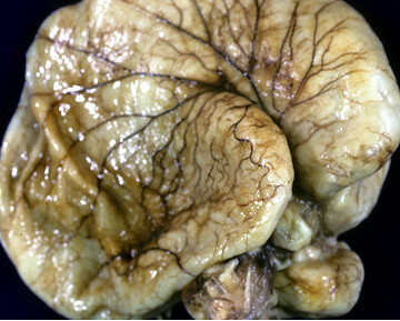
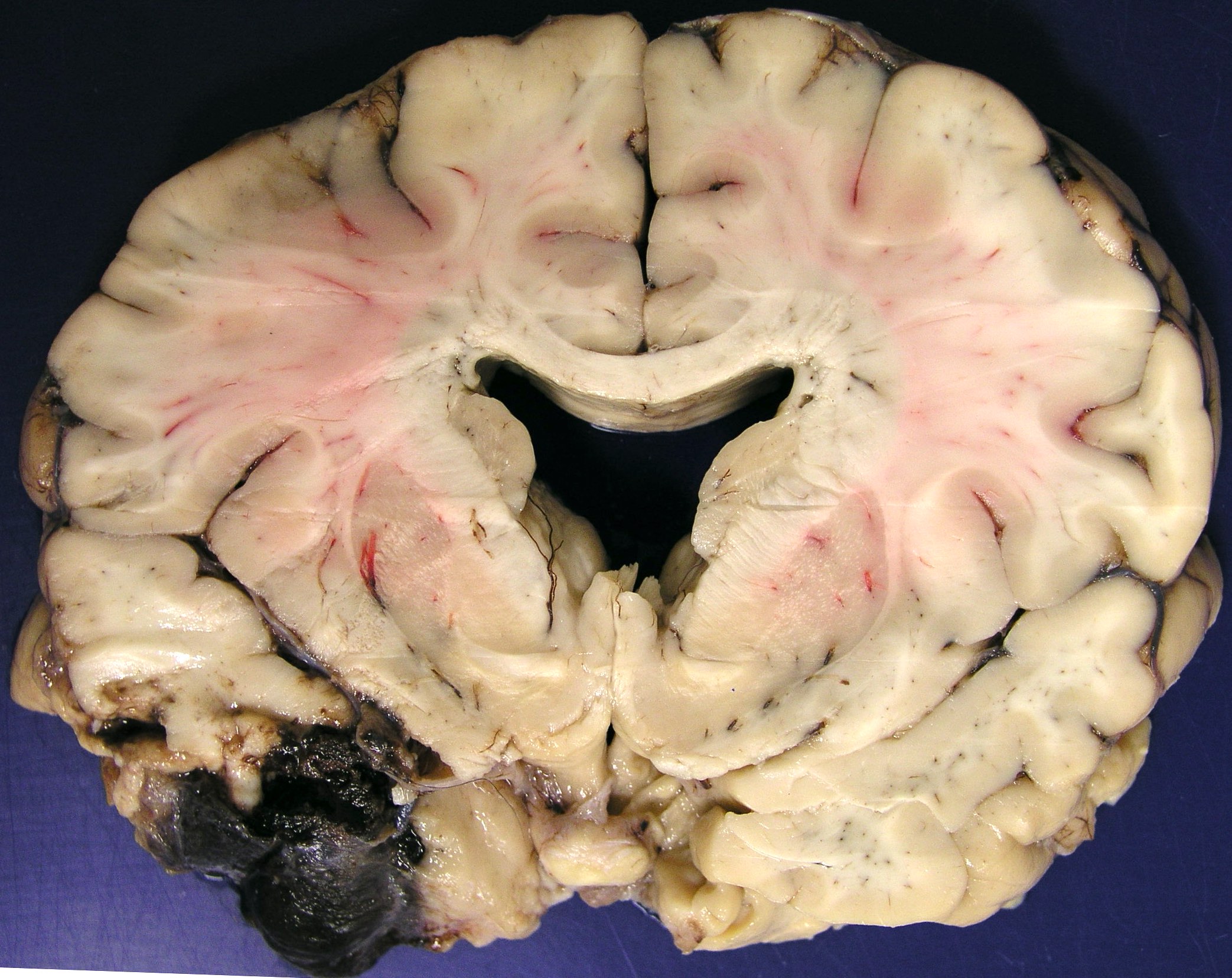
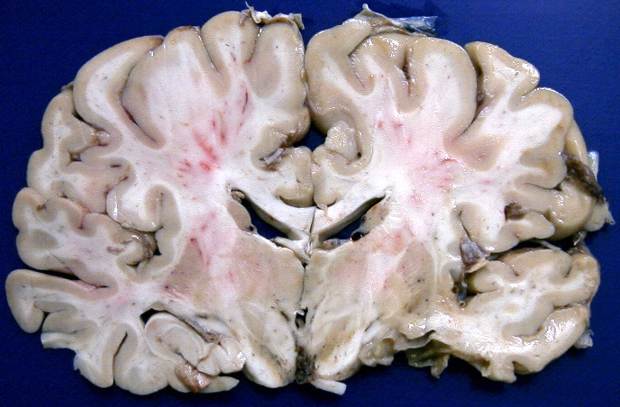
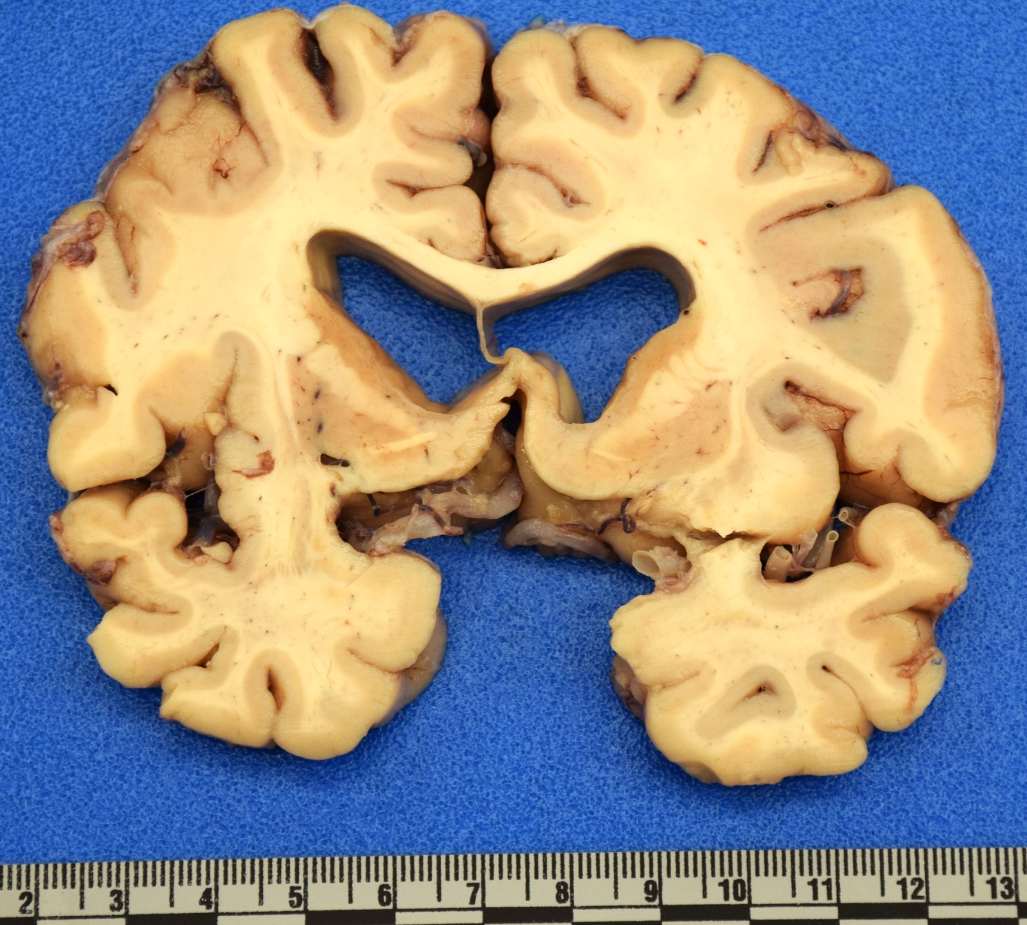
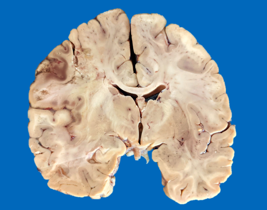
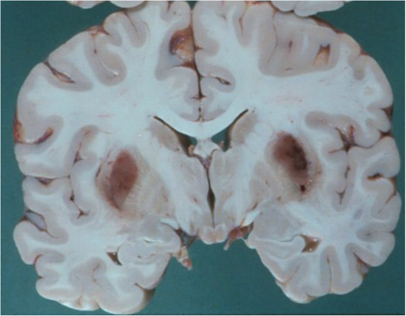
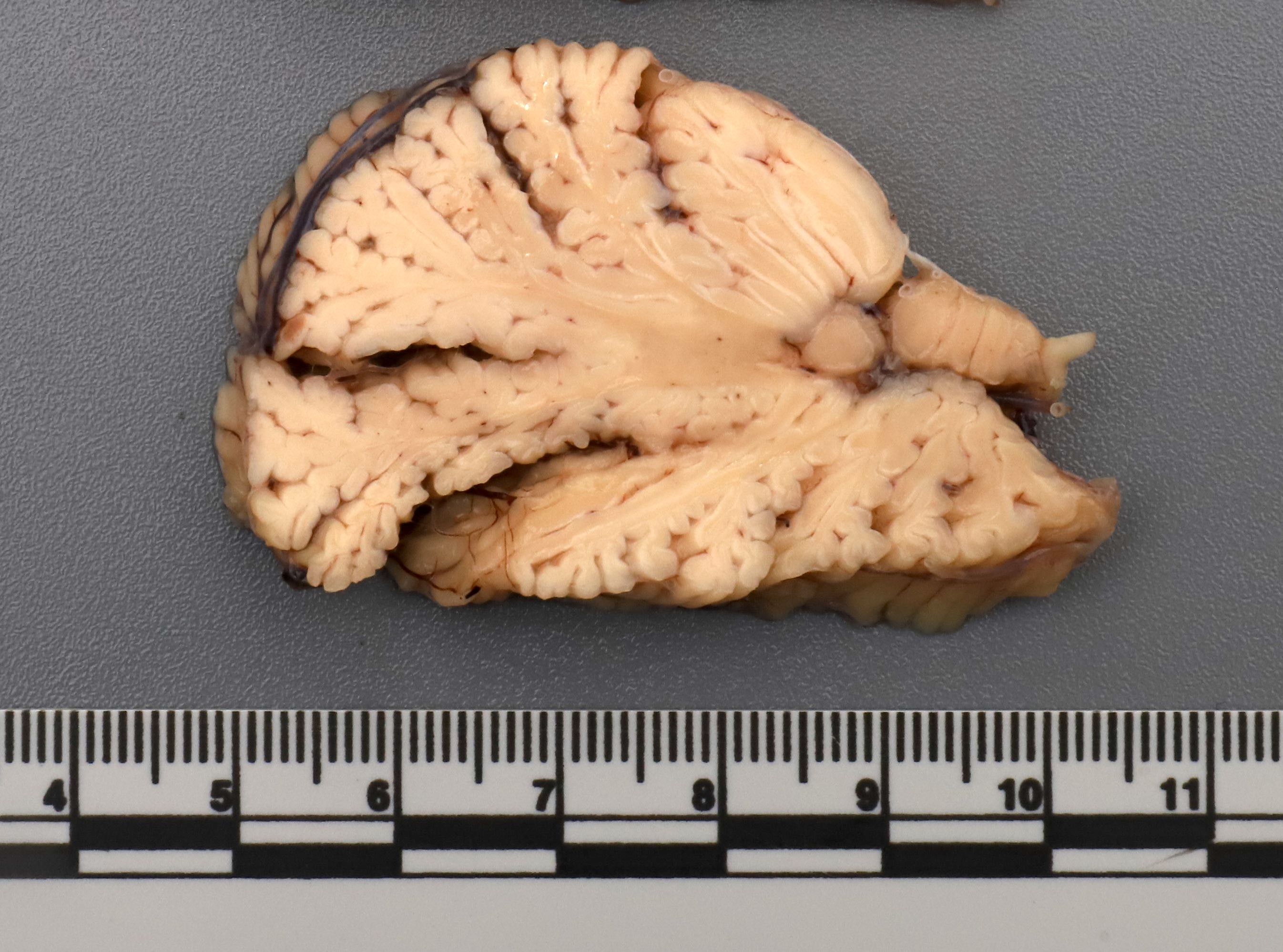
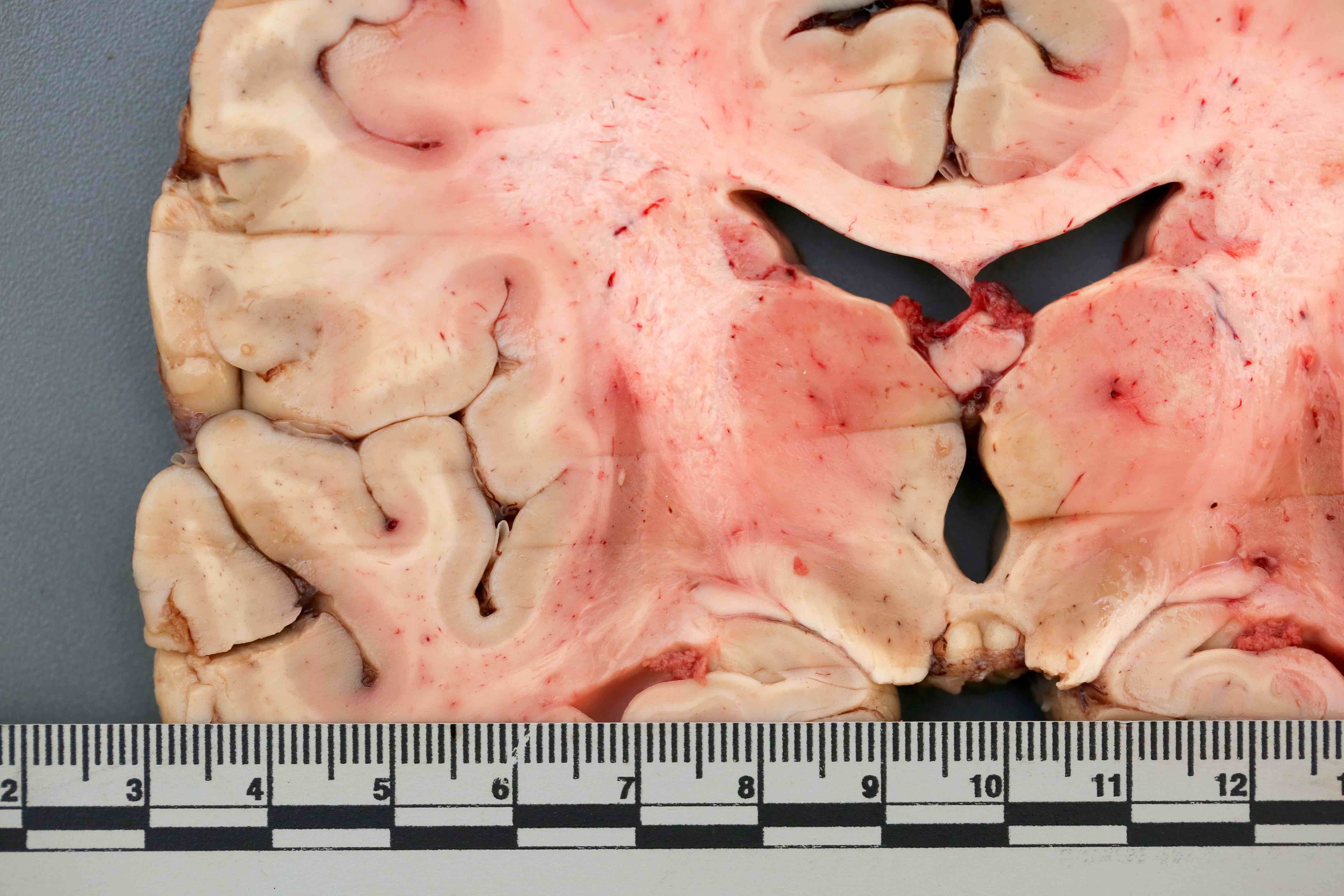
Brain, coronal section
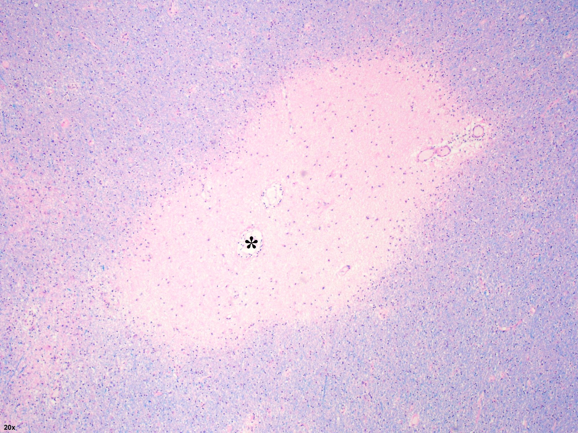
Contributed by Dennis K. Burns, M.D.
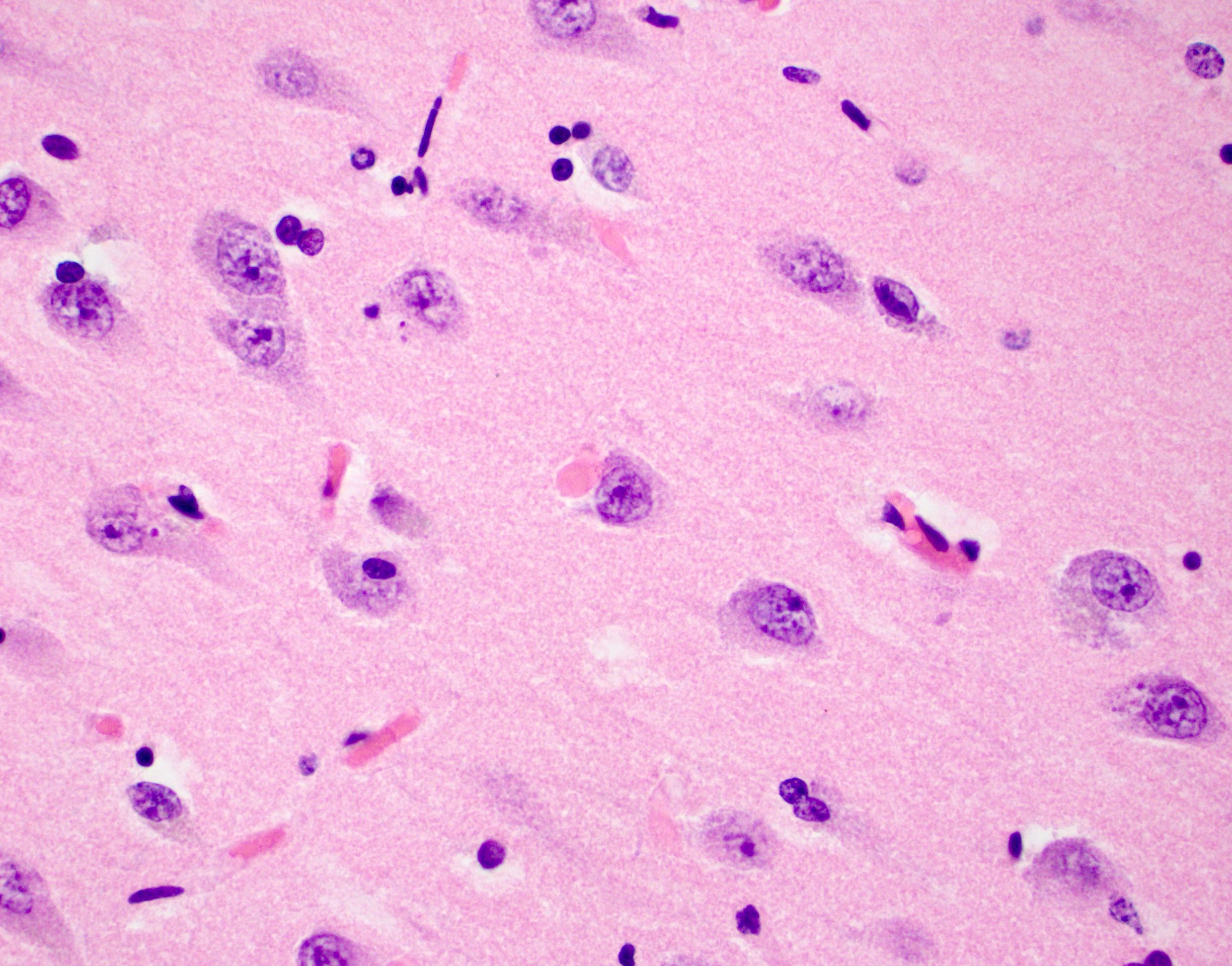
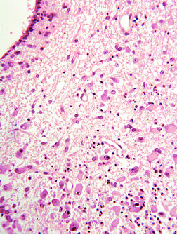
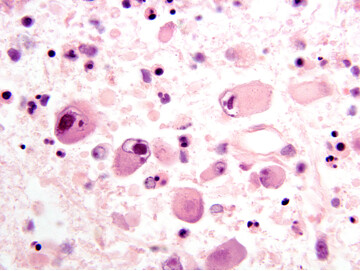
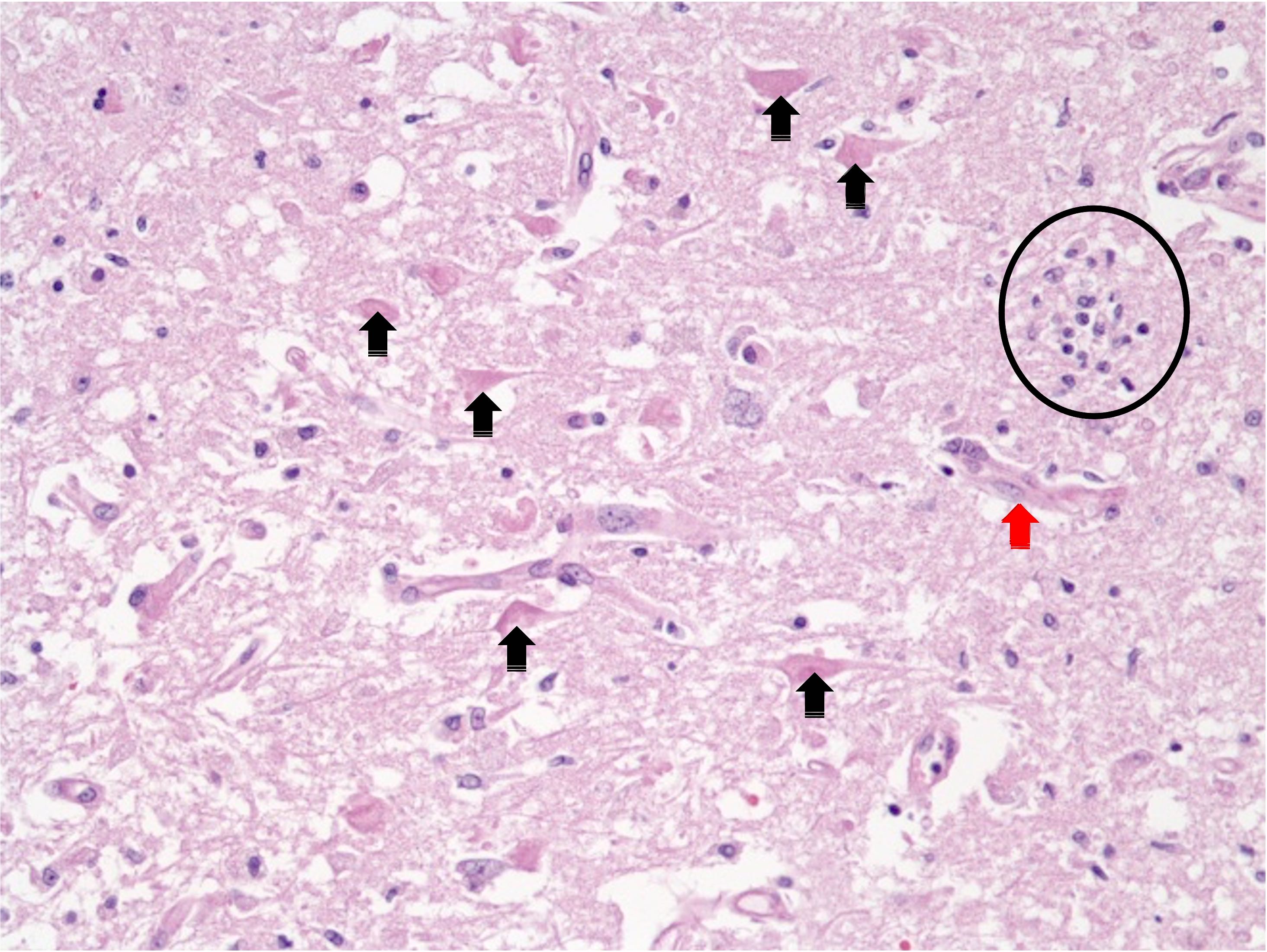
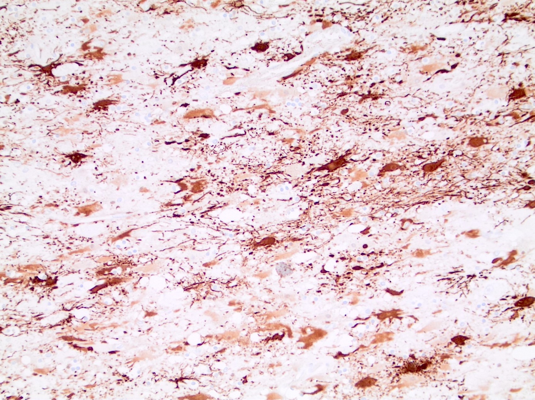
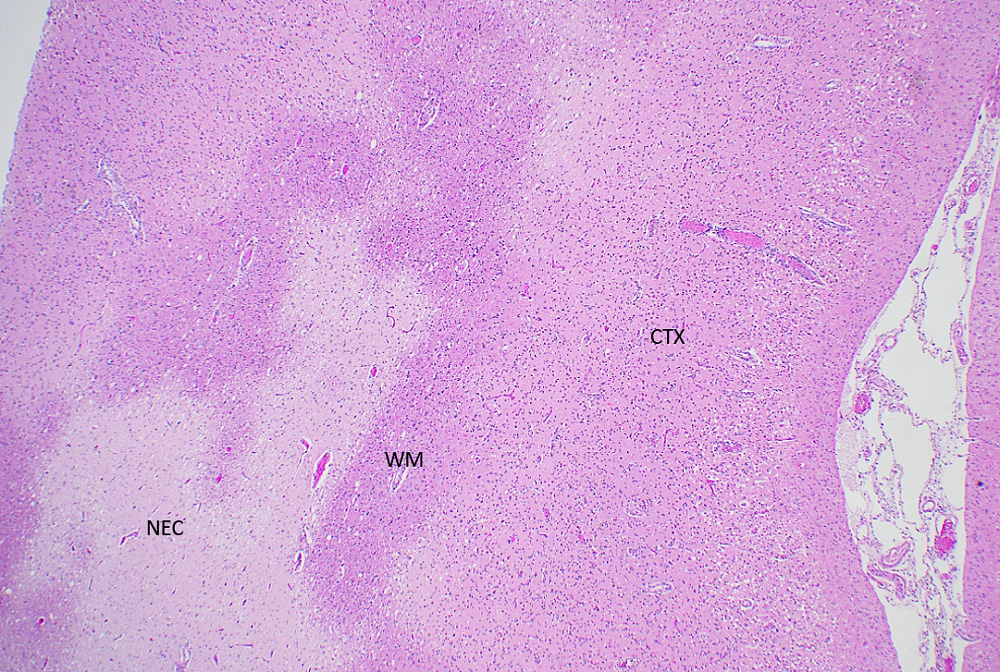
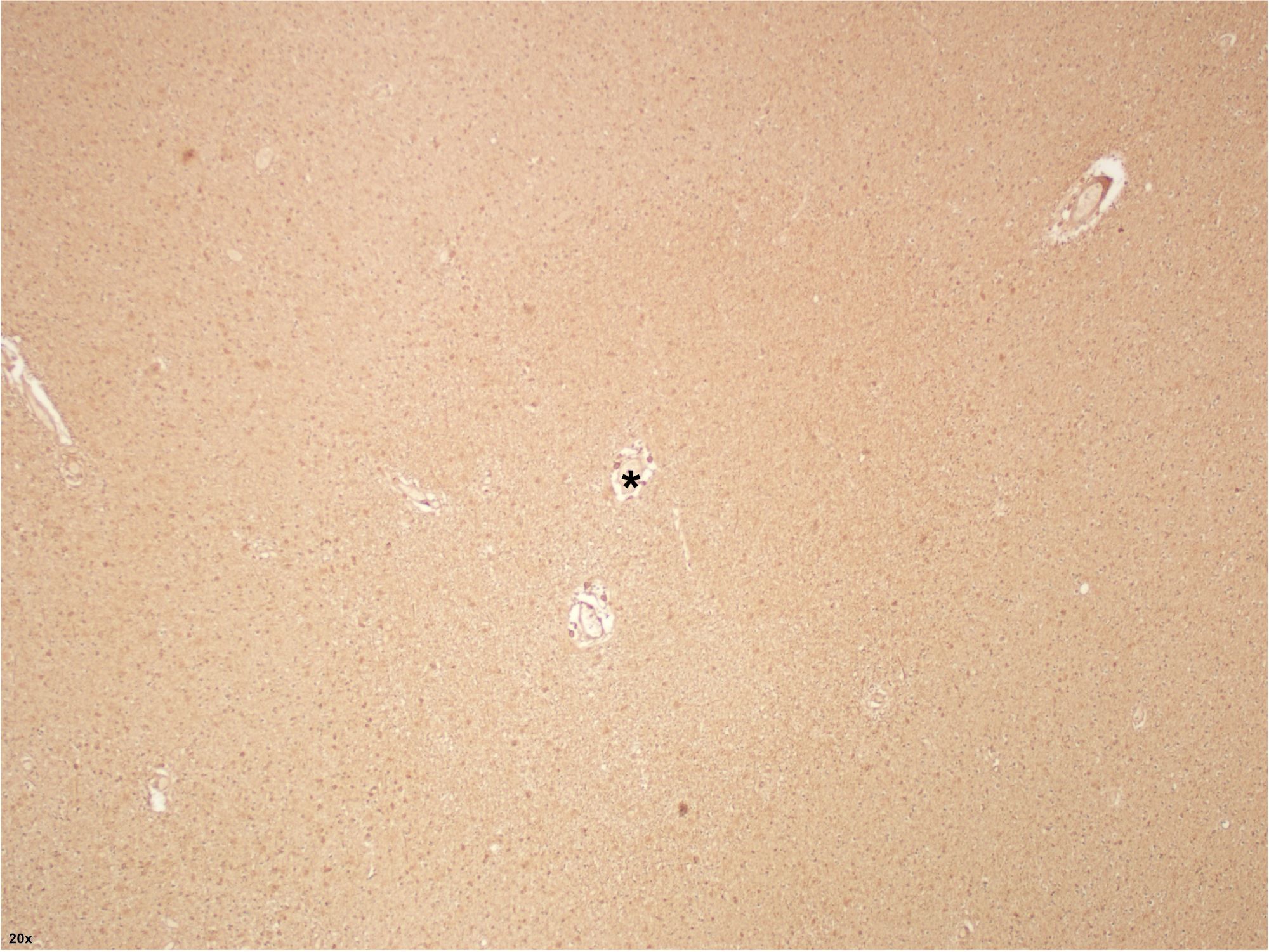
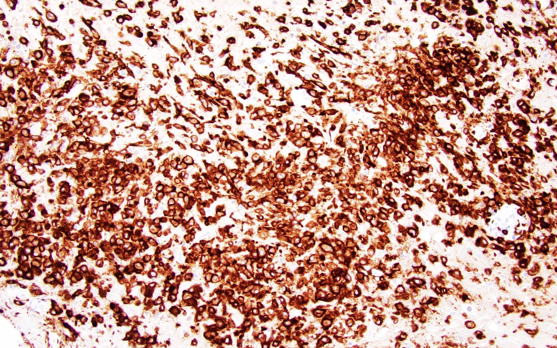
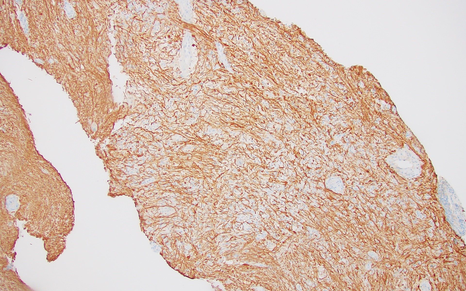
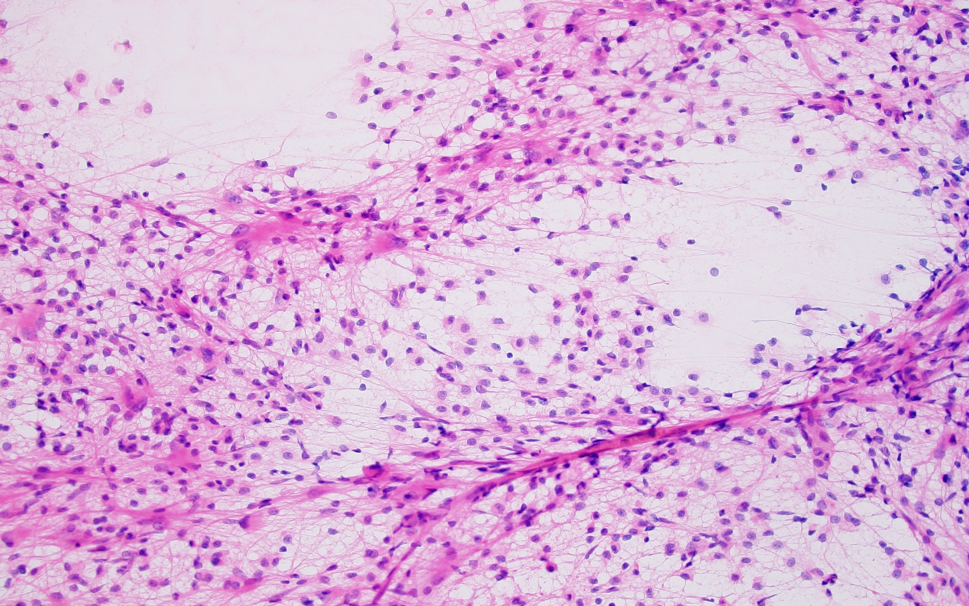
Paraffin section
Images hosted on other servers:
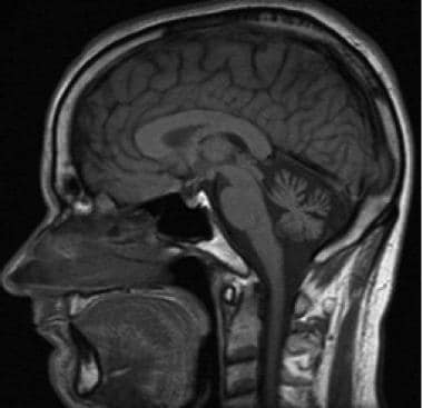
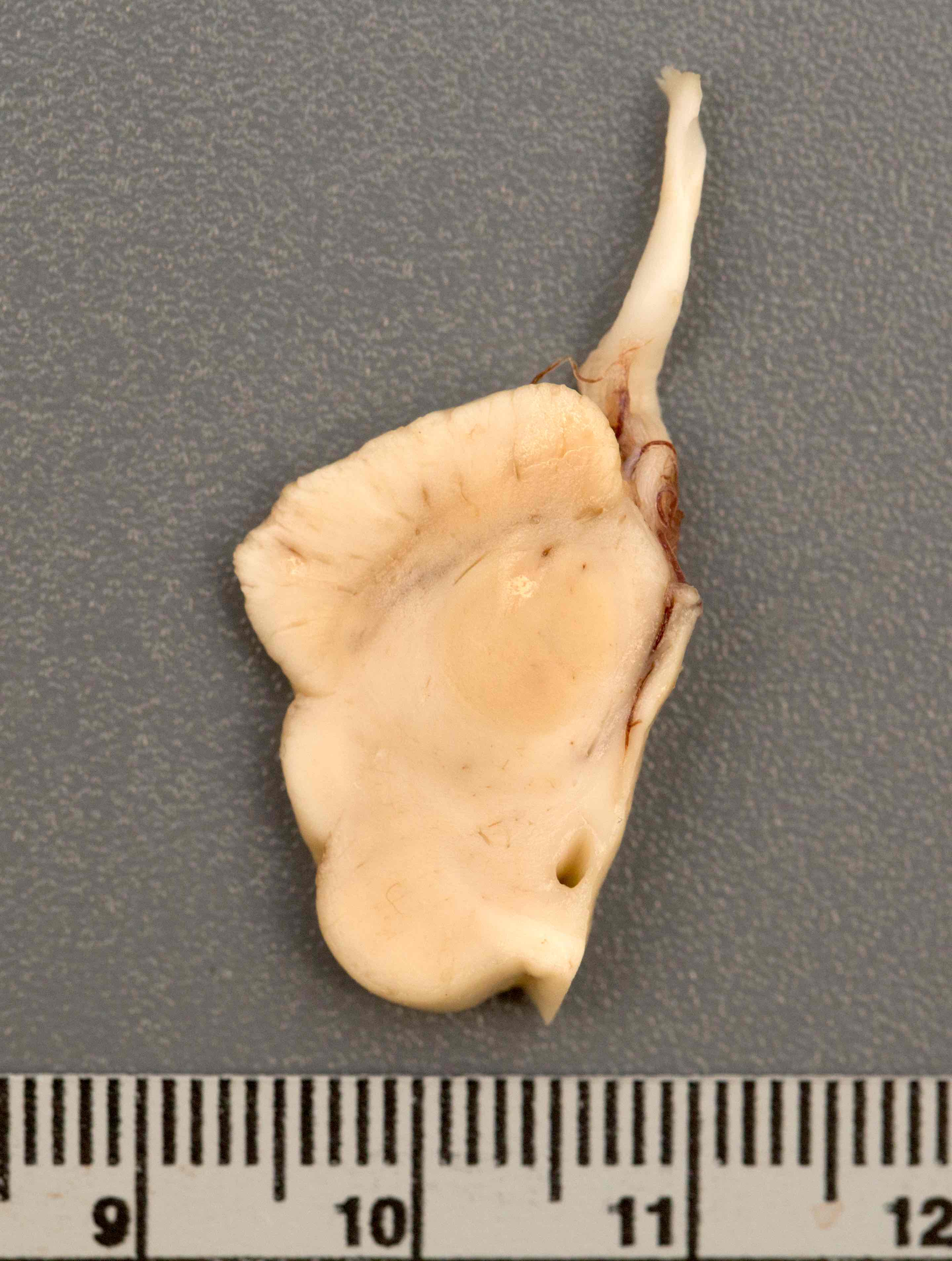
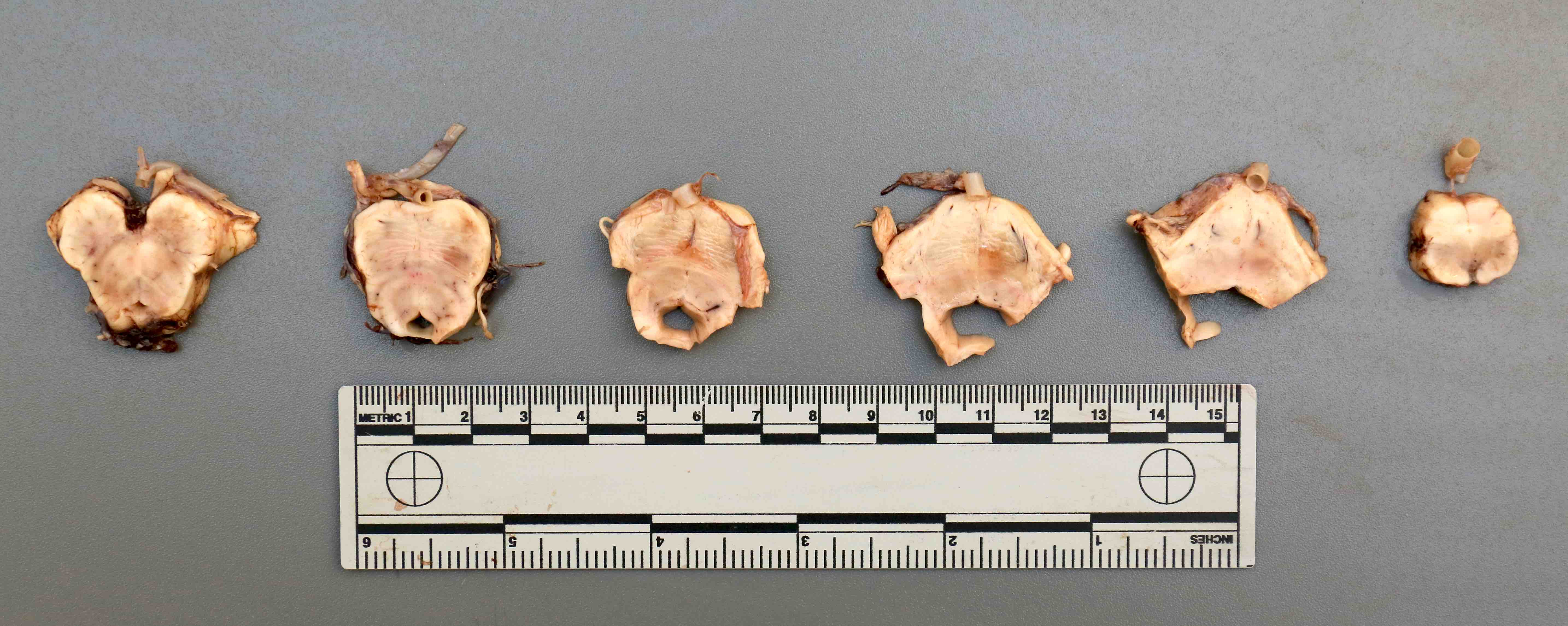
Marked diffuse cerebellar atrophy
Contributed by Kymberly A. Gyure, M.D.
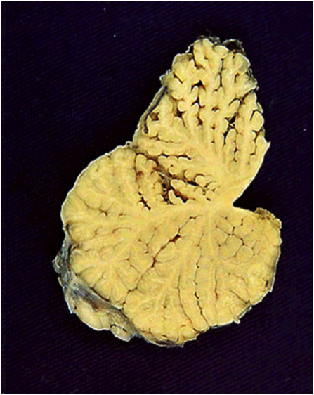
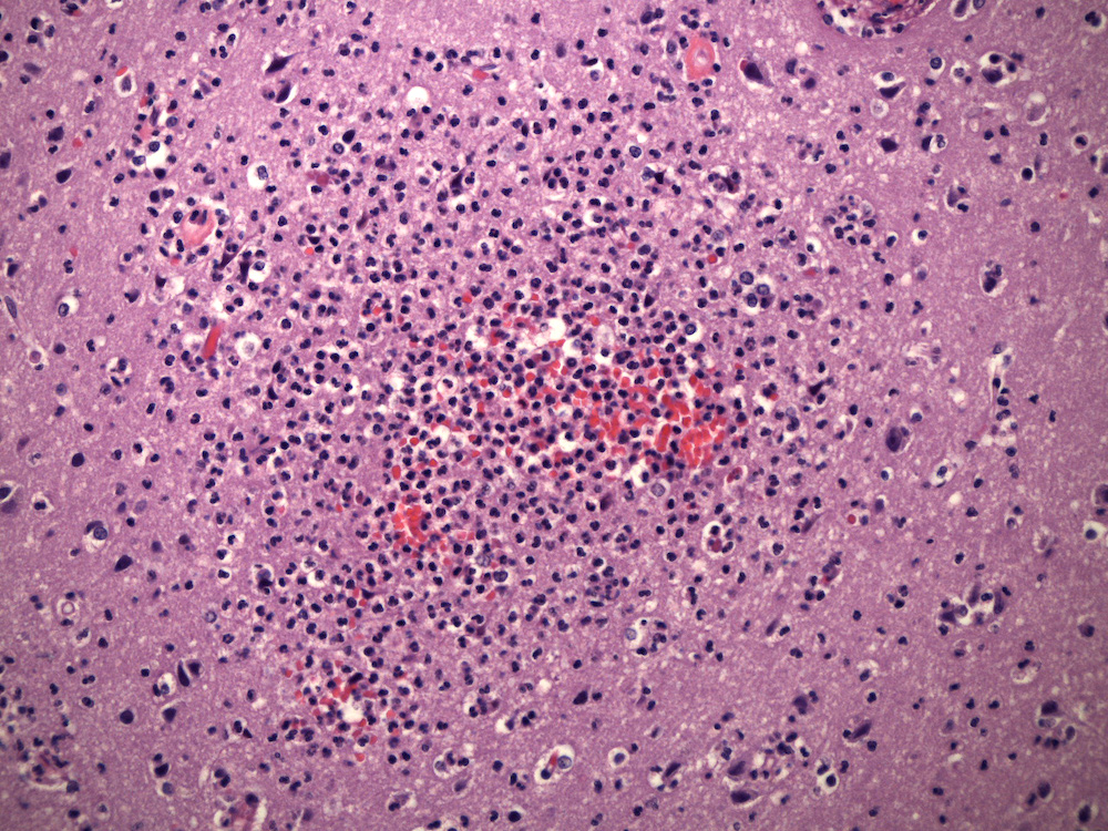
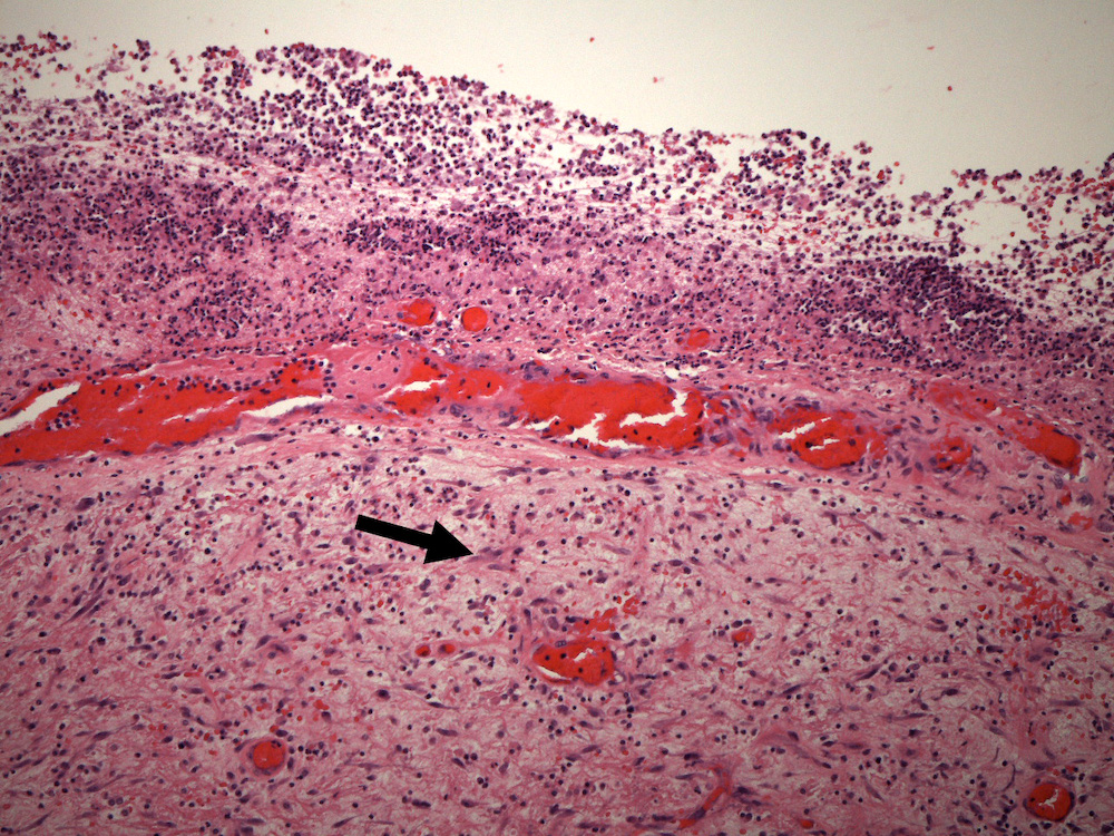
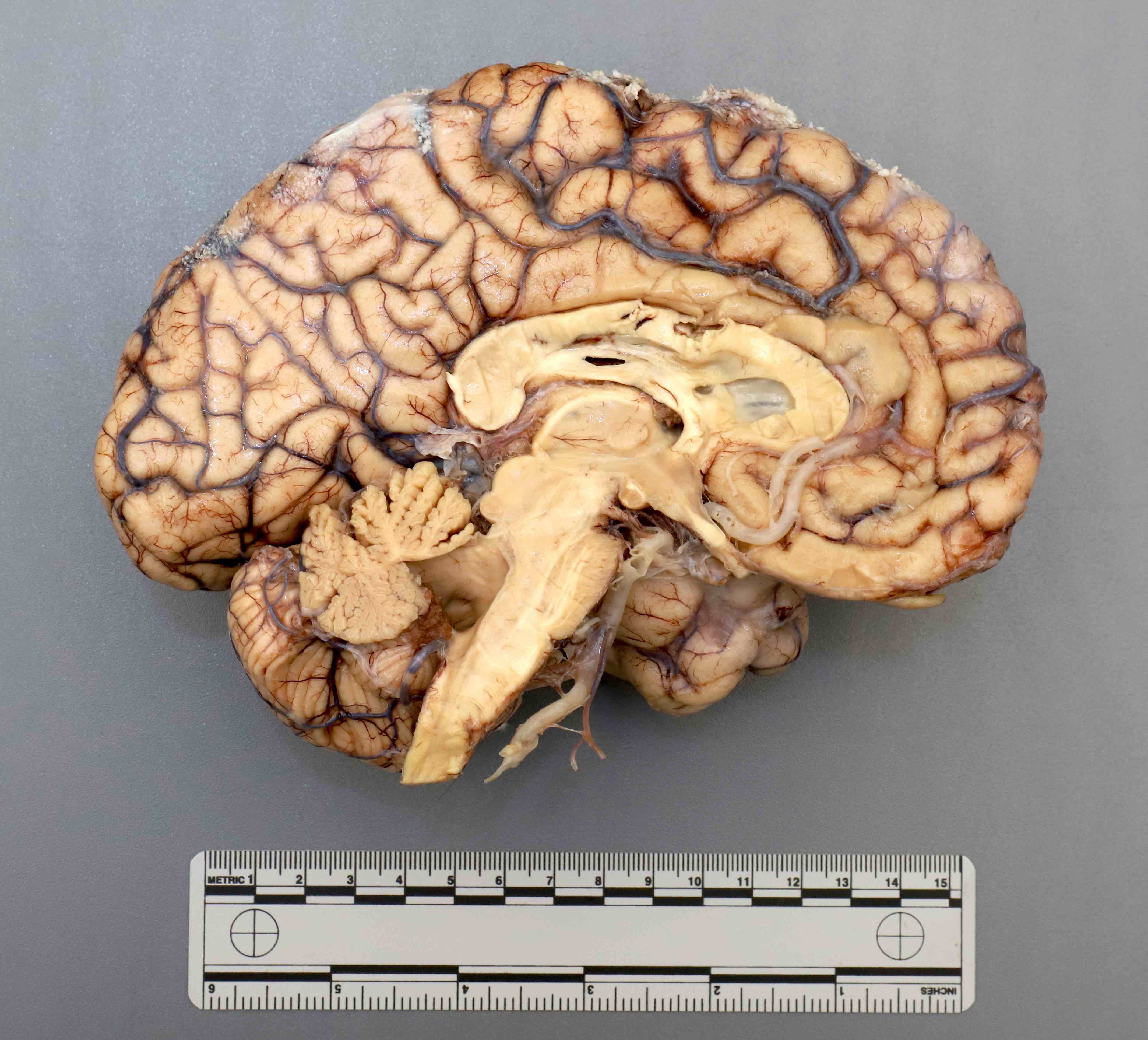
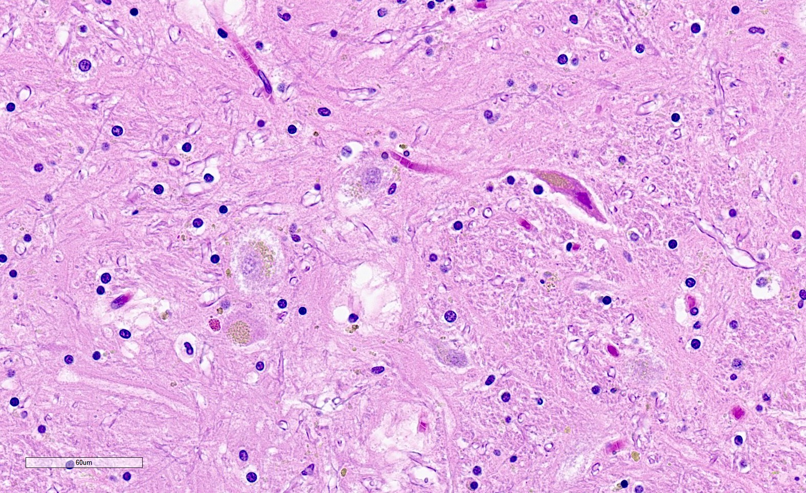
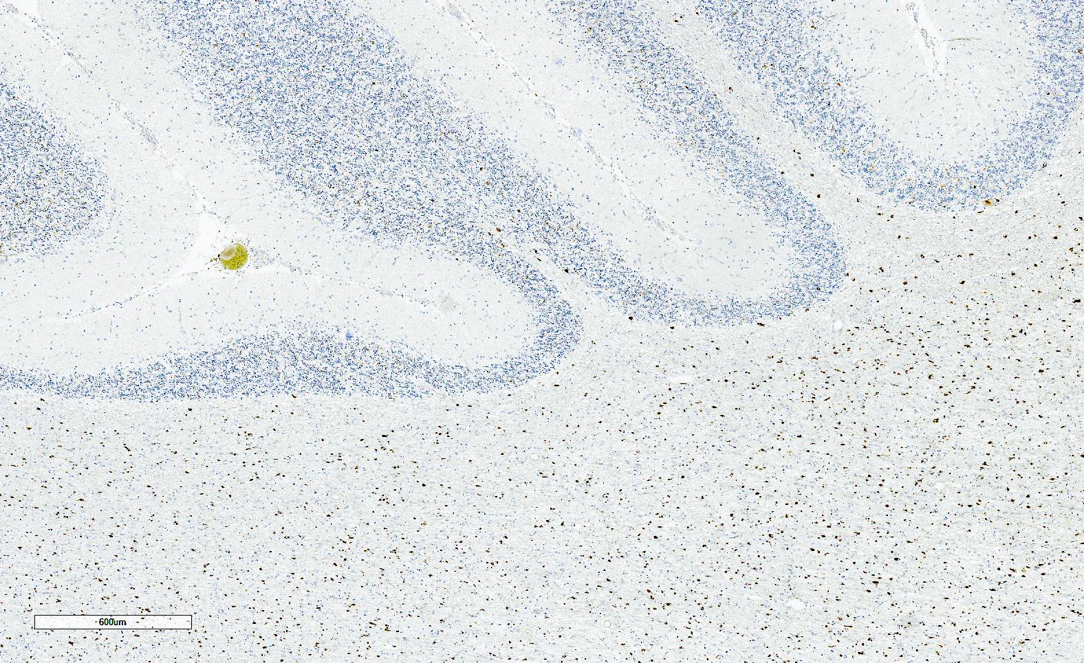
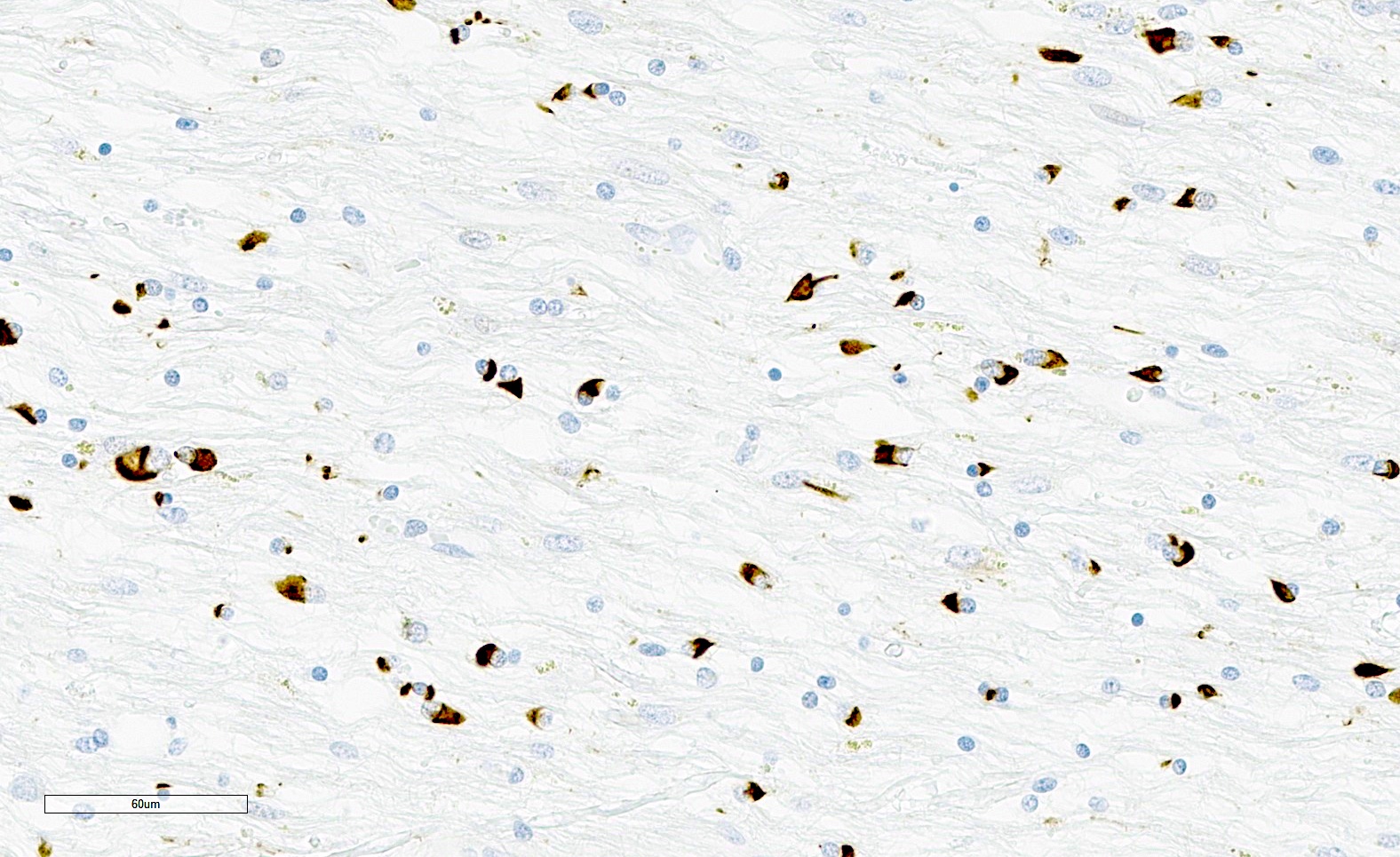
Alcoholic cerebellar degeneration
Contributed by Kymberly A. Gyure, M.D.

Alcoholic cerebellar degeneration
Images hosted on other servers:

Sampling for Braak staging

Stages of Braak NFT spread

CERAD neuritic plaque assessment

Neuropathologic change
Images hosted on other servers:

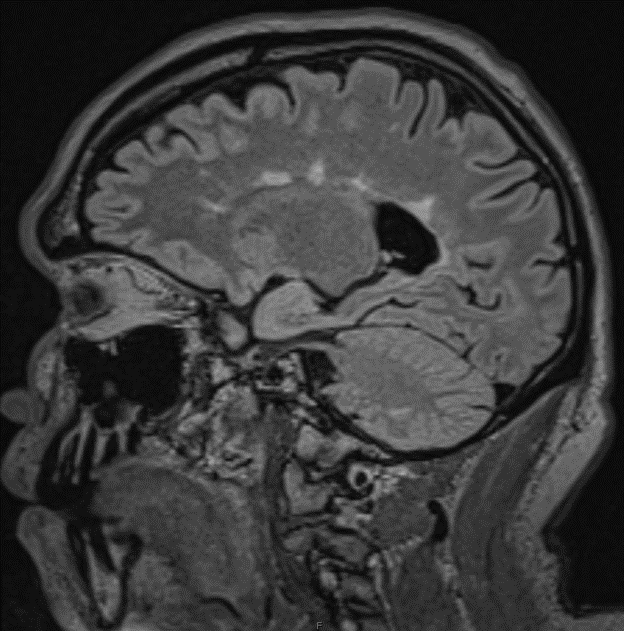
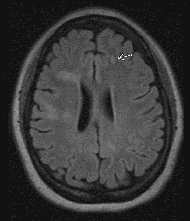
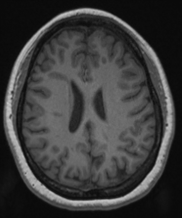
Cortical atrophy on MRI

Pittsburgh compound B PET imaging
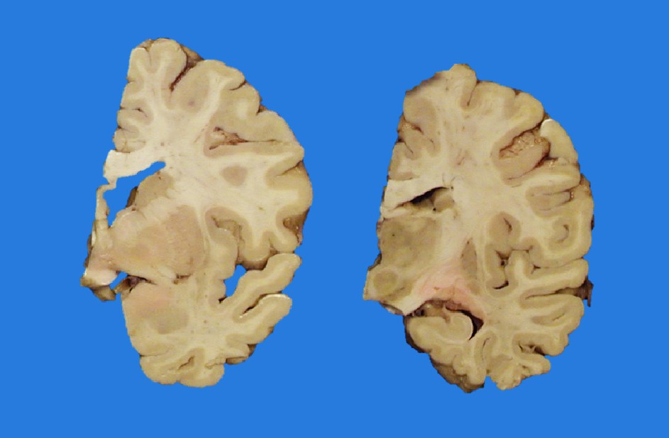
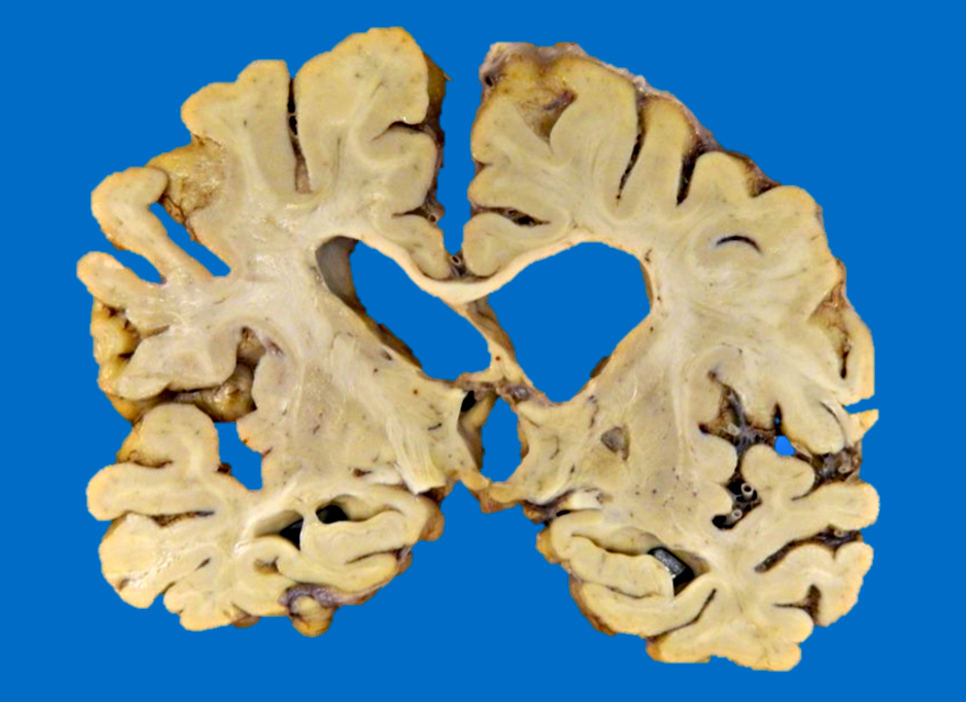
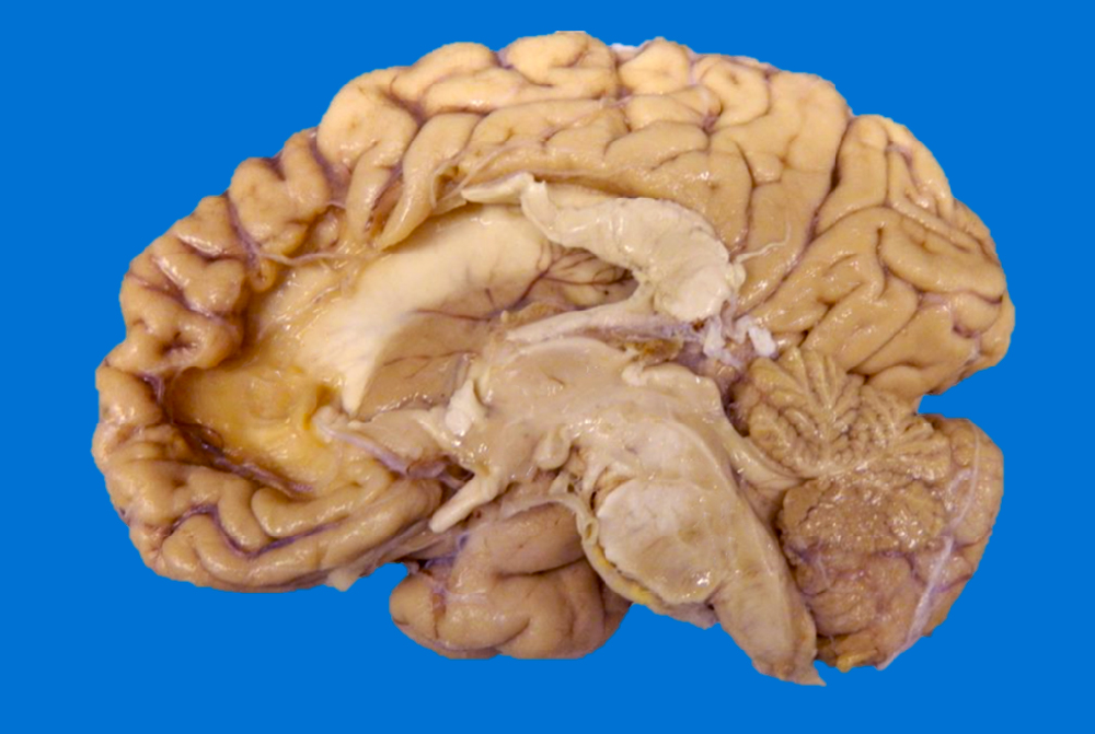
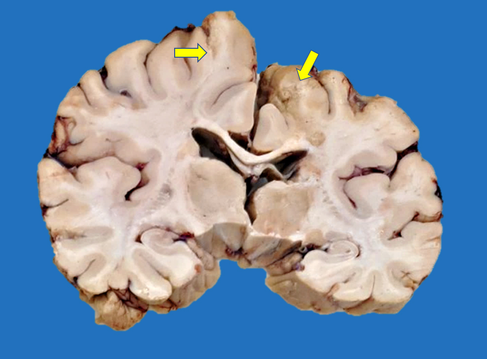
Contributed by Bartholomew White, M.D.
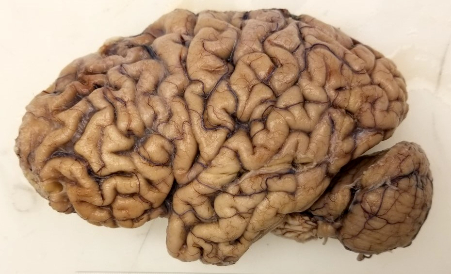
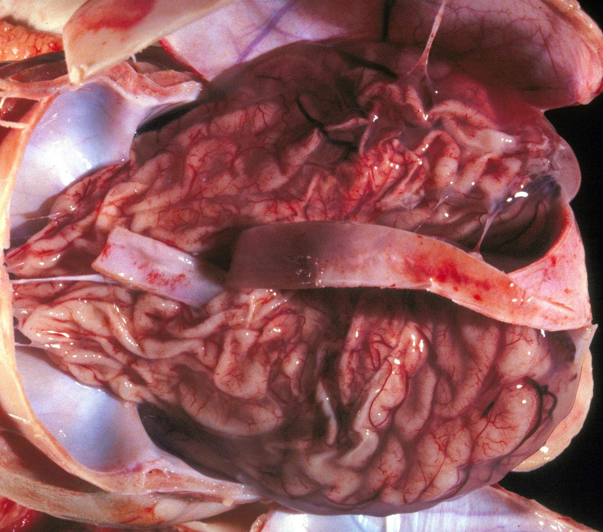
Cortical atrophy
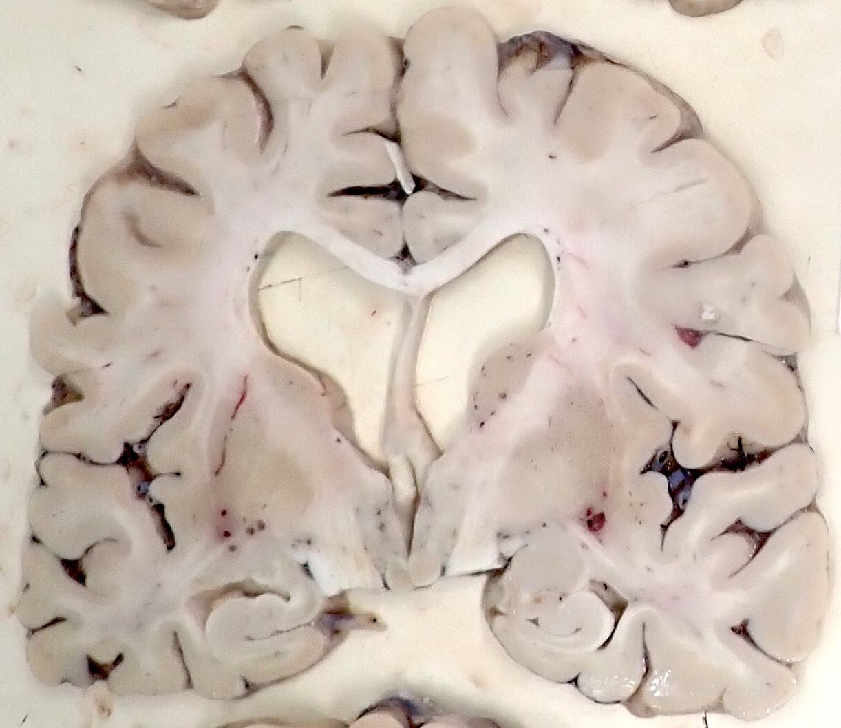
Ventricular dilation
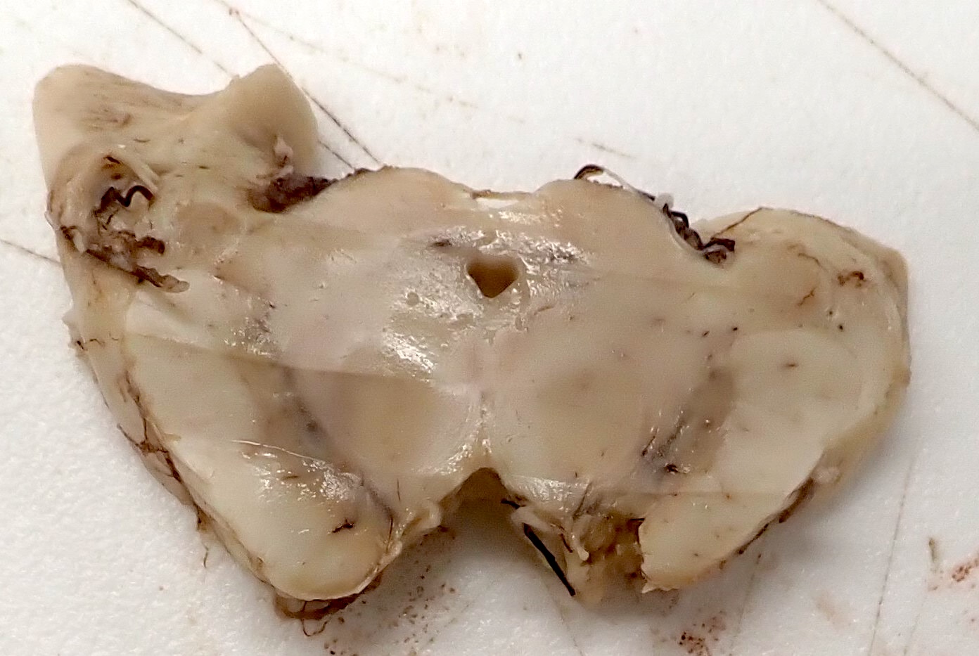
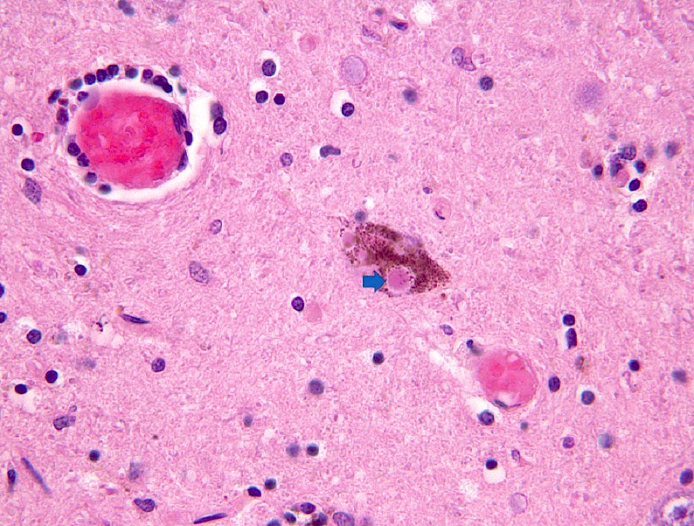
Substantia nigra and locus ceruleus
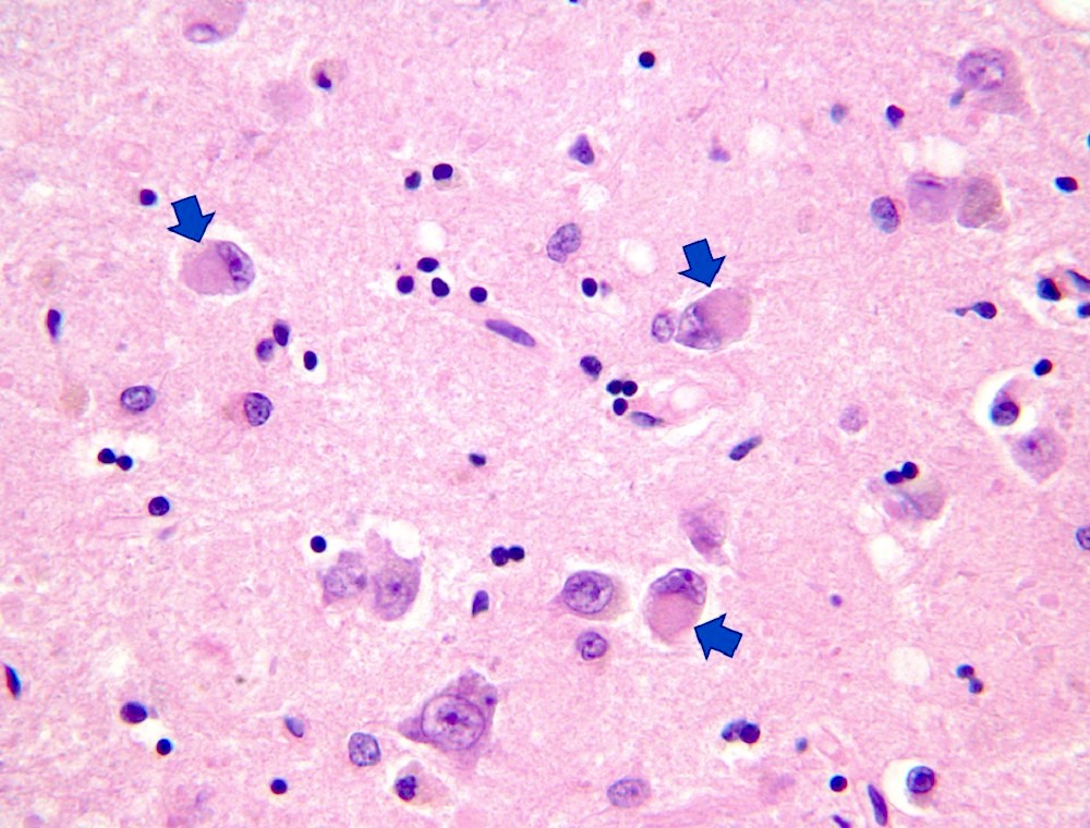
Contributed by Bartholomew White, M.D.
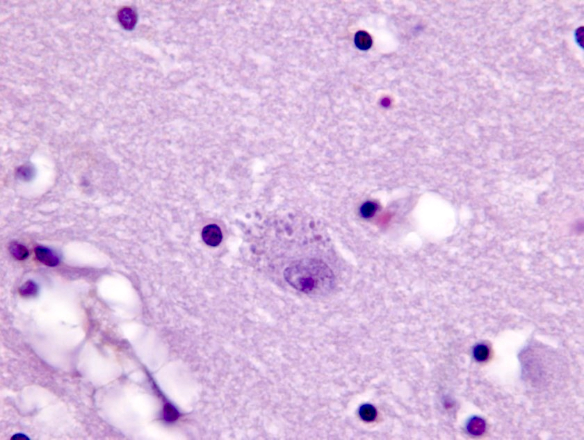
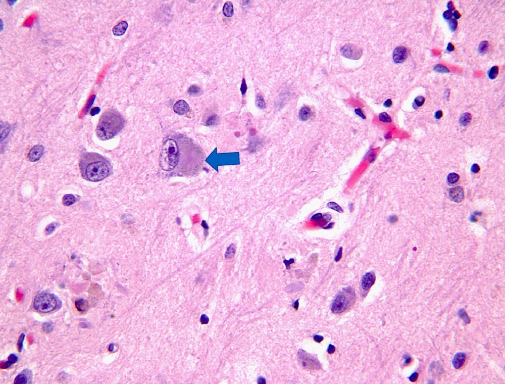
Granulovacuolar degeneration
Hirano bodies
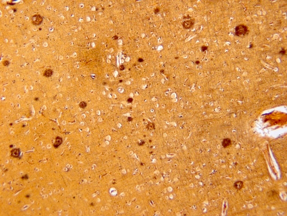
Diffuse plaque
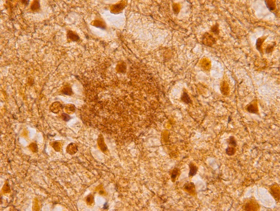
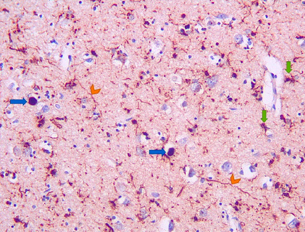
Neuritic plaque
Neurofibrillary tangle
Diffuse plaque
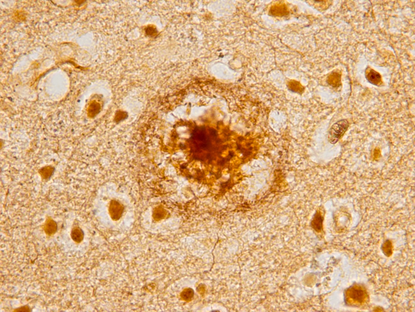
Neuritic plaque
Moderate neuritic plaques
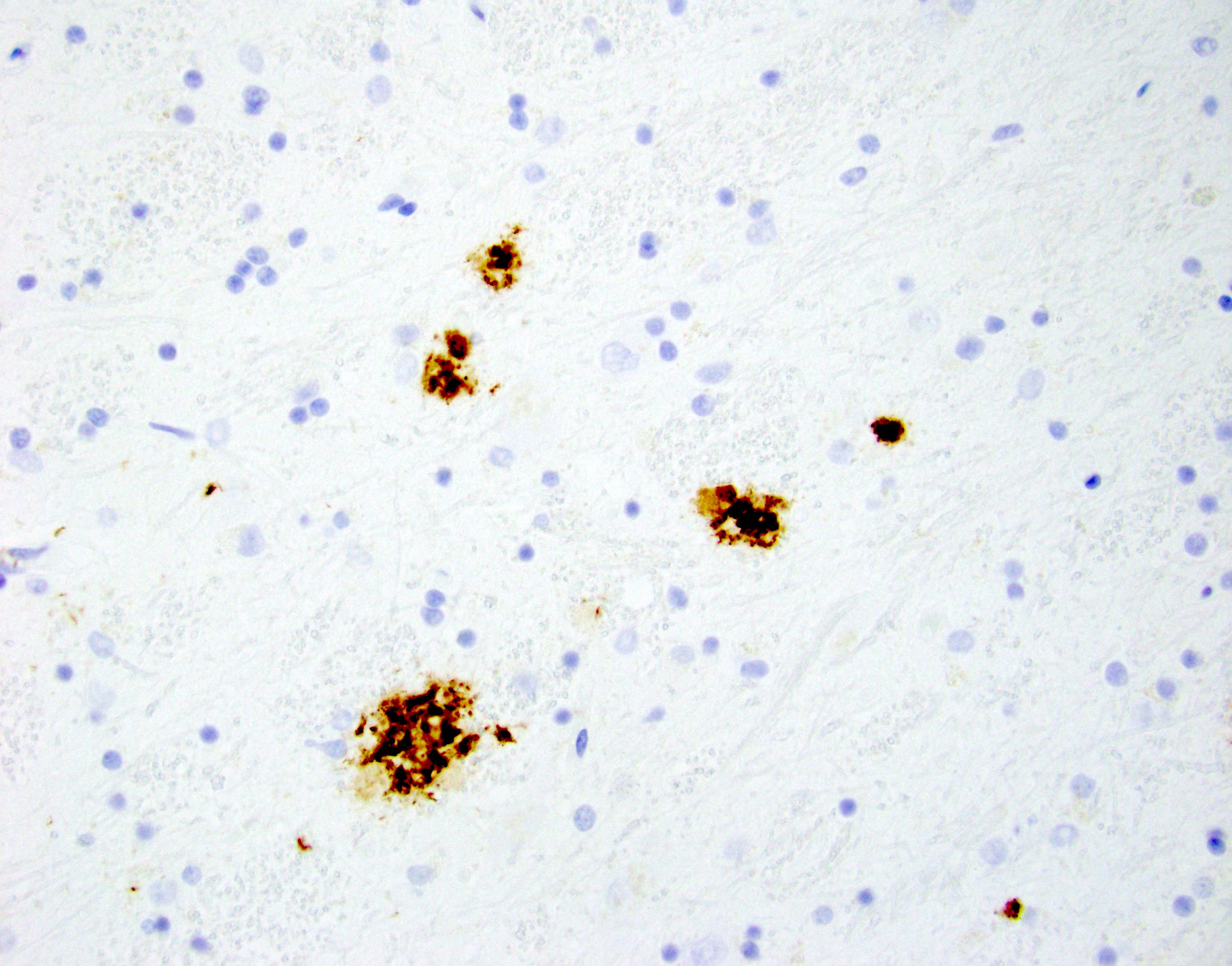
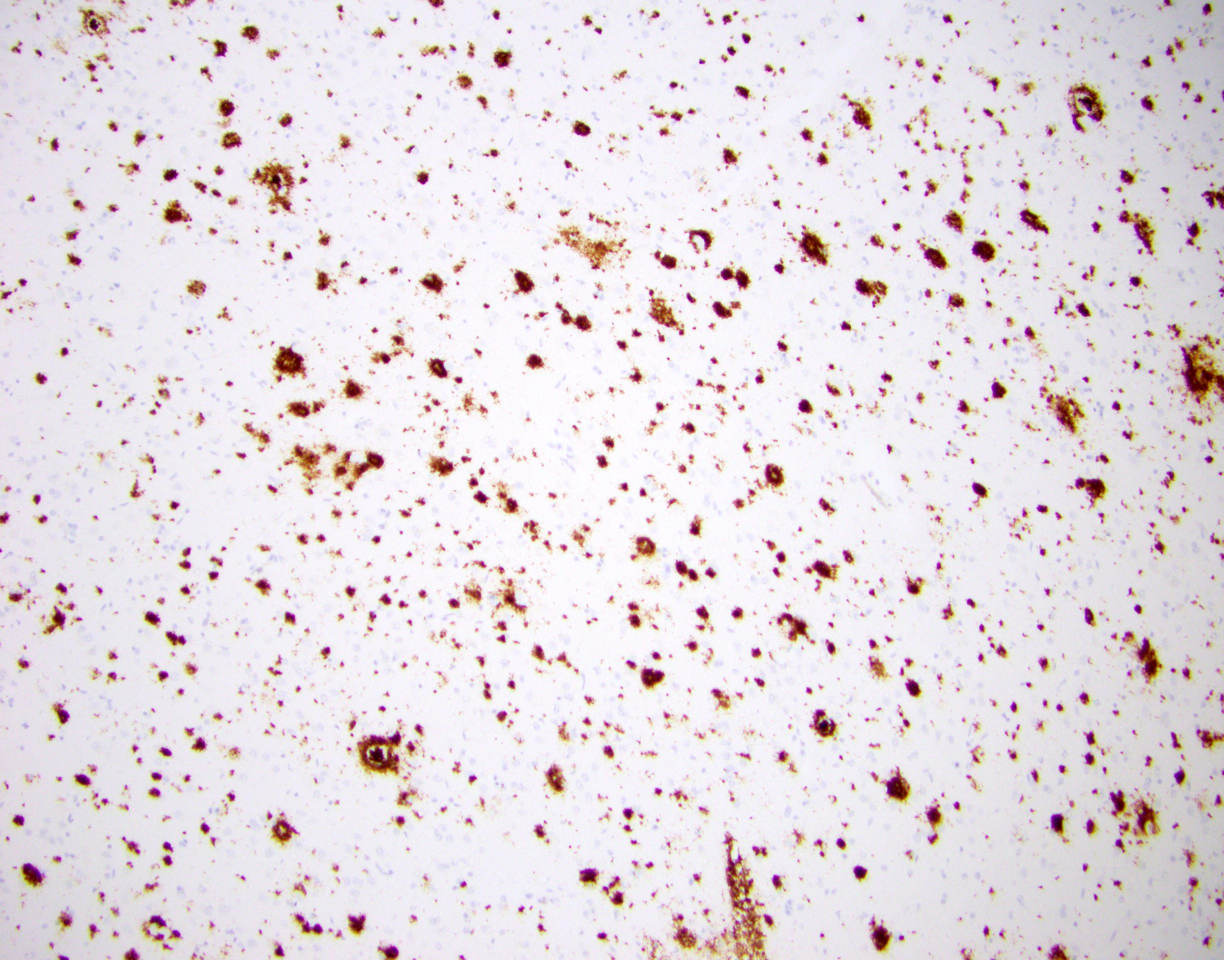
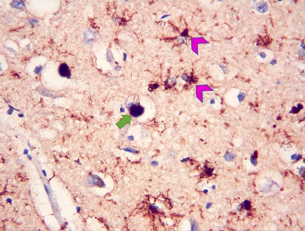
Beta amyloid aggregates
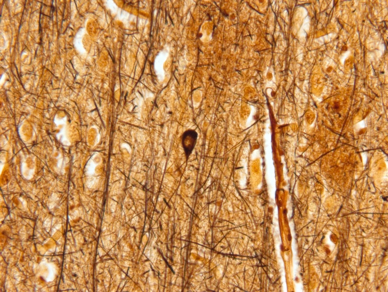
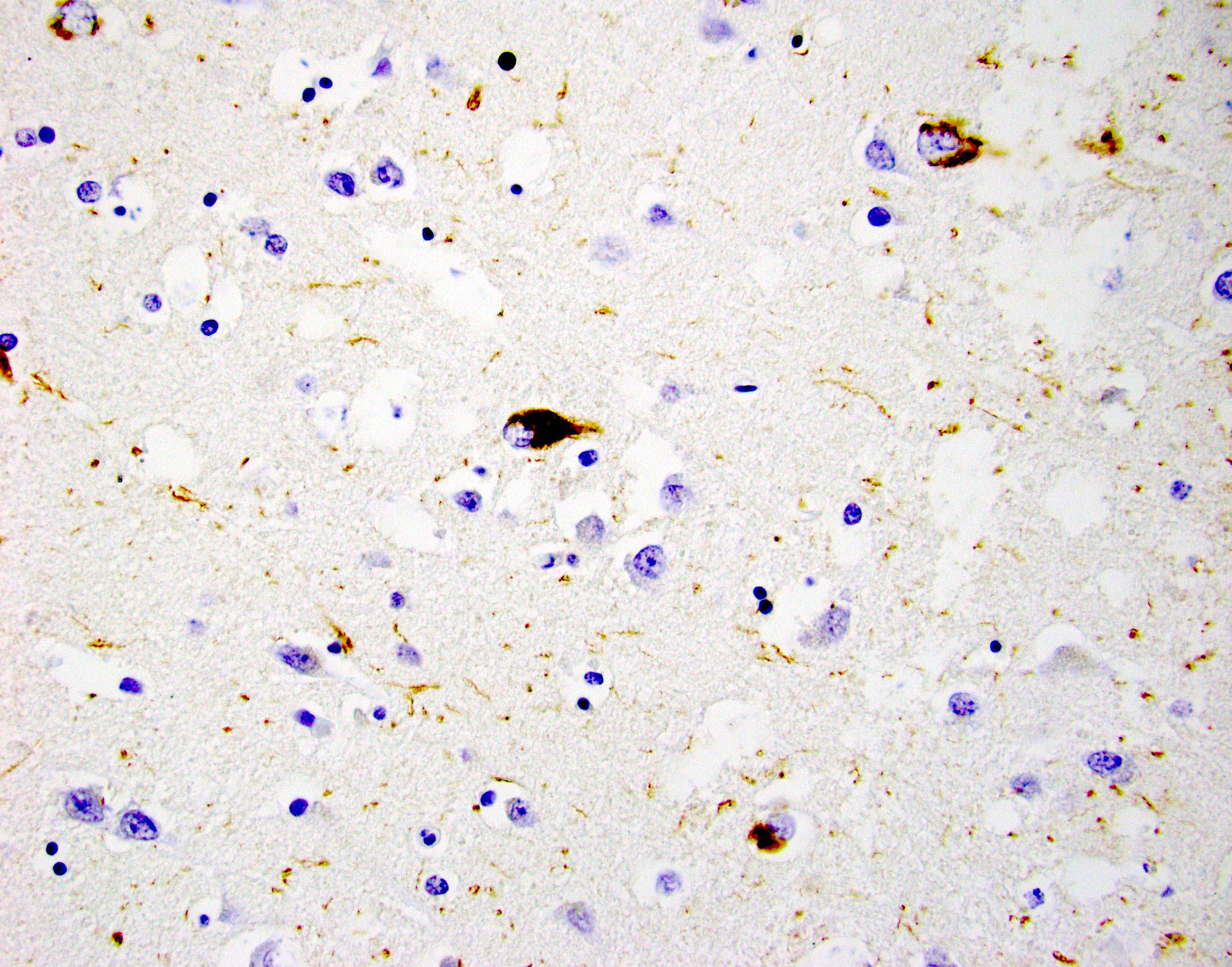
Neurofibrillary tangle
Contributed by Hannes Vogel, M.D.
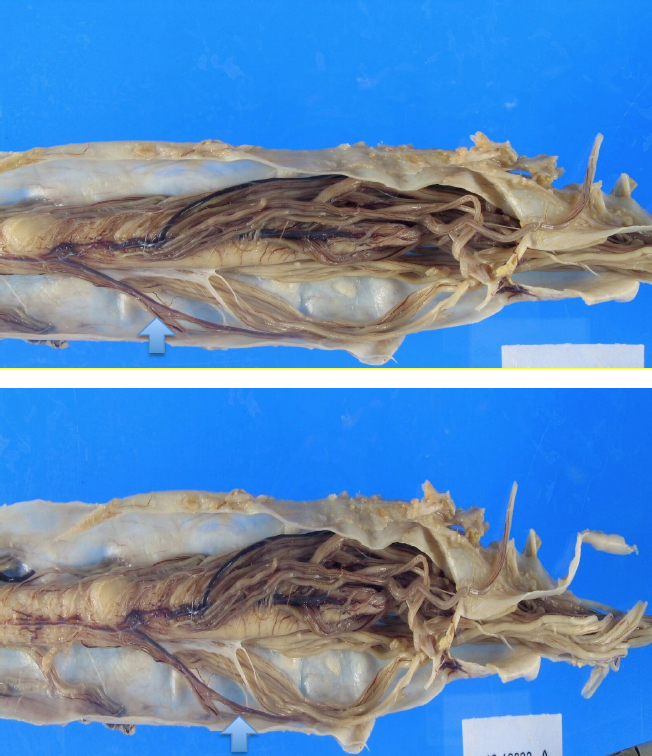
Spinal cord atrophy
Contributed by Eileen Bigio M.D. and Qinwen Mao, M.D. Ph.D.
Spinal nerve roots
ALS versus normal spine

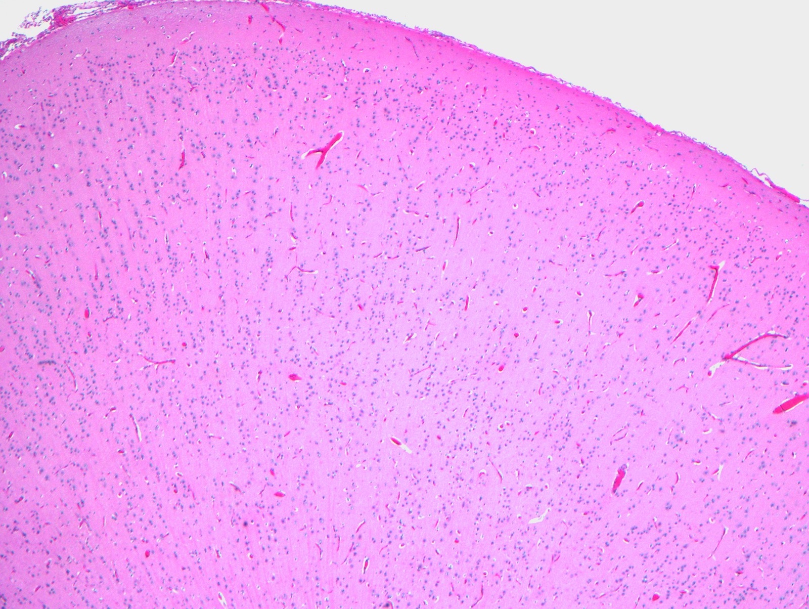
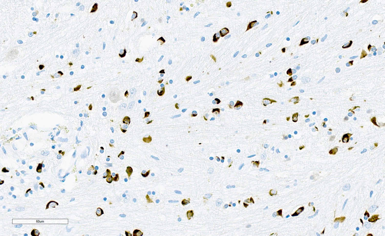
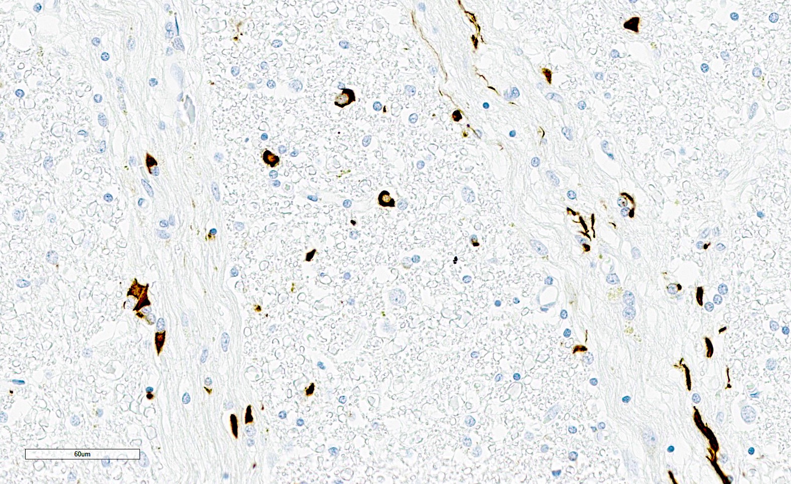
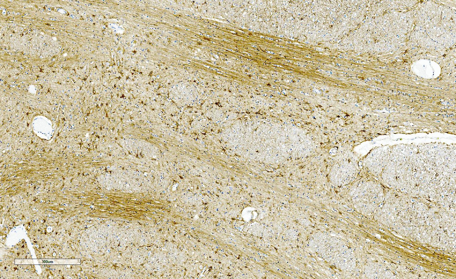
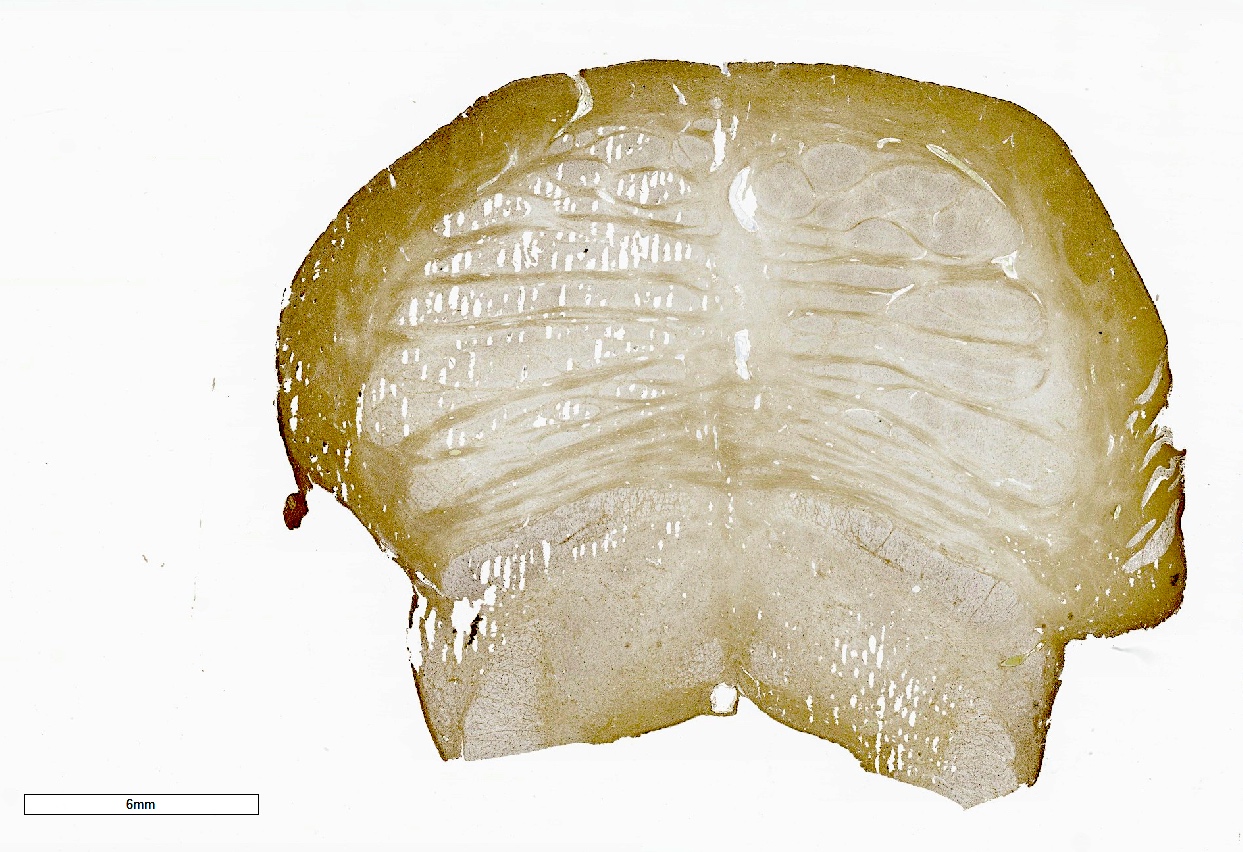
Corticospinal tract
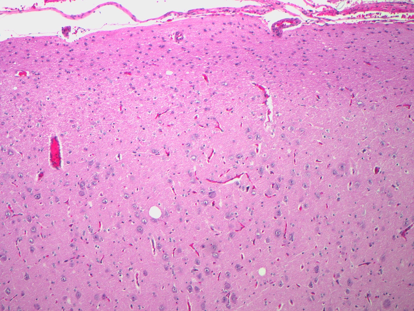
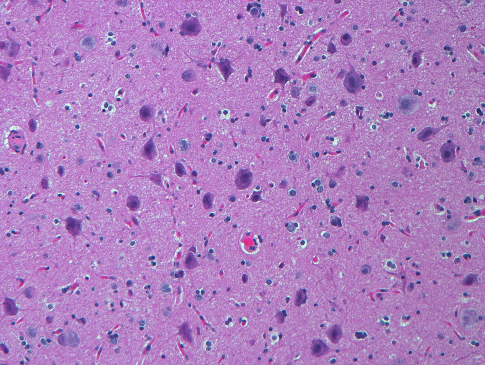
Motor cortex
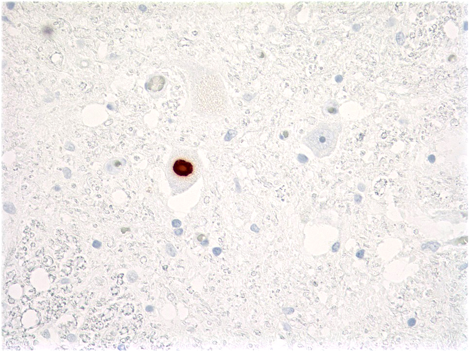
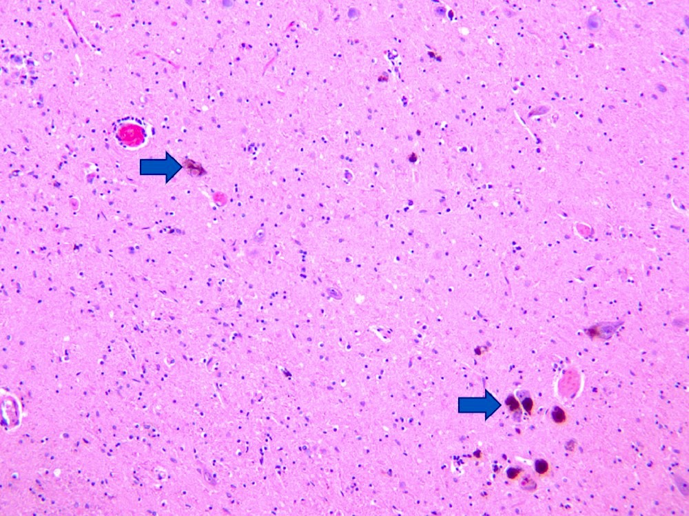
Bunina bodies
Lewy-like body
Contributed by Hannes Vogel, M.D., Matthew McCord, M.D., Christina Appin, M.D. and Missia Kohler, M.D.
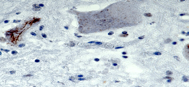
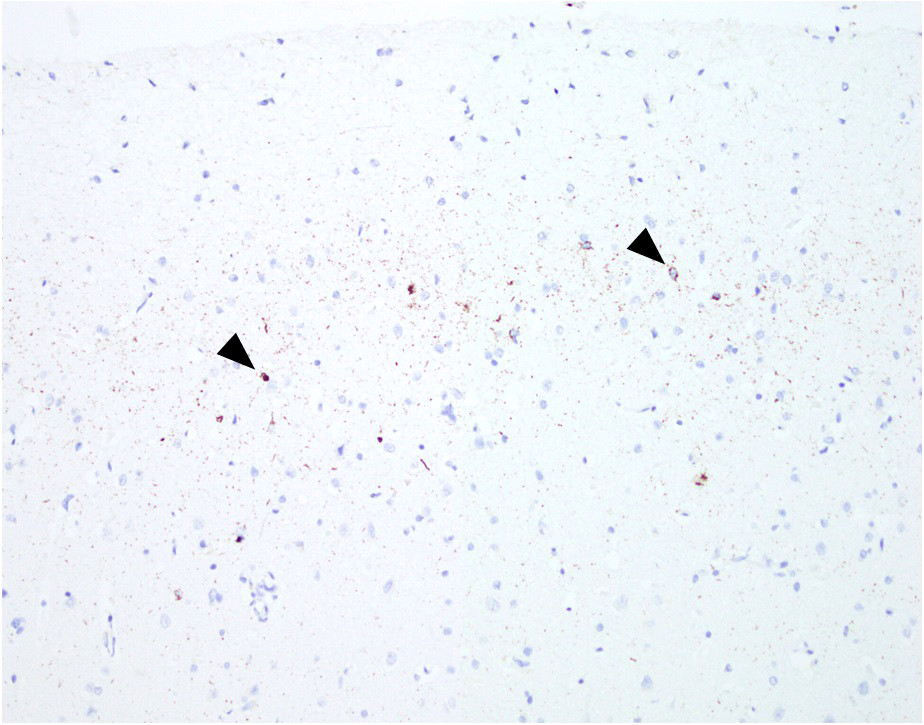
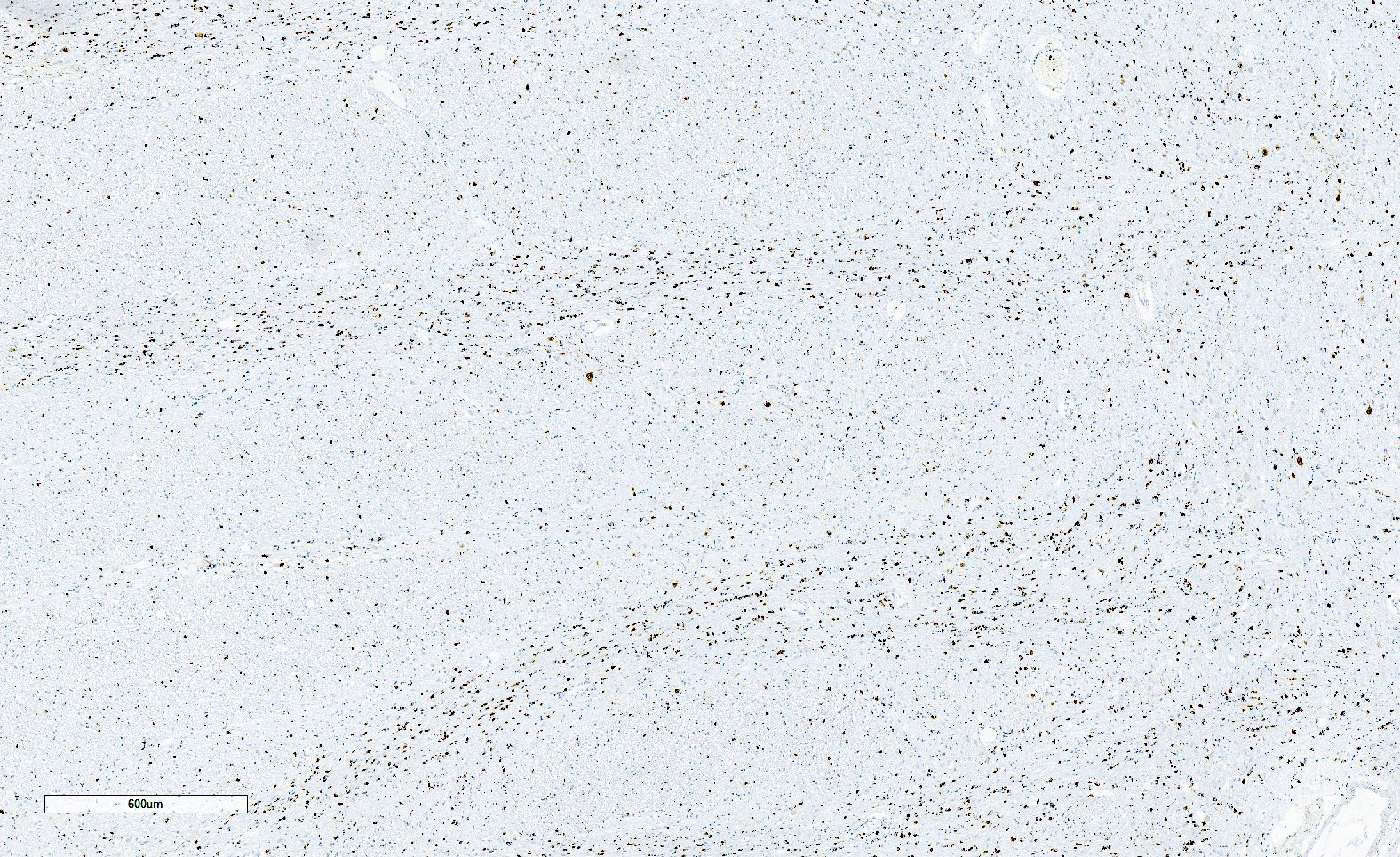
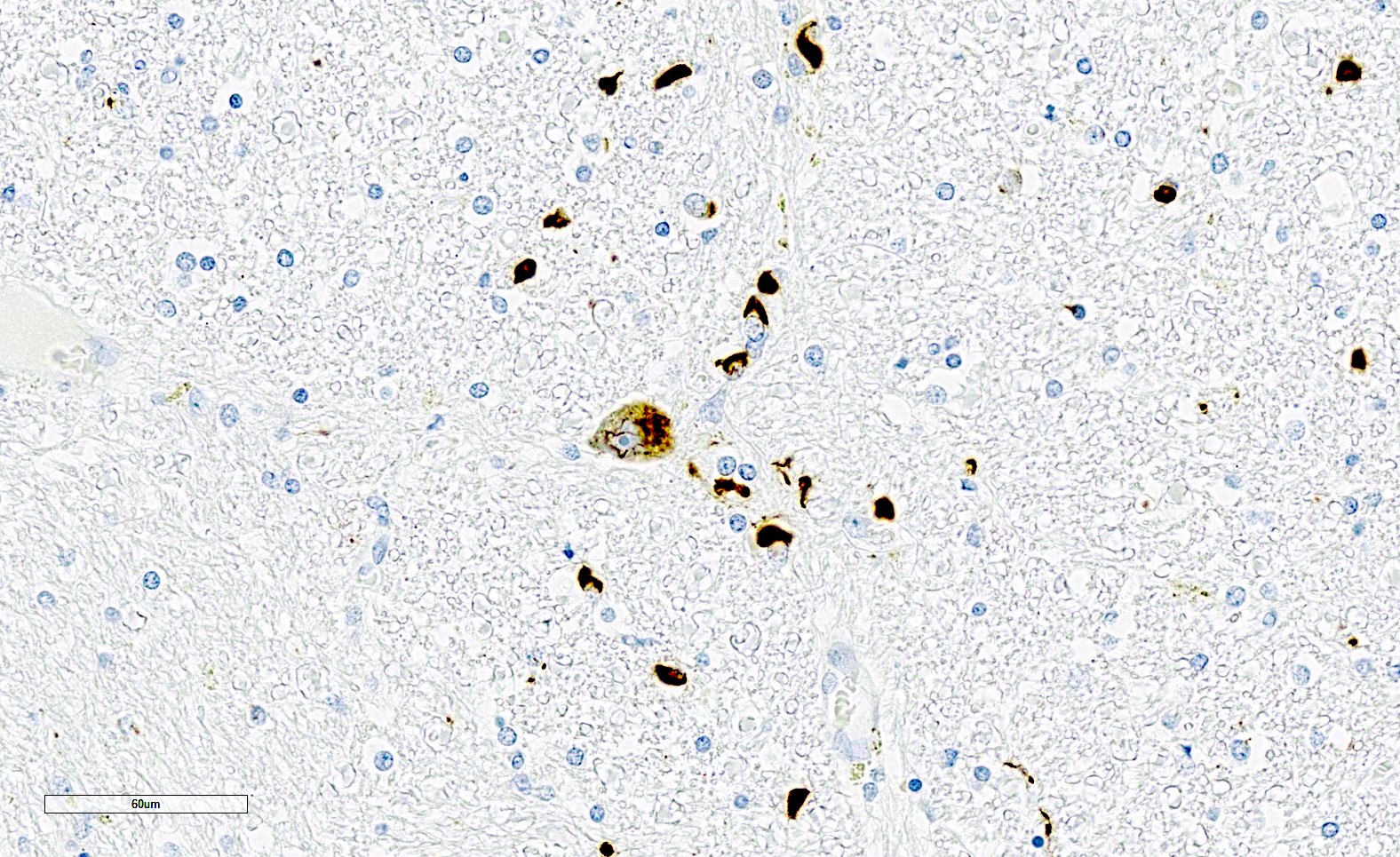
Inclusions immunoreactive for TDP-43
ALS / FTD overlap
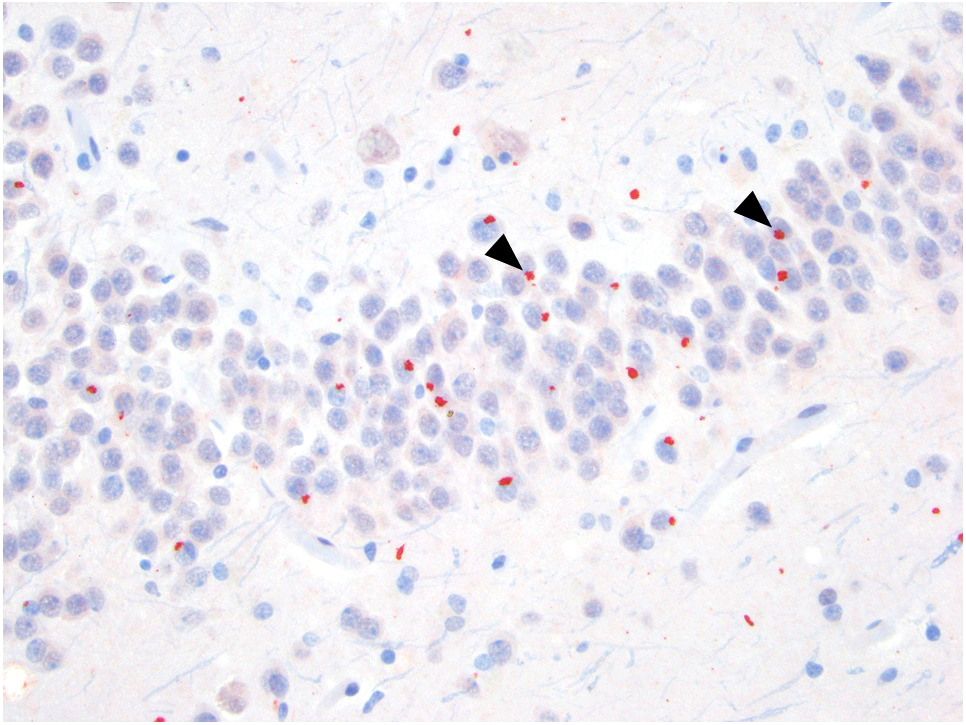
C9orf72 p62 positive inclusions
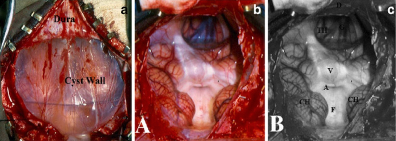
Contributed by Erdener Özer, M.D., Ph.D.
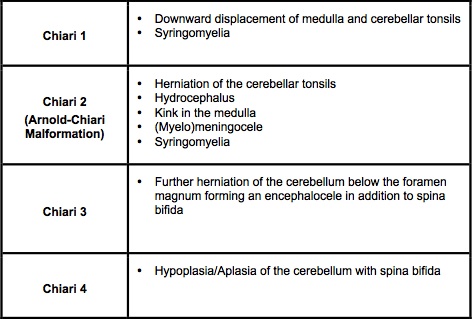
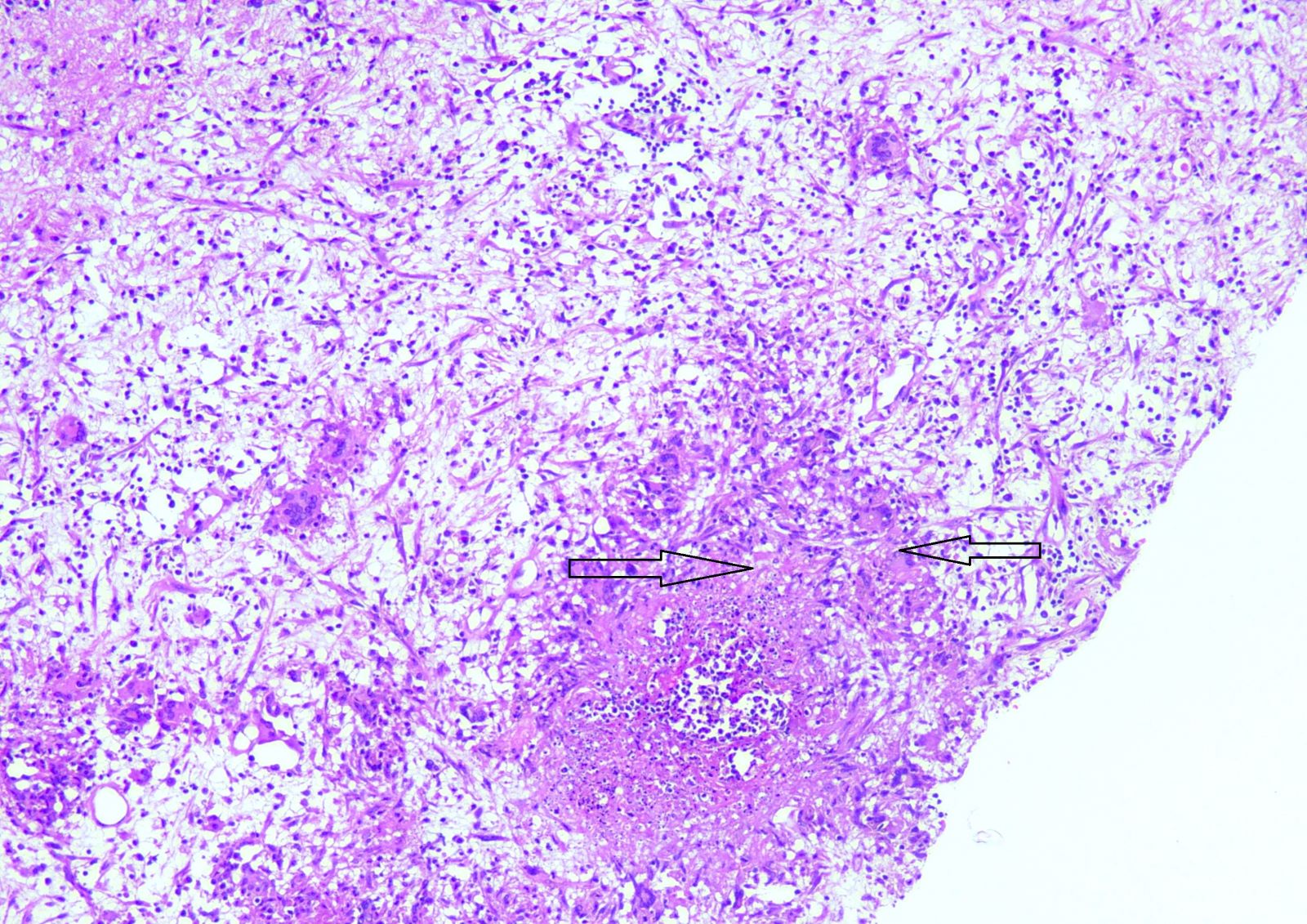
Types of Chiari malformations
Images hosted on other servers:

Sagittal FLAIR MRI scan
Contributed by Erdener Özer, M.D., Ph.D.
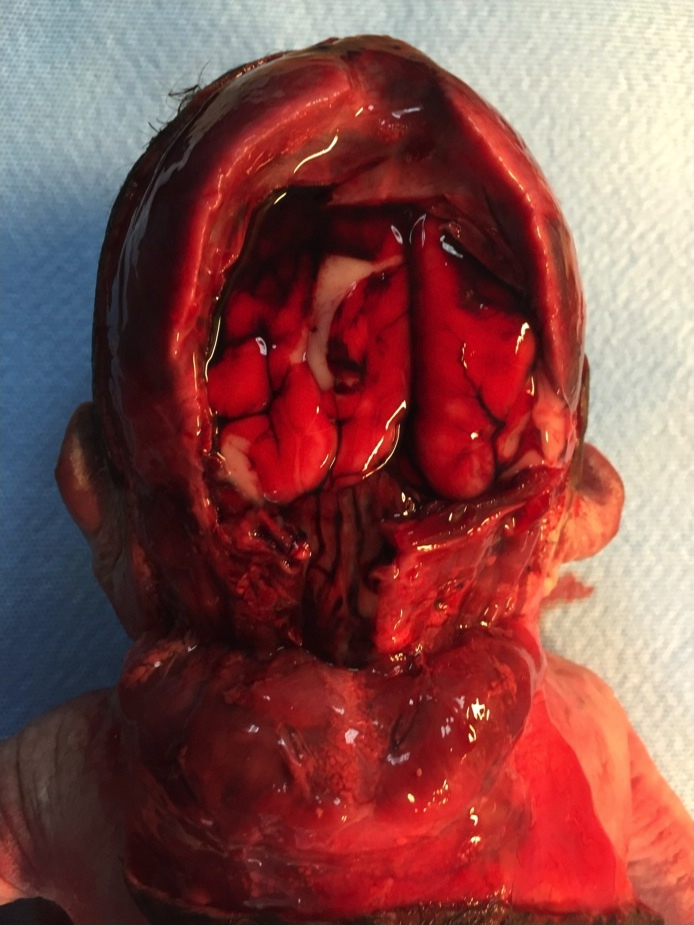
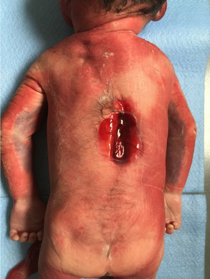
Chiari type II malformation
Images hosted on other servers:


CT / MRI scan of brain for neuroaspergillosis
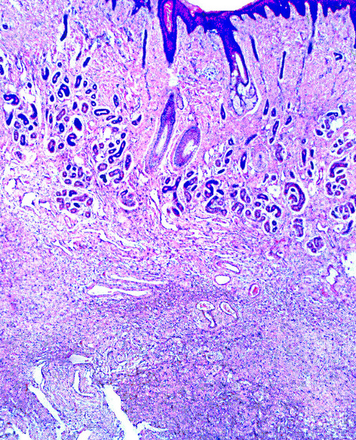
Contributed by Khurram Minhas, M.B.B.S.
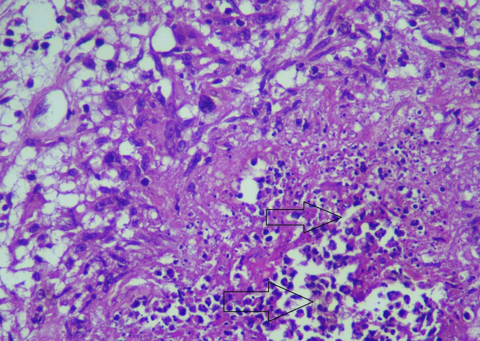
Necrotizing granulomatous inflammation
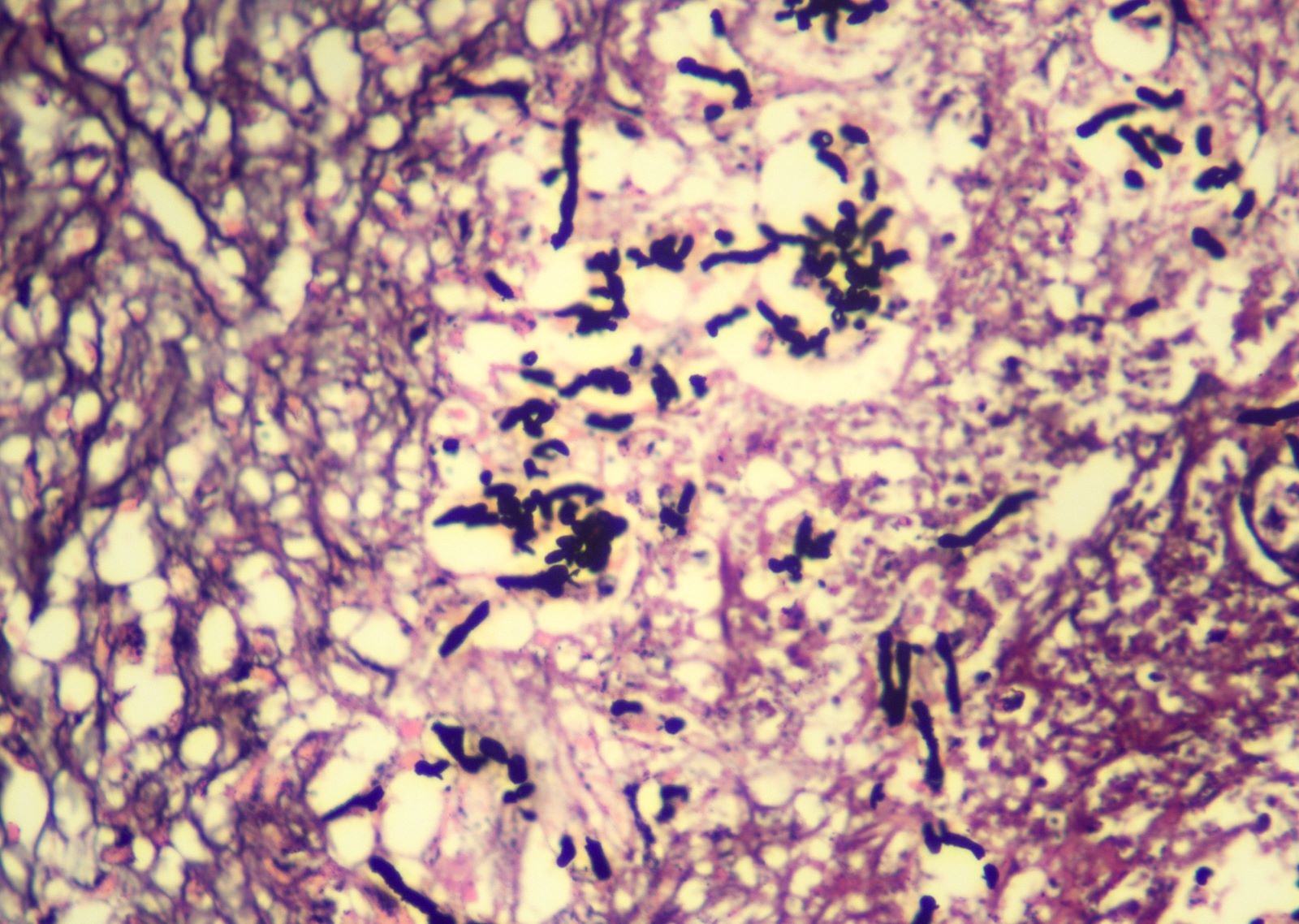
GMS stain
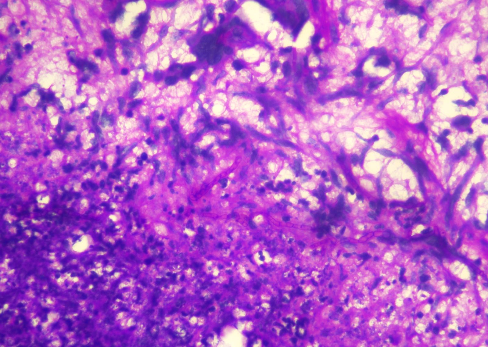
PASD stain
Images hosted on other servers:

Ventricular dilatation

Curvilinear high signal intensities
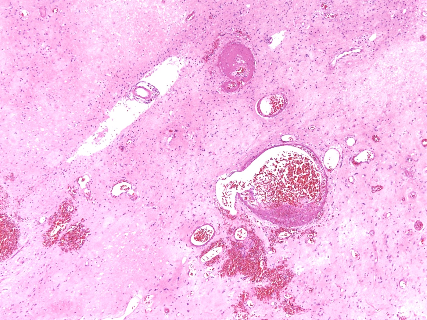
Contributed by Mark Cohen, M.D.
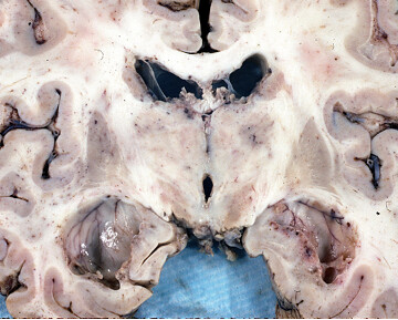
Coronal section

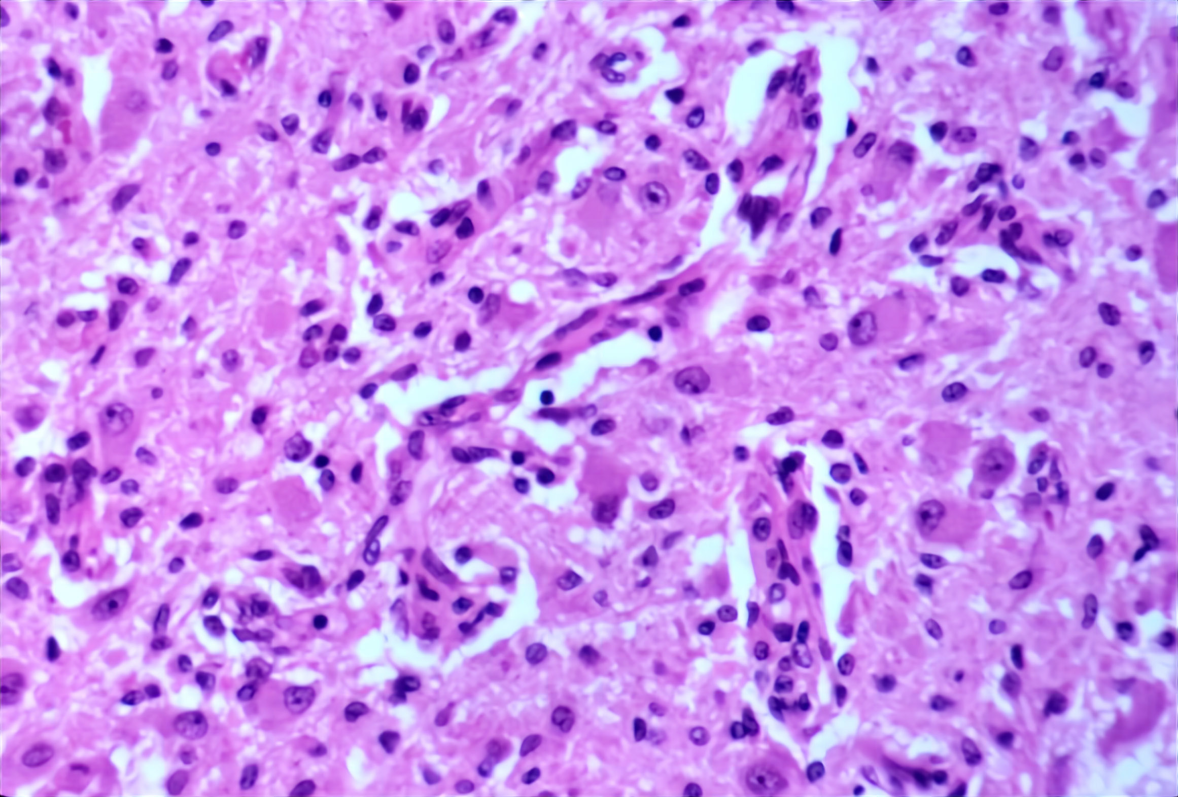
Inflammatory necrosis
Microencephaly with hydrocephalus
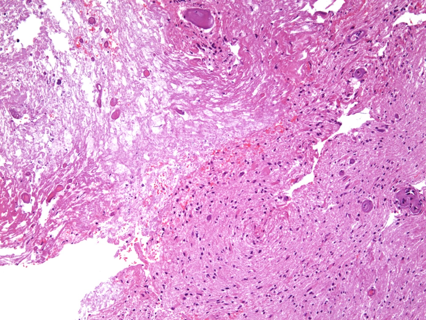
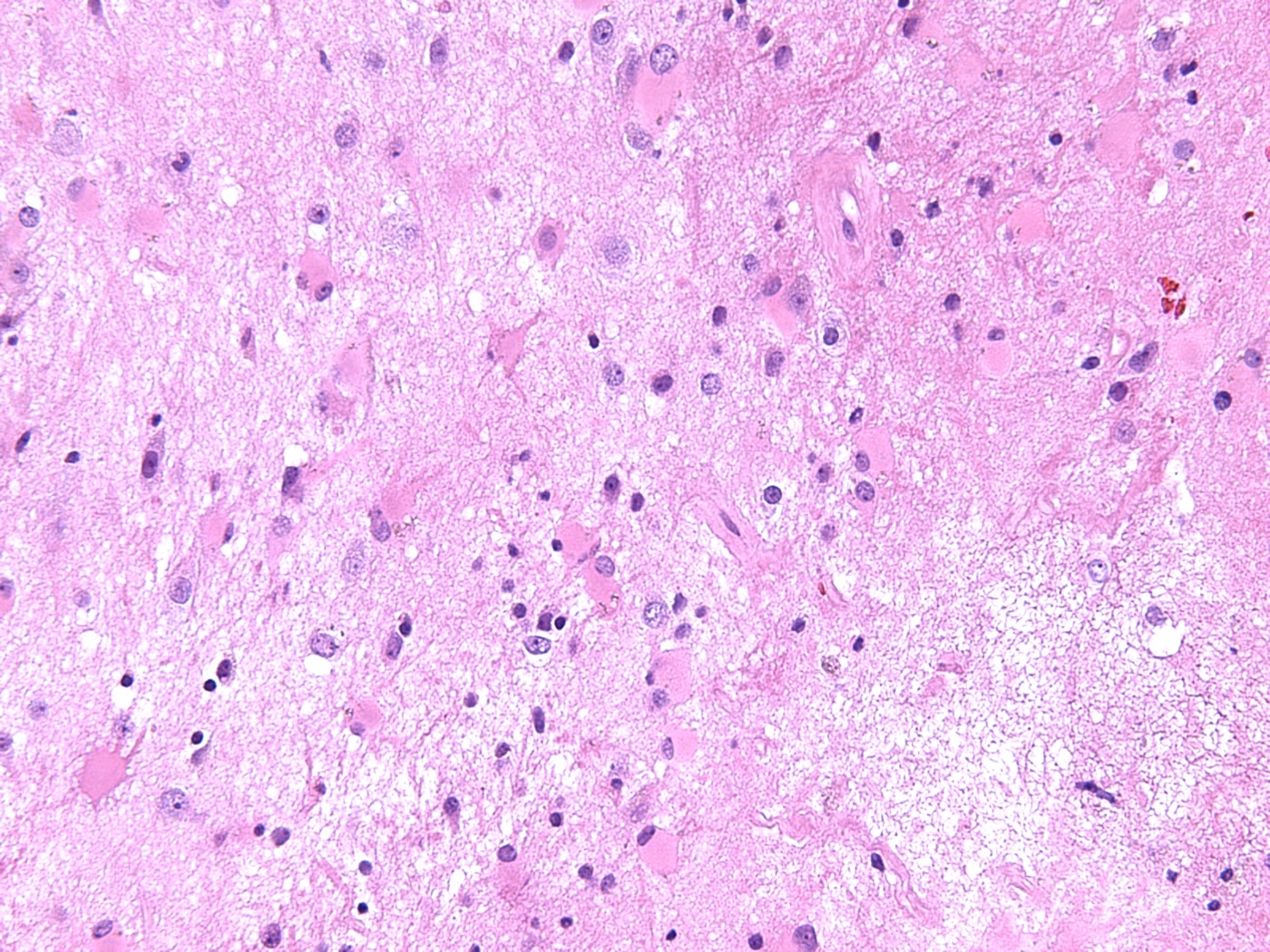
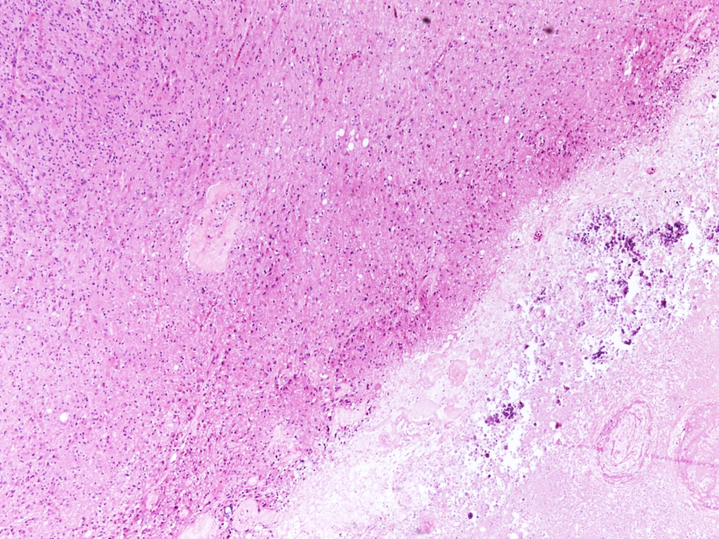
Contributed by Mark Cohen, M.D.
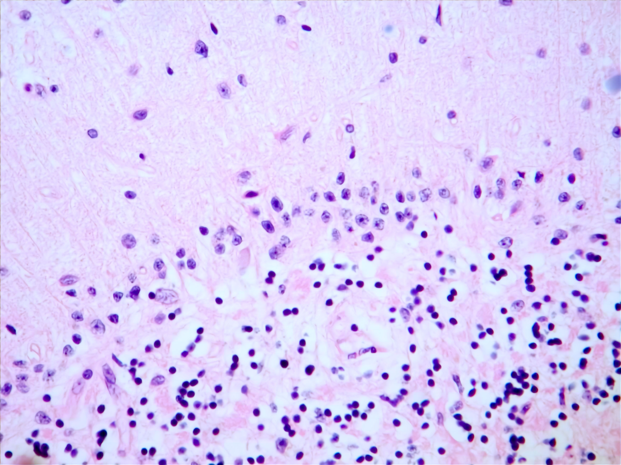
Low and high power, H&E
Images hosted on other servers:

16 hours after injury
Contributed by Kymberly A. Gyure, M.D.
Carbon monoxide injury
Images hosted on other servers:

Intracerebral hemorrhage and hemosiderosis

Arterial wall enhancement
Images hosted on other servers:

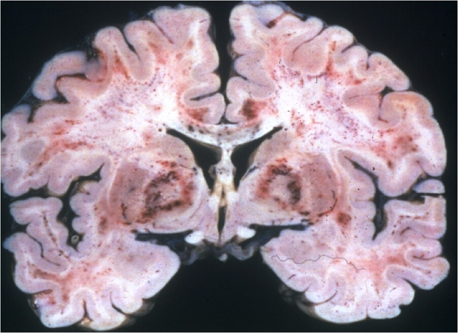
Temporoparietal lobar hemorrhage
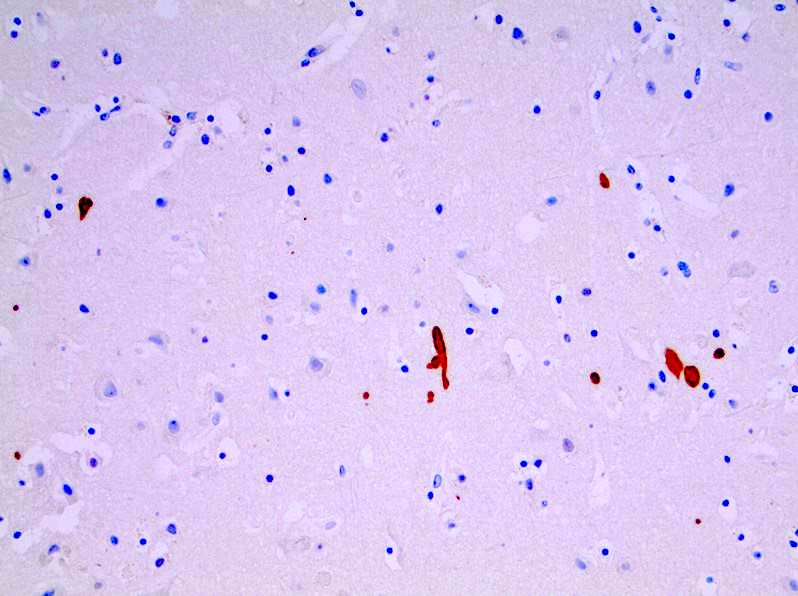
Contributed by Jared T. Ahrendsen, M.D., Ph.D. and Nicolas Kostelecky, M.D.
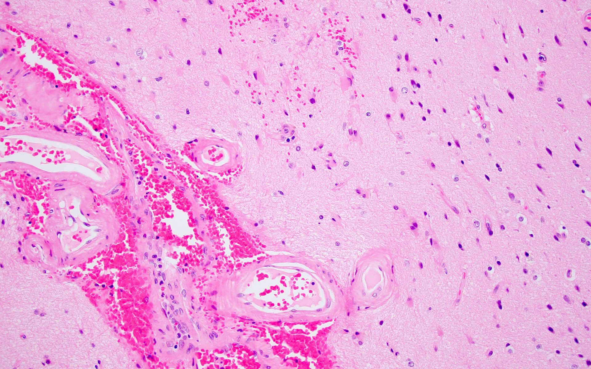
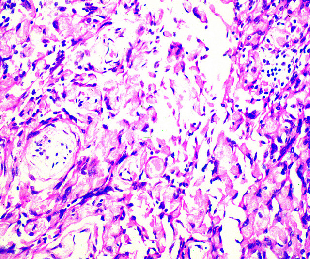
Small and medium sized blood vessels
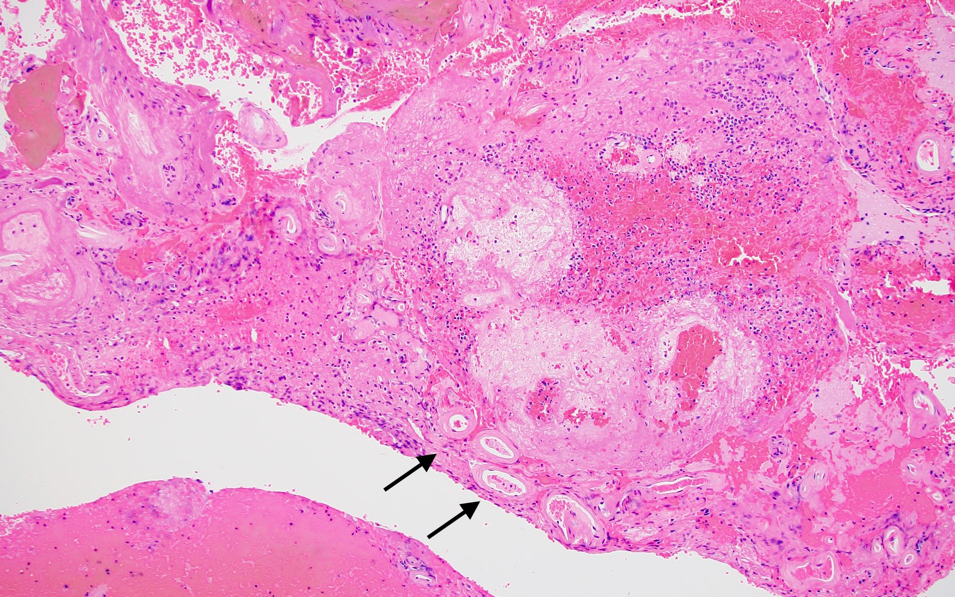
Hemorrhagic cerebral amyloid
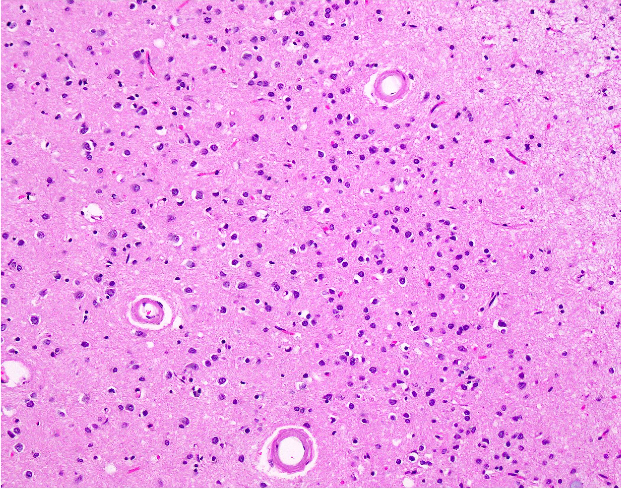
Cortical arterioles
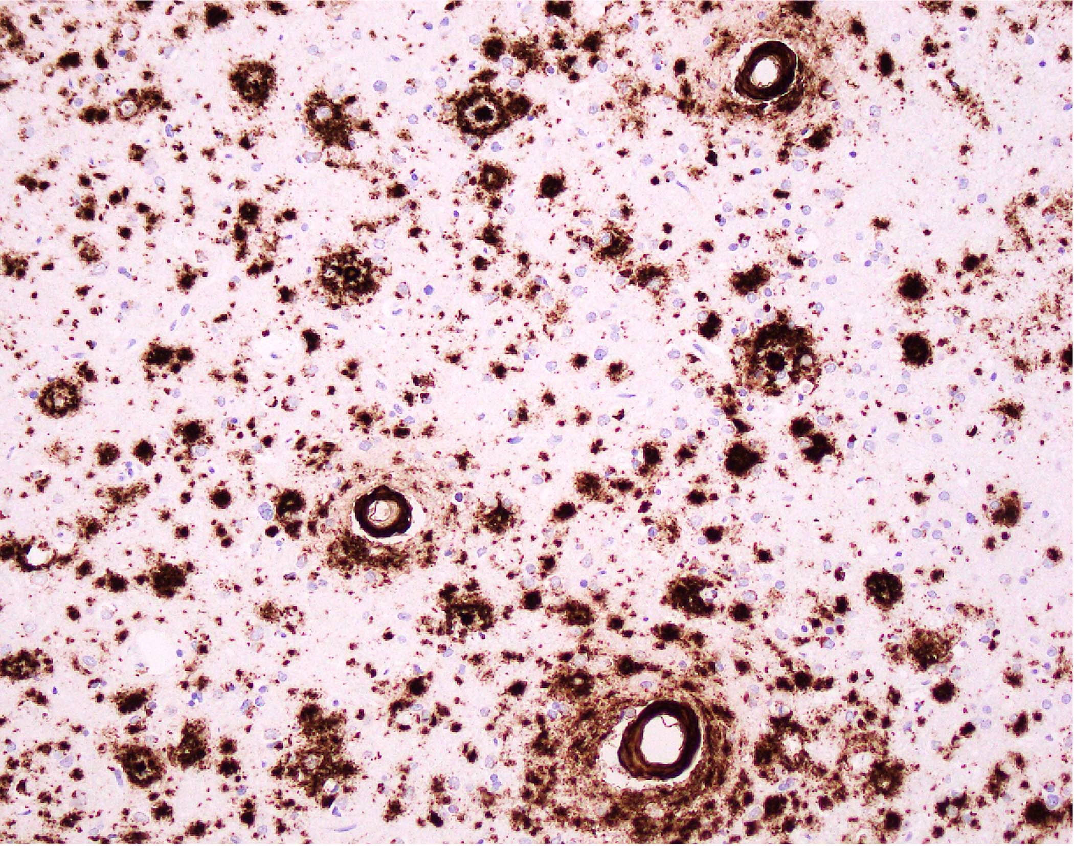
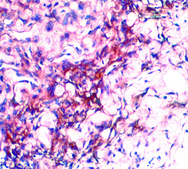
Beta amyloid IHC
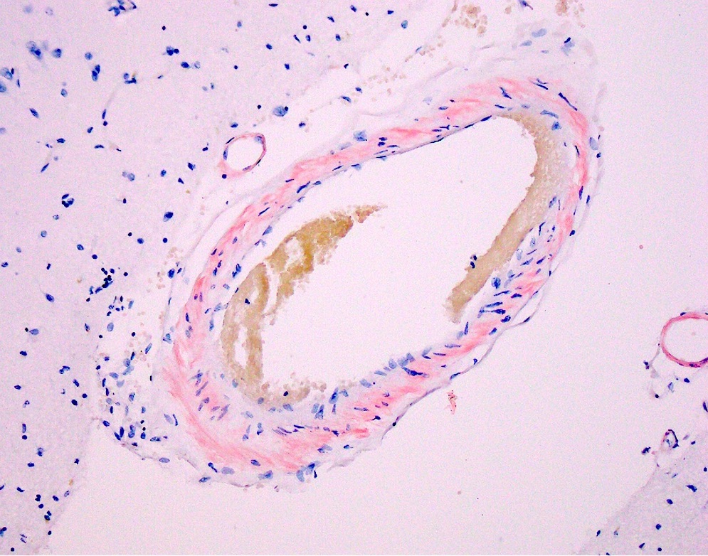

Congophilic leptomeningeal vessels
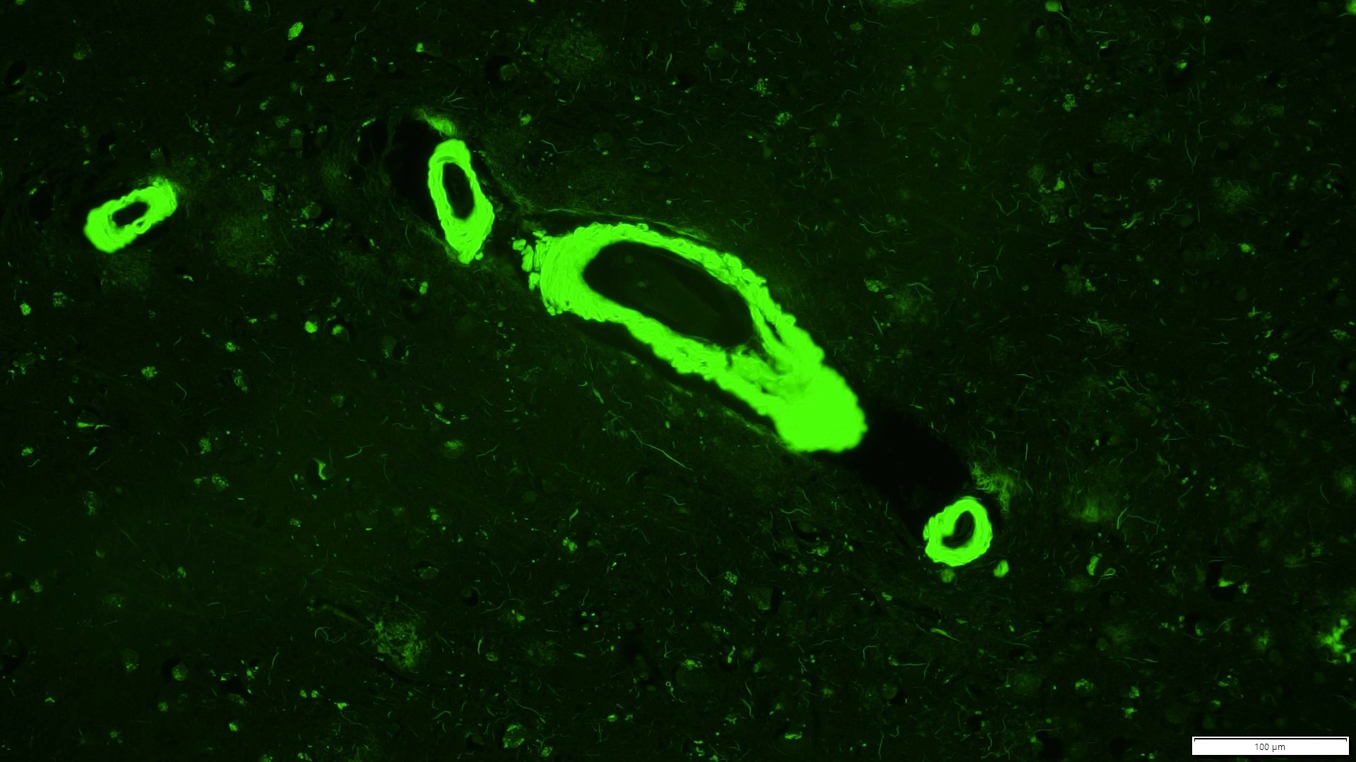
Bright green fluorescence of cortical arterioles
Images hosted on other servers:
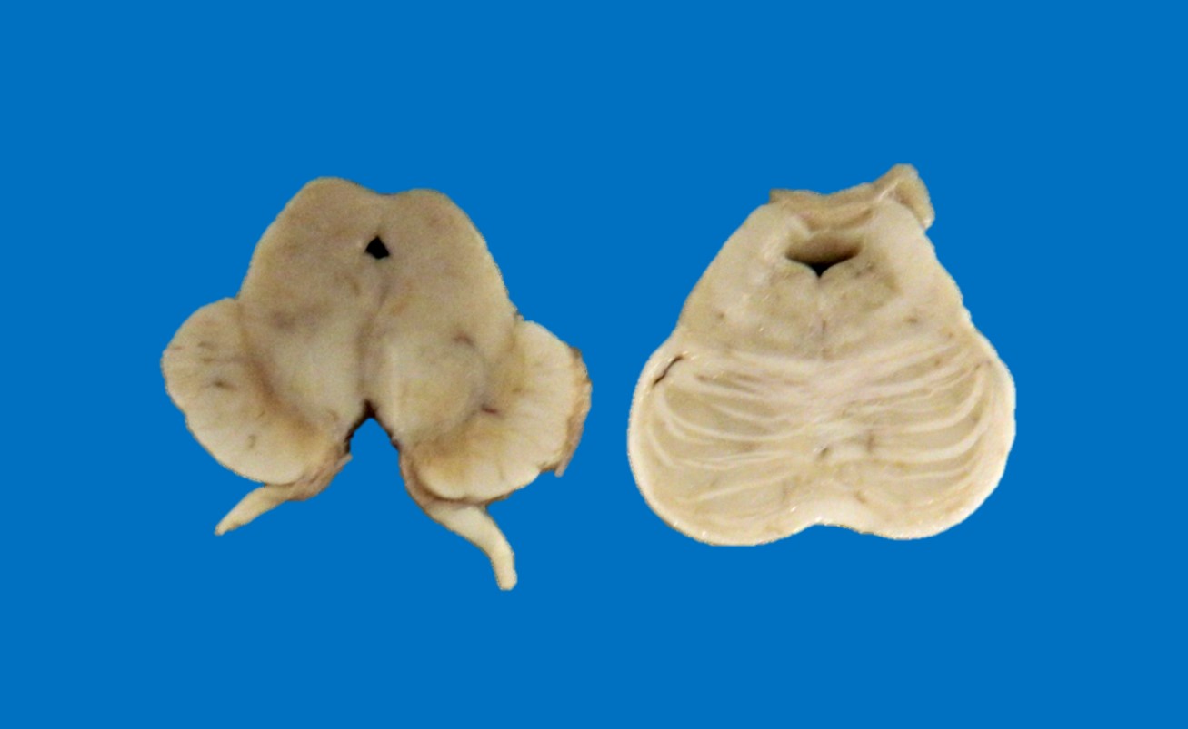
Variant DWS
with dysplasia
of pons and
cerebellum
Contributed by Jean-François Hirsch, M.D.
Dandy-Walker malformation
Images hosted on other servers:

Alpha synuclein and tau aggregation model

Alpha synuclein staging system
Images hosted on other servers:

Decreased dopamine transporter uptake

Low uptake in myocardial scintigraphy
Contributed by Javier Redding-Ochoa, M.D.
Brainstem pathology
Cerebrum
Contributed by Javier Redding-Ochoa, M.D.
Lewy bodies in amygdala
Poorly demarcated cortical Lewy body
Classical Lewy body
Synuclein pathology in amygdala
Neocortical alpha synuclein pathology
Substantia nigra neuron loss
Images hosted on other servers:

Micropolygyria marked by a focal small gyri

Transmantle sign, right superior frontal sulcus

Increased signal intensity

Cortical tubers of tuberous sclerosis
Images hosted on other servers:

Lissencephaly marked by absence of gyral formation

Area of gyral expansion

Resection showing disorganized architecture
Contributed by Richard Prayson, M.D.
IA pattern
IB pattern
IIA pattern
IIB pattern
Hyaline protoplasmic astrocytopathy
Nodular heterotopia
Cortical tuber
NeuN
Images hosted on other servers:

Cranial MRI

Symmetric patchy periventricular
Images hosted on other servers:

Stained sections of the white matter
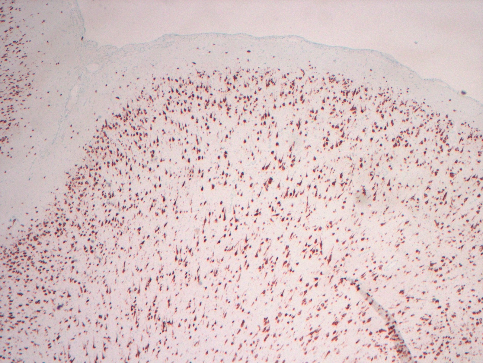
Contributed by Kymberly A. Gyure, M.D. and Palgun Nisarga, M.D.
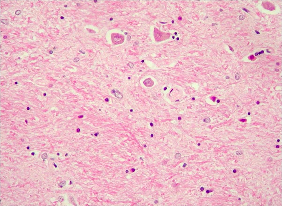
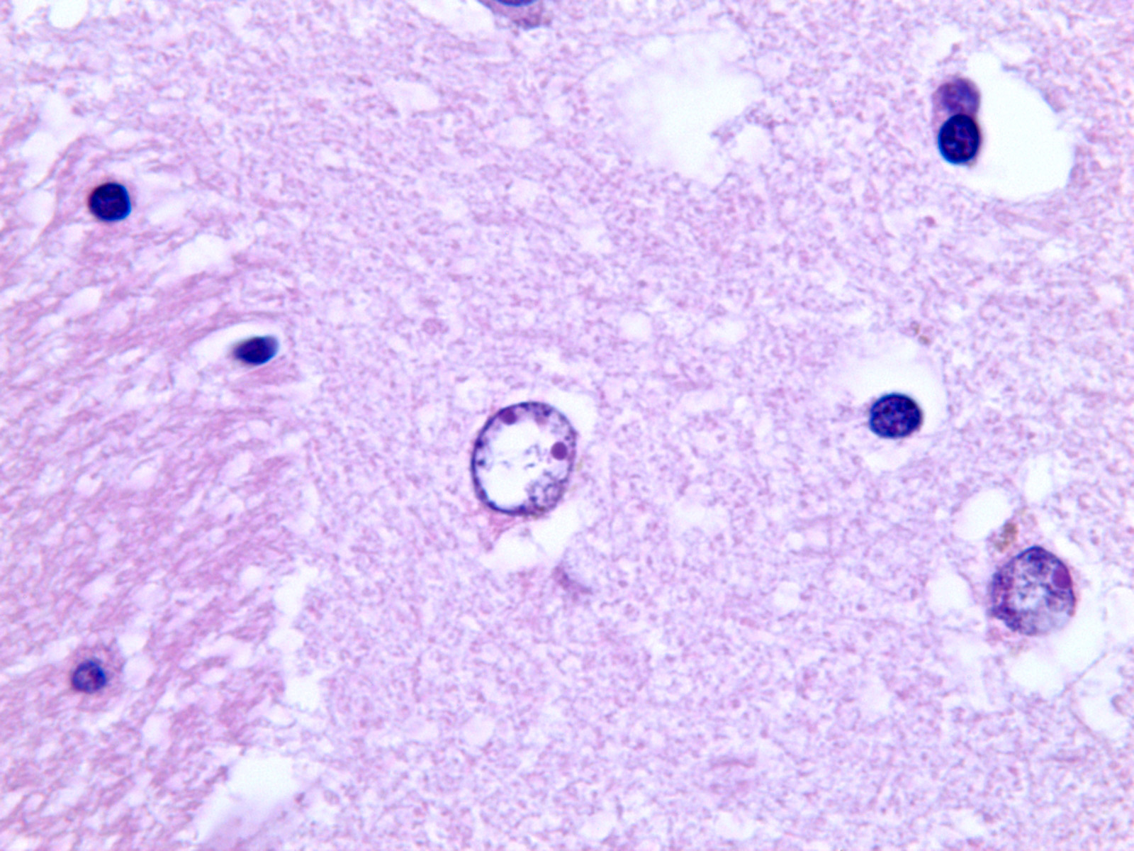
Alzheimer type II astrocyte
Images hosted on other servers:

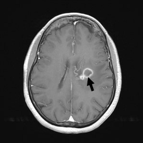
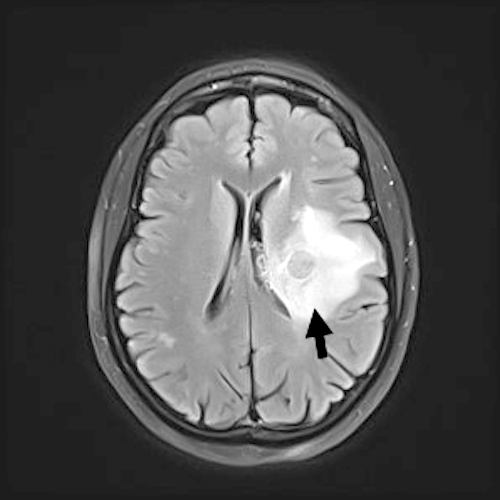
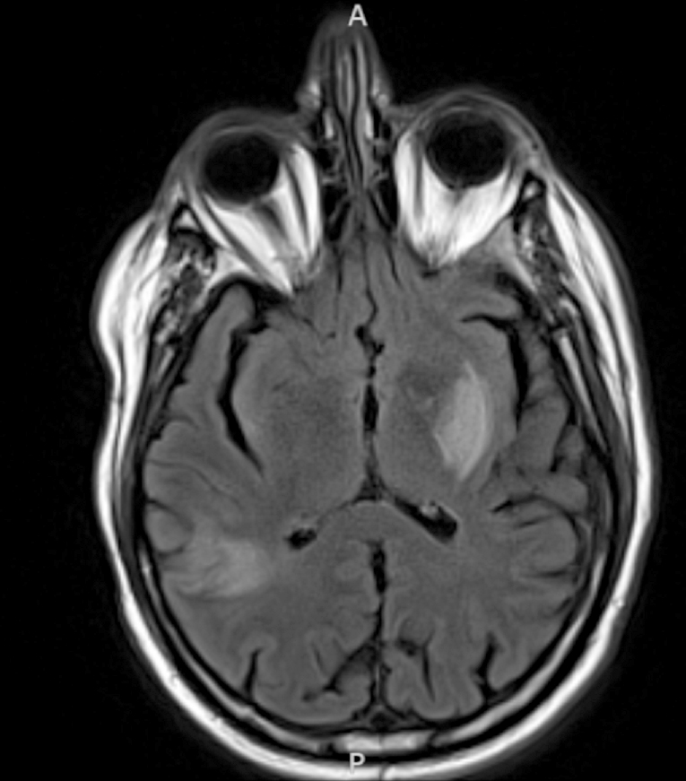
Axial FLAIR sequences

Increased T2 signal

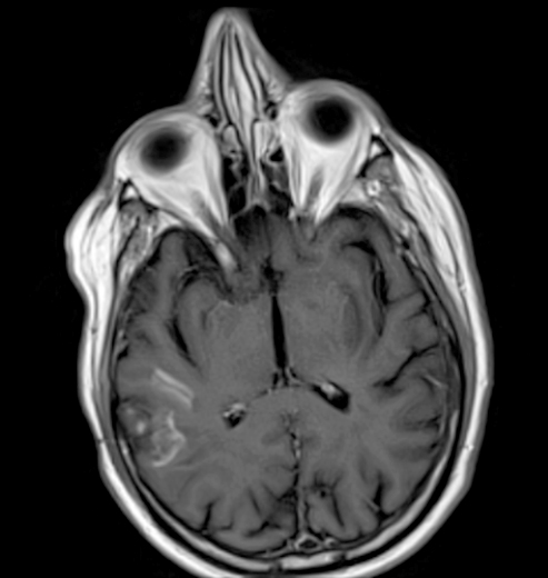
Axial gadolinium enhanced T1 sequences
Images hosted on other servers:

Cerebral cortical gray matter

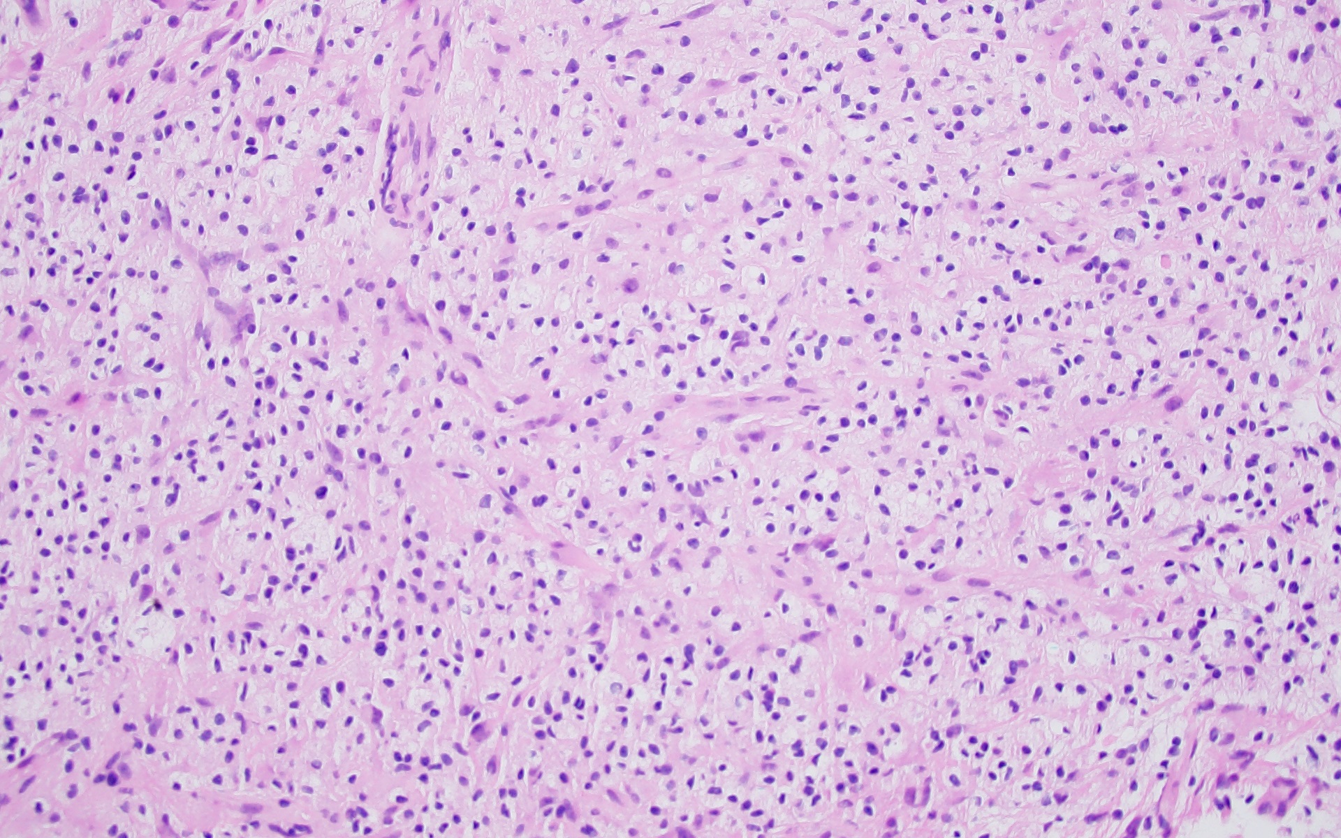
Perivascular inflammation: lymphocytes and macrophagess

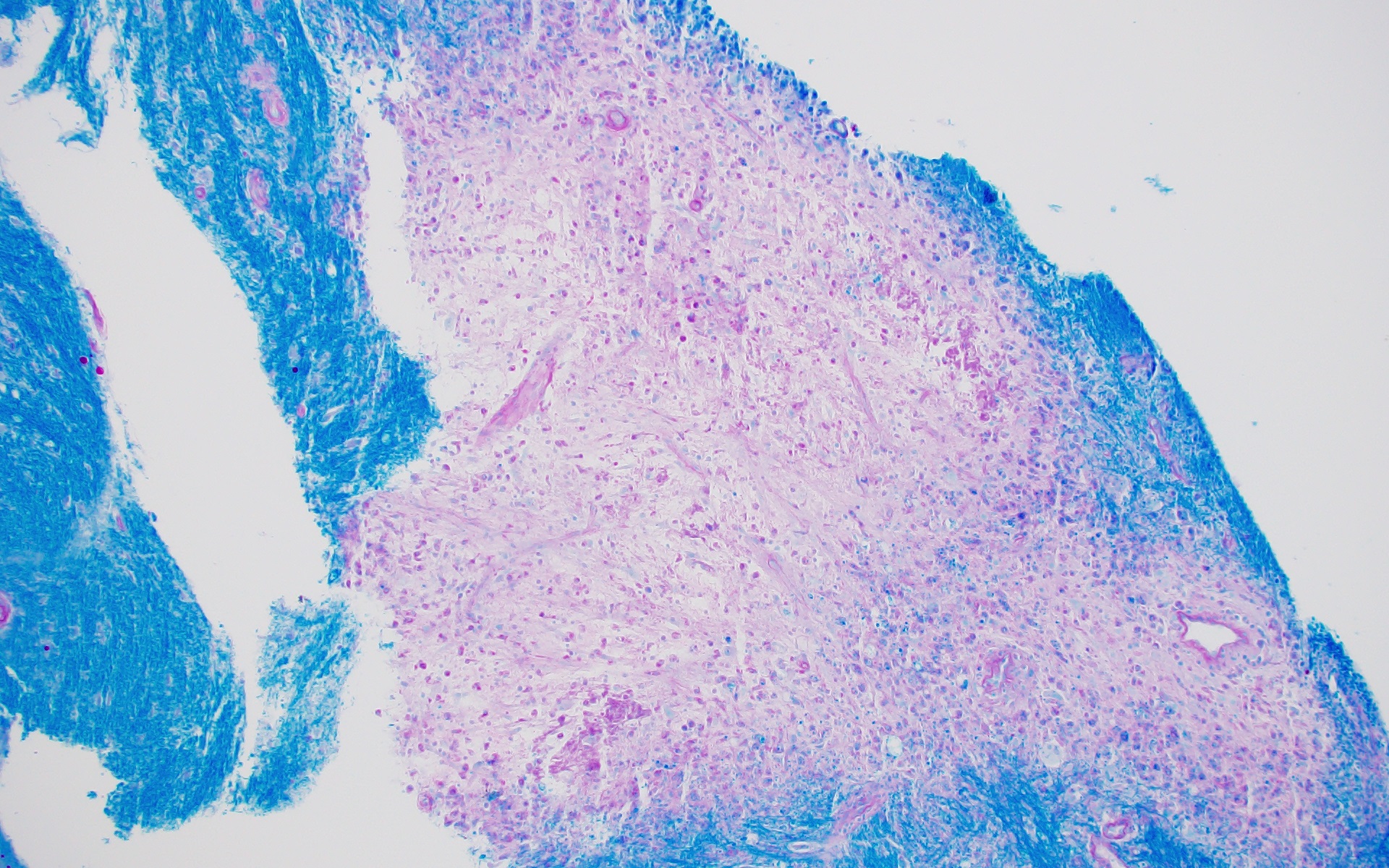
Gradual loss of myelination

Marked loss of axons

Intense macrophage infiltration
Contributed by Serguei Bannykh, M.D.
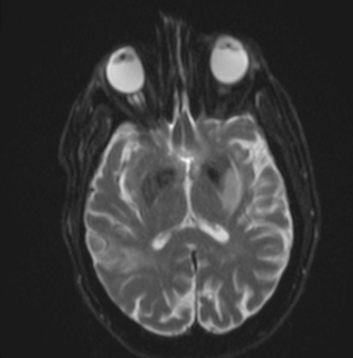
Temporal lobe involvement with restricted diffusion
Brain involvement in HSV encephalitis
Brain edema in HSV encephalitis
Images hosted on other servers:

Brain MRI in acute HSV1 encephalitis

Asymmetric medial temporal hyperintensities

FLAIR hyperintensities

Bilateral FLAIR

Abnormal enhancement

Hemorrhages in both temporal lobes

Bilateral symmetric cortical swelling

High signal intensity lesions
Contributed by Serguei Bannykh, M.D.
Acute HSV encephalitis with hemorrhagic transformation
Medial temporal lobe involvement
Subacute neonatal HSV encephalitis
Chronic HSV encephalitis
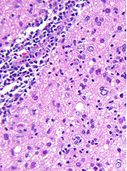
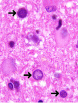
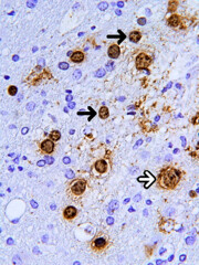
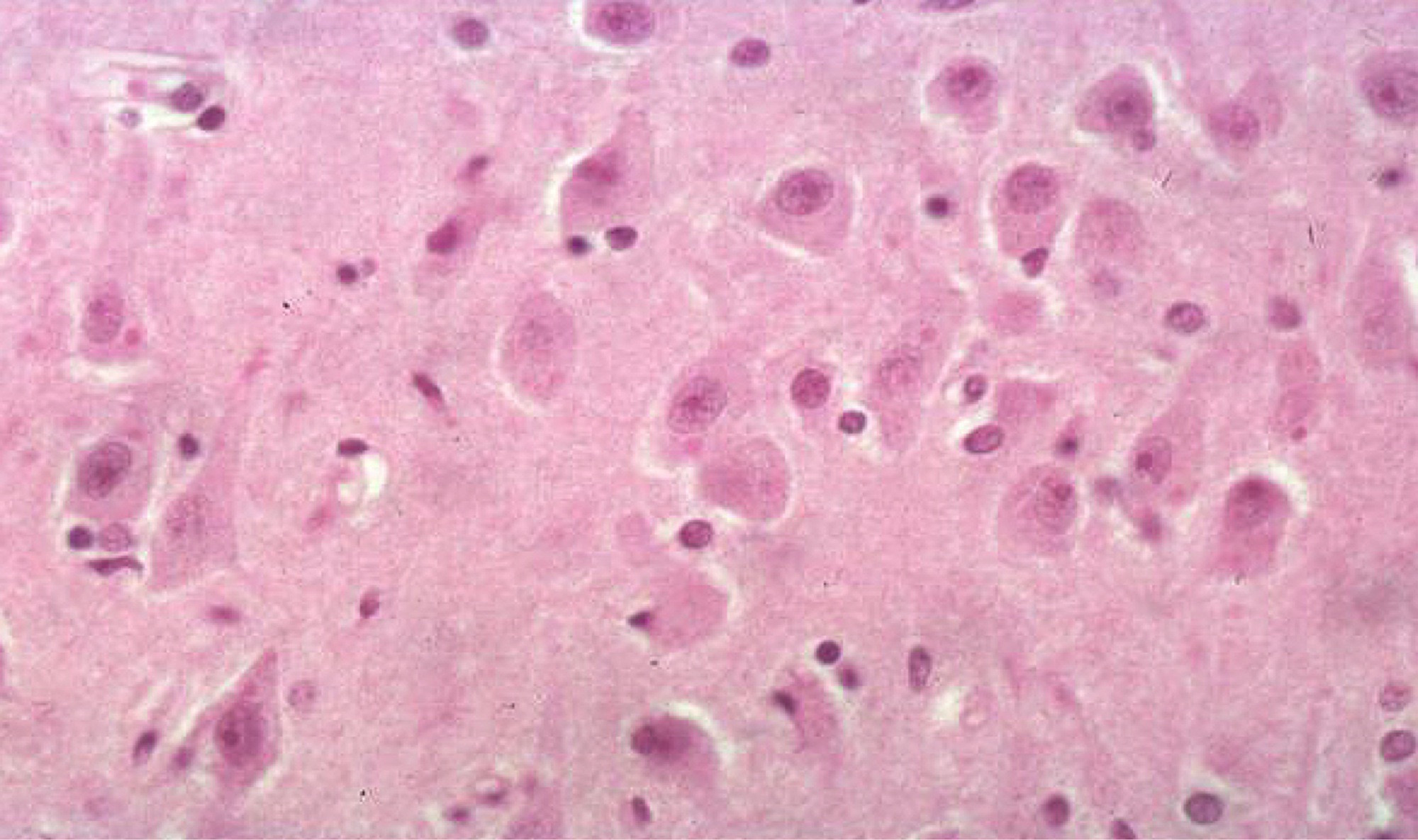
Contributed by Mahzad Azimpouran, M.D. and Serguei Bannykh, M.D.
Red neurons, microglial nodule

Occasional neuronal intranuclear inclusions
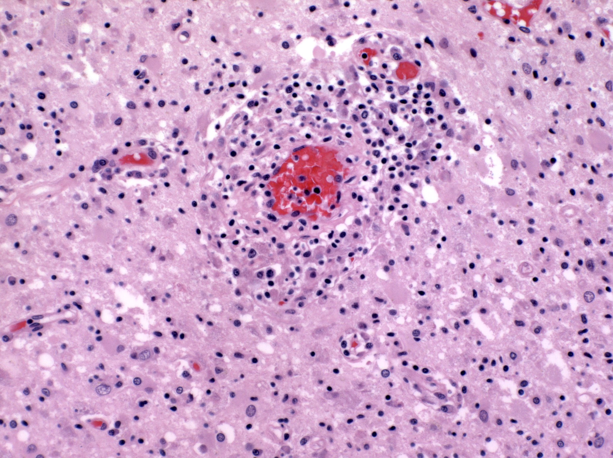
Predominantly lymphocytic inflammation
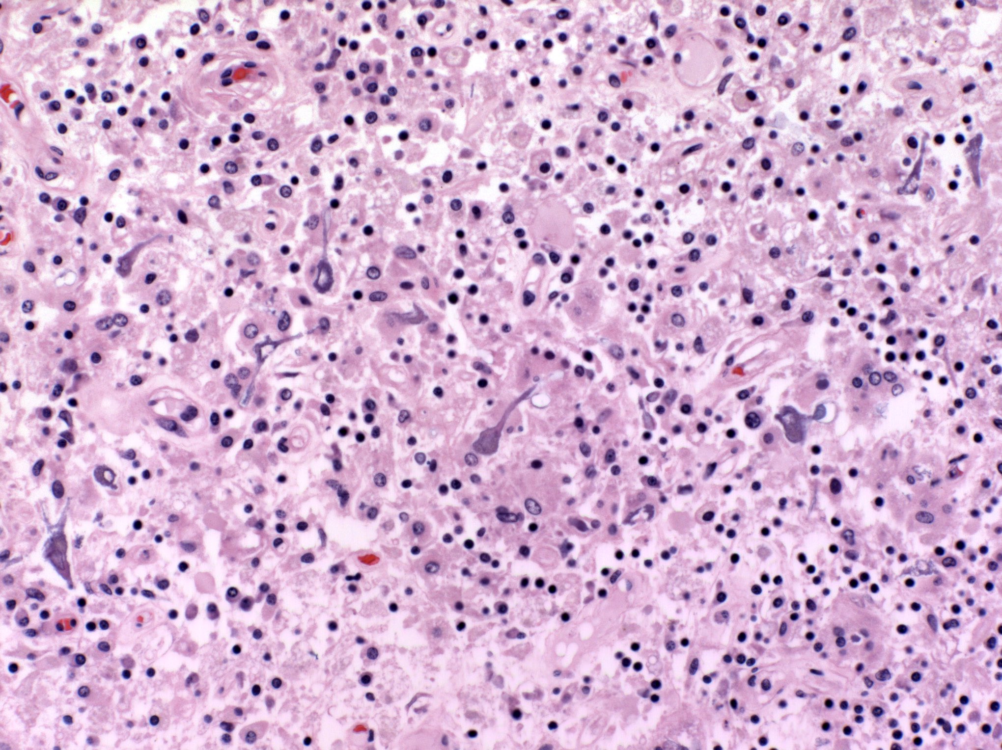
Calcific changes in neurons
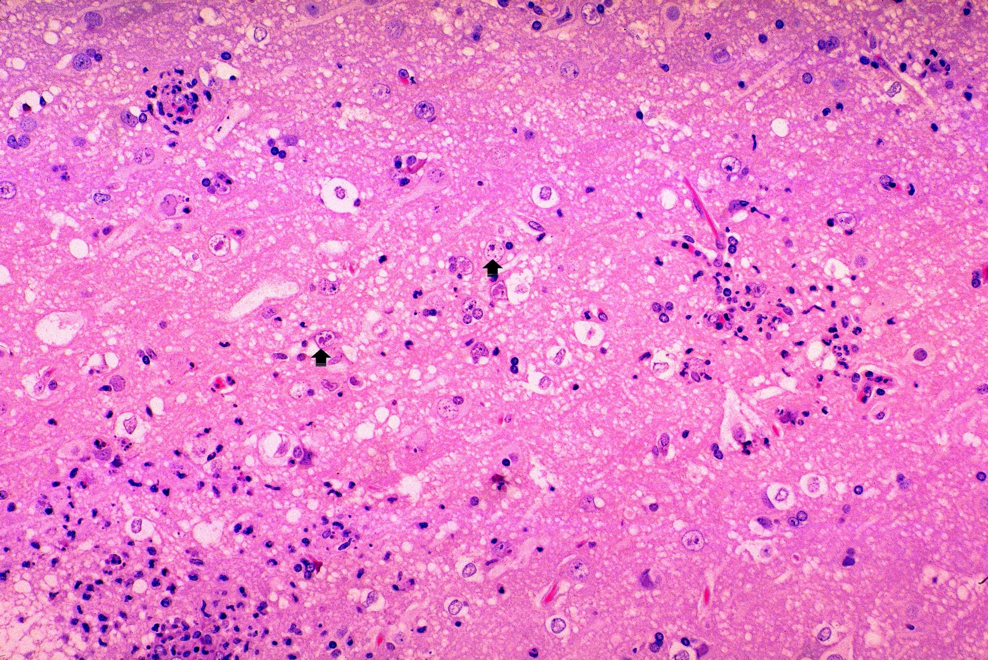
Intranuclear inclusions
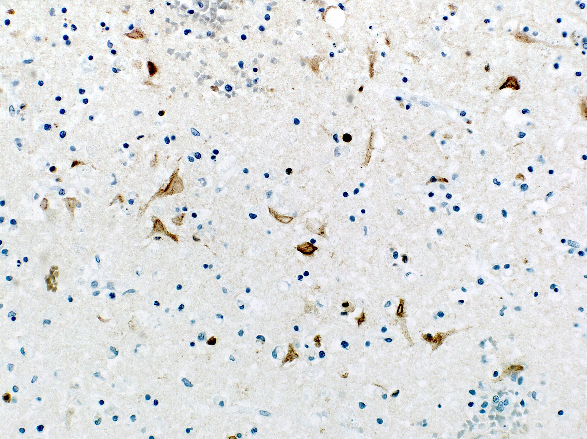
IHC staining for HSV1
Images hosted on other servers:

Herpes nucleocapsids within nucleus of a neuron
HSV encephalitis
Images hosted on other servers:

Pathogenic effects of mHTT

Clinical disease progression

Vonsattel grading; gross pathology
Images hosted on other servers:

Caudate volume HDL2 and HD versus normal
Contributed by Jared T. Ahrendsen, M.D., Ph.D.
Vonsattel grade 3 atrophy
Images hosted on other servers:

Vonsattel grading
Contributed by Jared T. Ahrendsen, M.D., Ph.D.
Caudate, neuronal loss
Caudate, reactive gliosis
Ubiquitin positive inclusions
Images hosted on other servers:

Stroke pathophysiology spatiotemporal relationships

Cell pathophysiology summary

CNS vascular territories
Contributed by Javier Redding-Ochoa, M.D. and Thomas Zaikos, M.D., Ph.D.
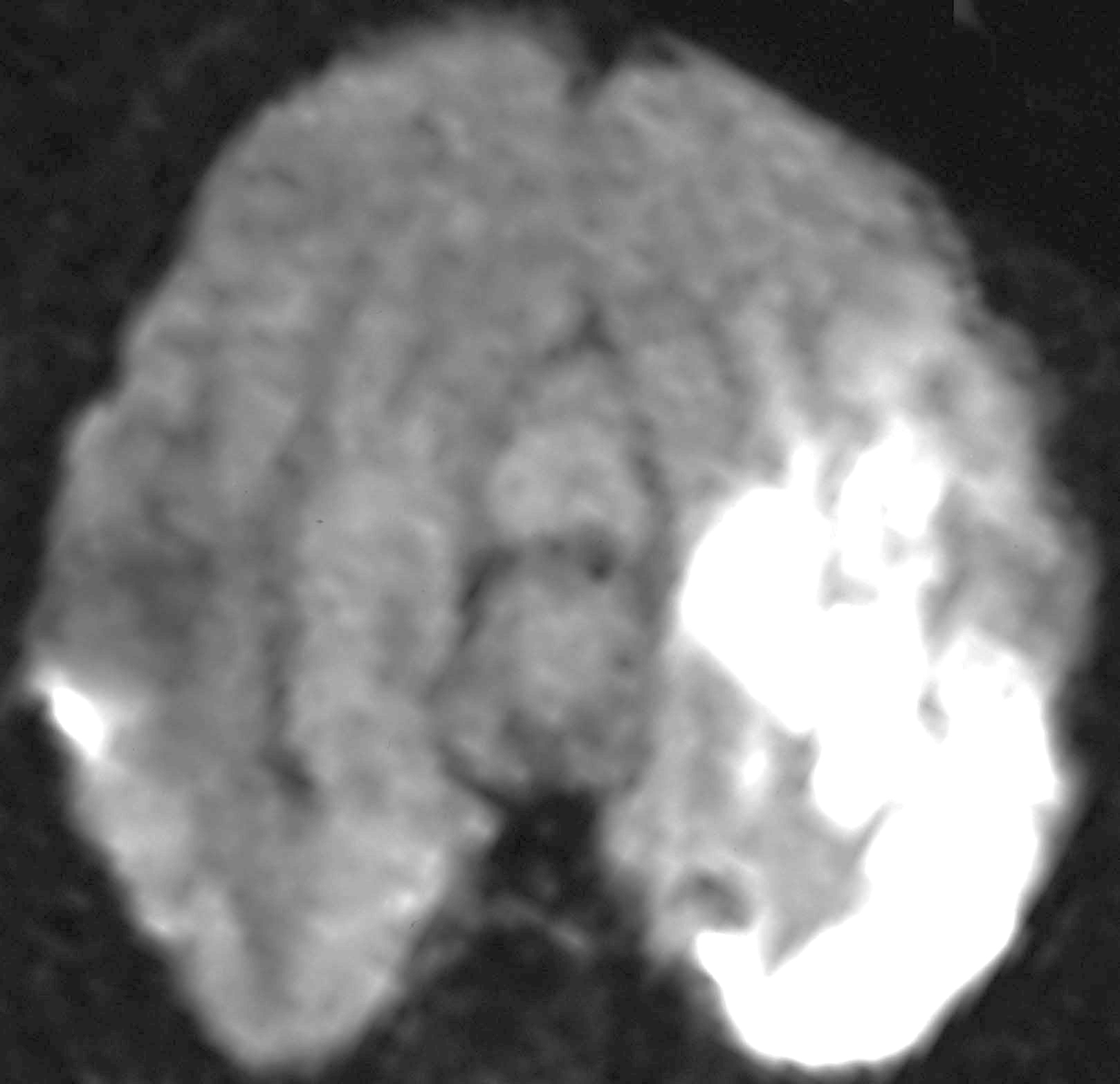
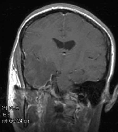
Recent infarcts (T1 postcontrast MRI)
Recent infarcts (diffusion weighted MRI)
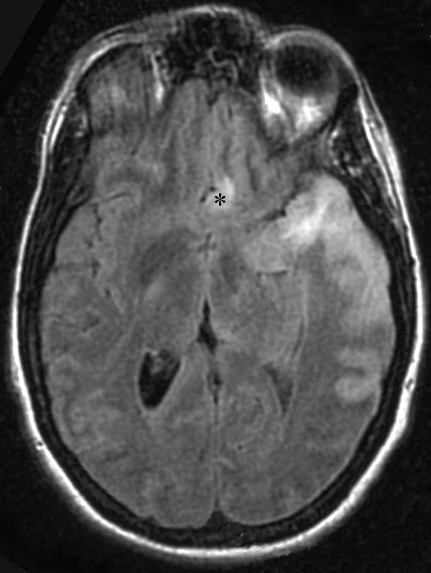
Recent infarcts (T2 FLAIR MRI)
Remote infarct (MRI FLAIR)
Contributed by Javier Redding-Ochoa, M.D., Koping Chang, M.D. and Olga Pletnikova, M.D.
Subacute infarct
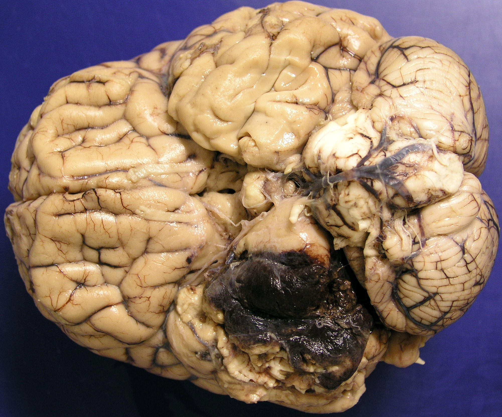
Bilateral occipital infarcts
Chronic infarct
Lacunar infarct
Chronic ACA infarct
Watershed infarcts
Contributed by Javier Redding-Ochoa, M.D.
Geographic necrosis
Acute infarct
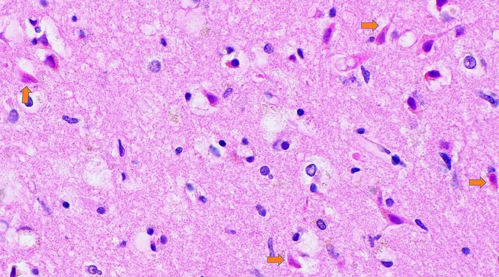
Red neurons
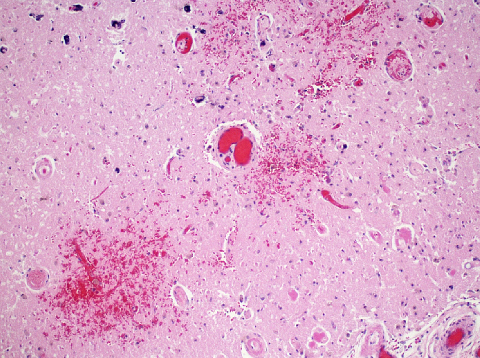
Acute infarct with focal hemorrhage
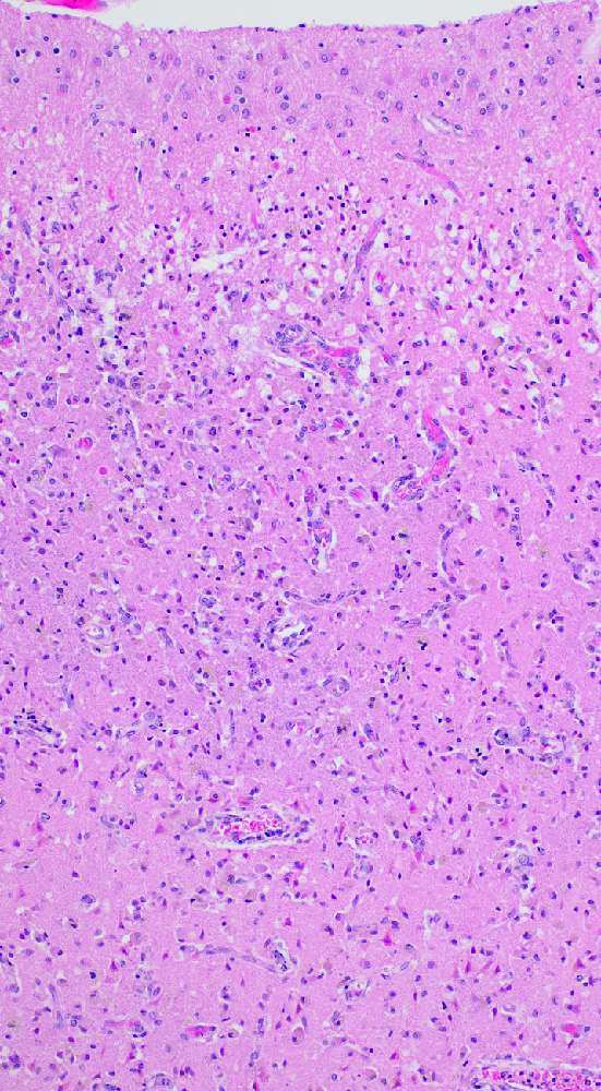
Subacute infarct
Endothelial reaction (subacute infarct)
Lacunar infarct
Macrophage infiltration
Neuronal ferrugination
Axonal balloons
Brain old infarcts versus cerebral abscess
Brain infarcts tutorial
Images hosted on other servers:

Marked loss of posterior white matter
Images hosted on other servers:

Krabbe disease

Vanishing white matter disease
Images hosted on other servers:
Peroxisomal

X linked adreno-leukodystrophy: gliosis and inflammation
Lysosomal:

Krabbe disease: globoid cells

Metachromatic leukodystrophy: loss of myelin

Metachromatic
leukodystrophy:
brown metachromasia
in peripheral nerve
Other:

Pelizaeus-Merzbacher disease: loss
of myelin

Pelizaeus-
Merzbacher
disease: cerebellar
degeneration

Alexander disease, Rosenthal
fibers

Canavan disease
Images hosted on other servers:
Peroxisomal:

X linked adreno-leukodystrophy: trilamellar lipid products
Lysosomal:

Metachromatic leukodystrophy: storage of sulfatides
Images hosted on other servers:

Cystic renal dysplasia

Pulmonary hypoplasia
Images hosted on other servers:

Cystic renal dysplasia
Contributed by Mark Cohen, M.D.
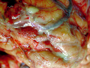
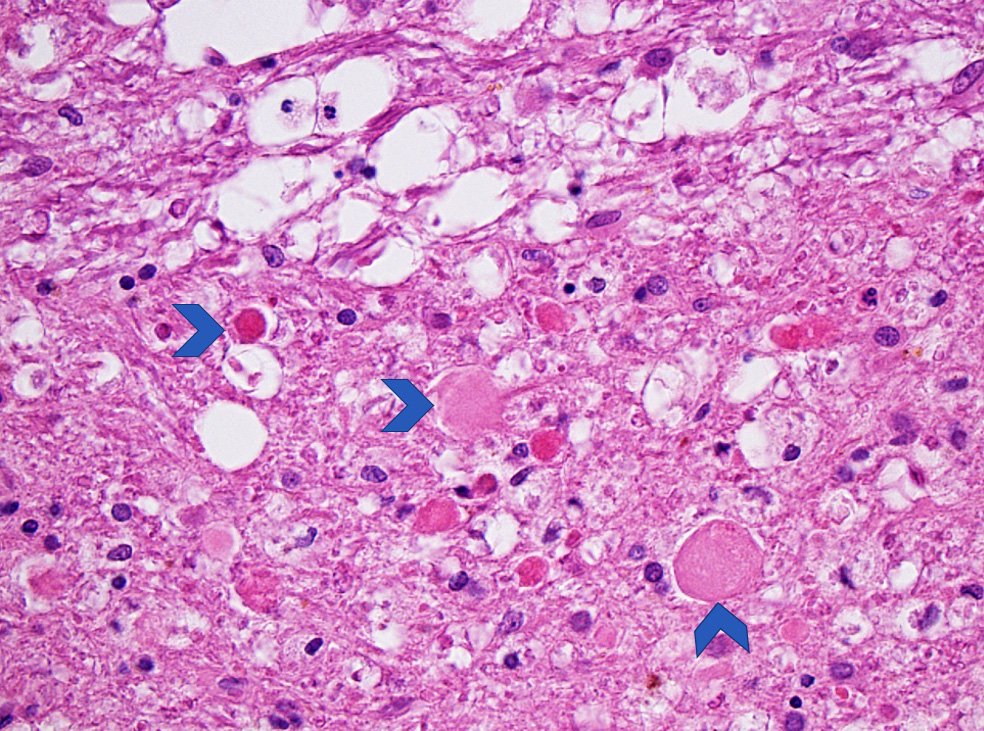
Streptococcus - dilated cerebral veins associated with the thick purulent exudate within the leptomeninges
Tuberculous meningitis
Images hosted on other servers:

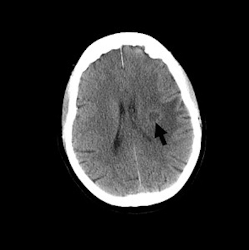
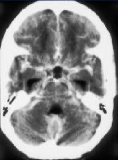
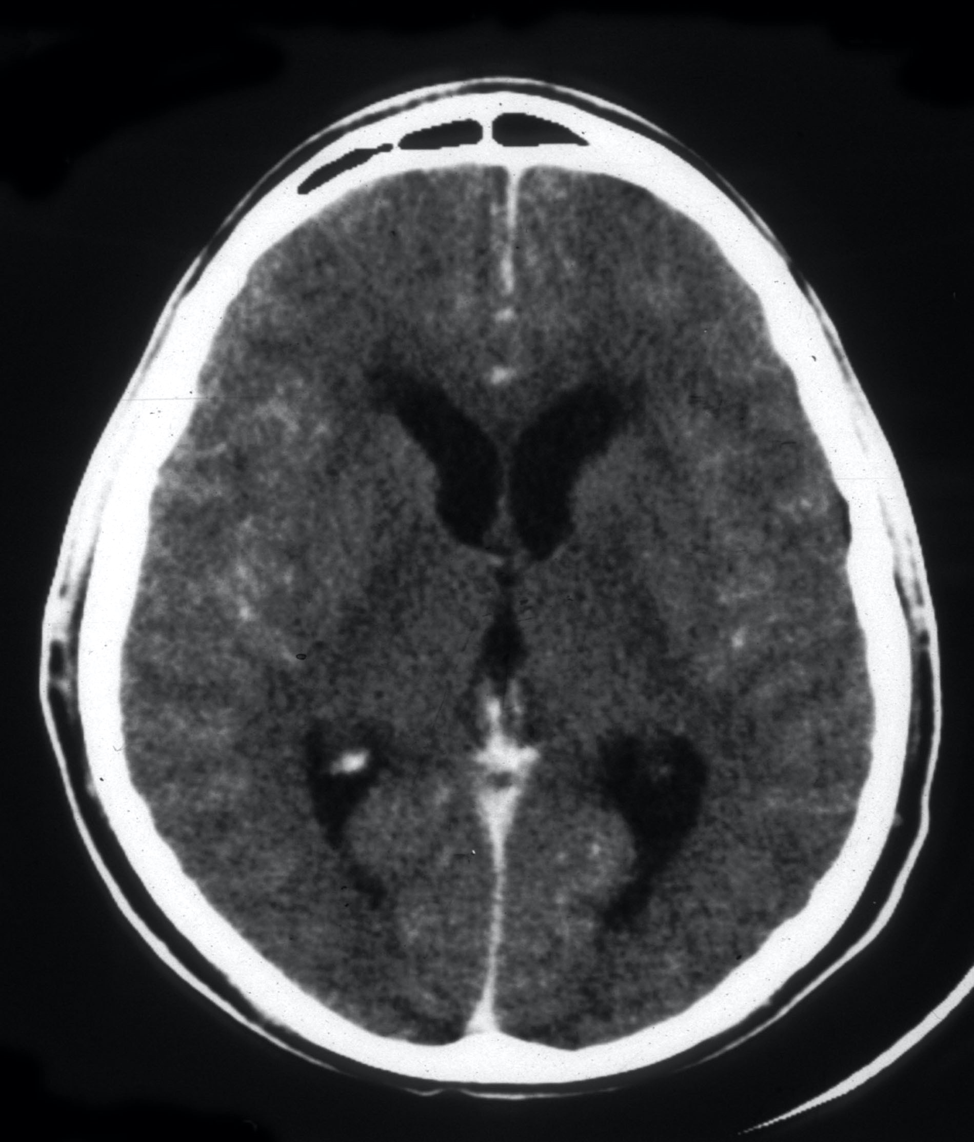
Hypodensity in bilateral putamen

Bilateral symmetrical hypodensities

Involves bilateral frontal lobes

Lesions show restriction of diffusion
Contributed by Kymberly A. Gyure, M.D.
Methanol
Images hosted on other servers:

Toxic leukoencephal-opathy

Neurotoxicity
Contributed by Kymberly A. Gyure, M.D.

Methotrexate toxicity
Contributed by Jared T. Ahrendsen, M.D., Ph.D. and Pouya Jamshidi, M.D.
Dawson fingers
Inactive MS lesion
Images hosted on other servers:

Plaques in spinal cord

Tumefactive MS
Contributed by Rachel A. Multz, M.D. and Jared T. Ahrendsen, M.D., Ph.D.
Subcortical and periventricular plaques
Subcortical plaques
Periventricular plaques
Contributed by Rachel A. Multz, M.D. and Jared T. Ahrendsen, M.D., Ph.D.
Chronic demyelination plaque
Partial remyelination
Active MS lesion
Active MS lesion
Contributed by Jared T. Ahrendsen, M.D., Ph.D.
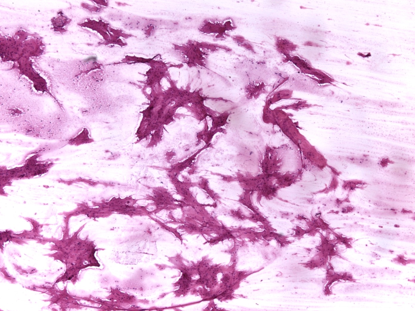
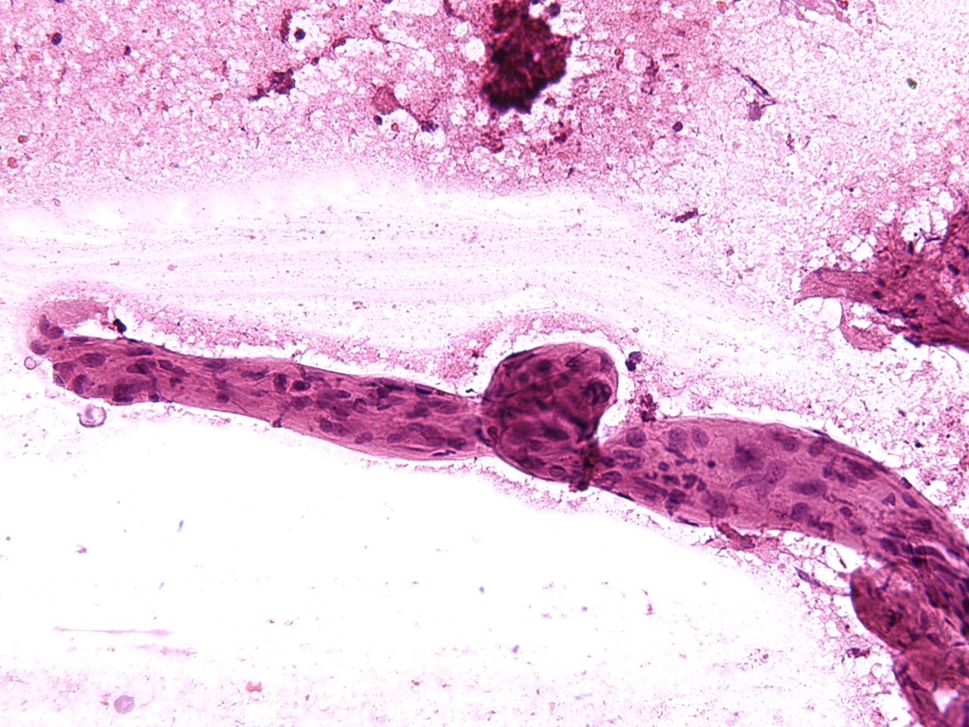
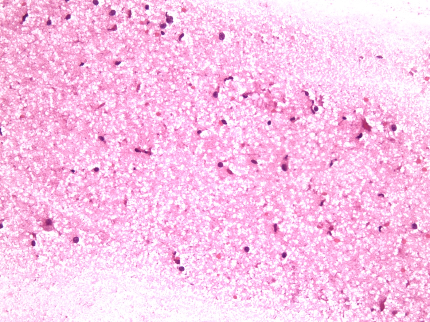
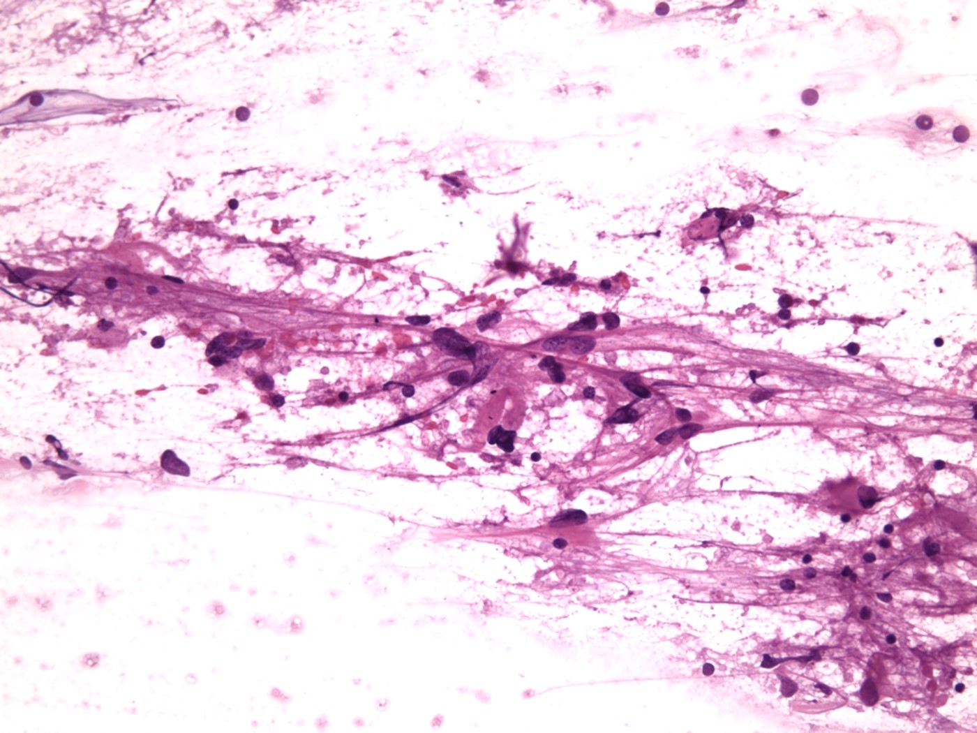
Smear preparation of demyelination lesion
Images hosted on other servers:

Bilateral putaminal atrophy

Hot cross bun sign

Hot cross bun sign and cerebellopontine atrophy

MRI findings
Images hosted on other servers:

Clinical signs of MSA-C
Contributed by Emile Pinarbasi, M.D., Ph.D.
Cerebellar atrophy
Discoloration of the putamen
Depigmentation of substantia nigra
Pontine atrophy
Contributed by Emile Pinarbasi, M.D., Ph.D.
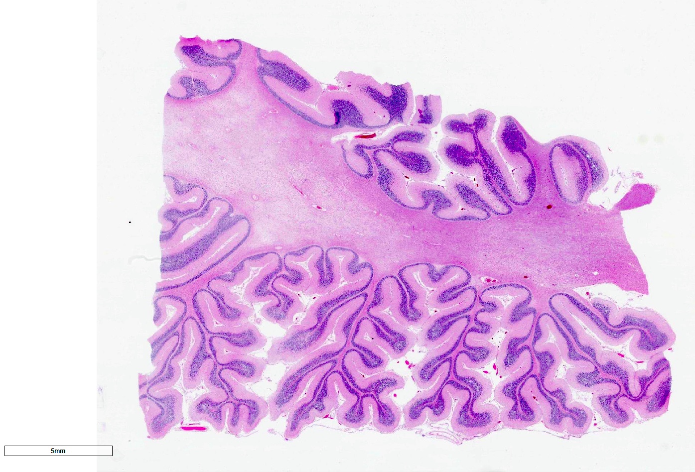
Cerebellar atrophy with prominent white matter loss
Lipofuscin accumulation in putamen
Alpha-synuclein positive GCIs
Flame and sickle shaped GCIs
Alpha-synuclein GCIs and neuropil threads
Gliosis in pontine crossing fibers
GCIs in pontine crossing fibers
Filamentous intraneuronal inclusions
Images hosted on other servers:

Glial cytoplasmic inclusions
Contributed by Mark R. Wick, M.D.

Sacral
Contributed by Mark R. Wick, M.D.
Skin
Skin, EMA

Various images
Images hosted on other servers:

Small lesion
Images hosted on other servers:

Myelin stain
Images hosted on other servers:

Infective mechanism of N. fowleri
Images hosted on other servers:

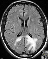
CT scan

MRI
Contributed by Mohammad Khurram Minhas, M.B.B.S.
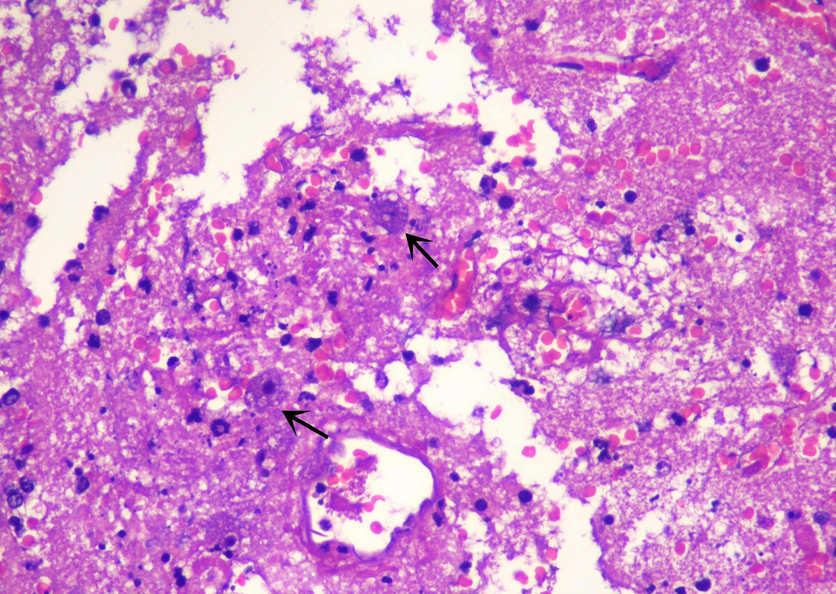
Prominent nucleoli
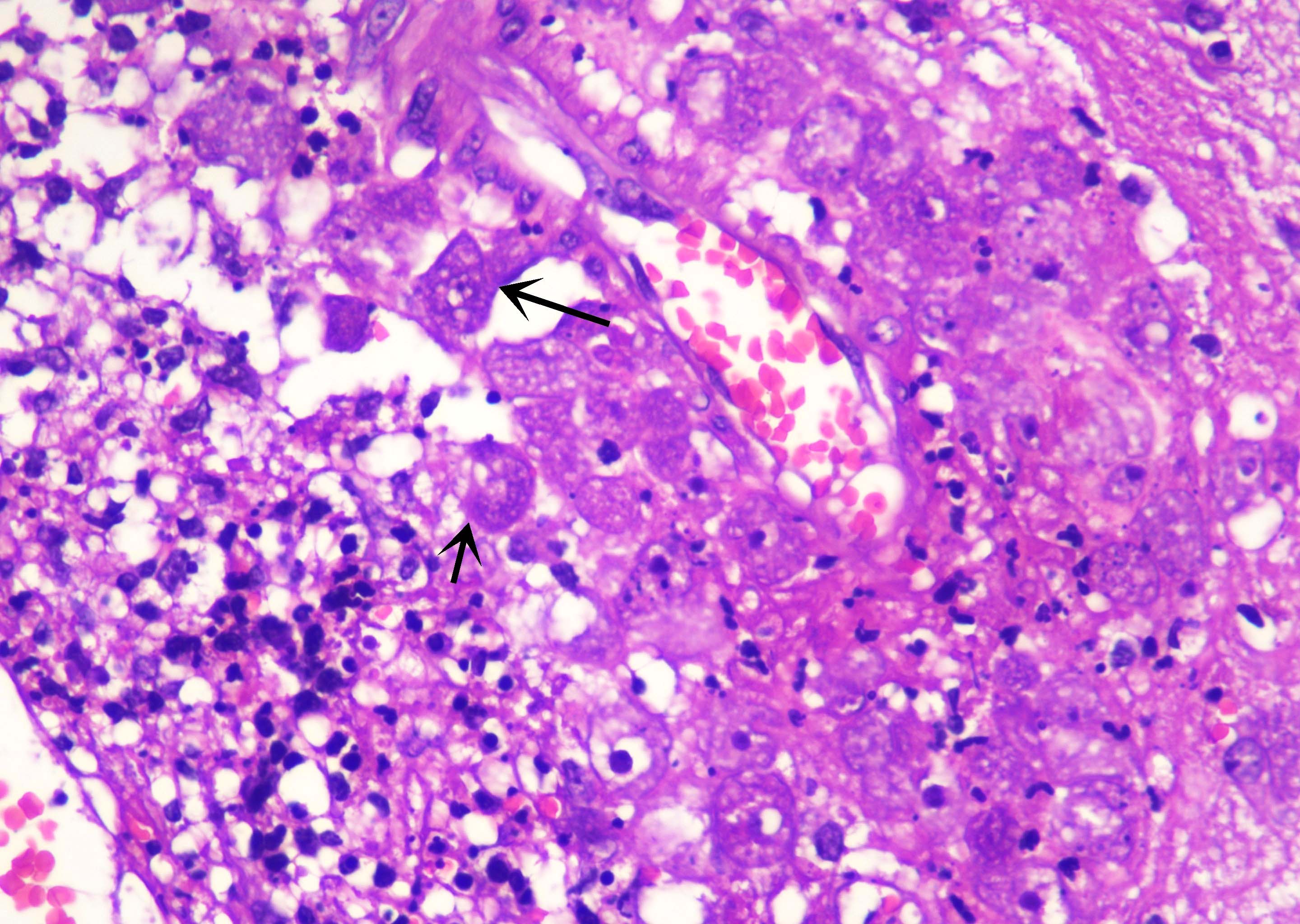
Acute necrotizing inflammation
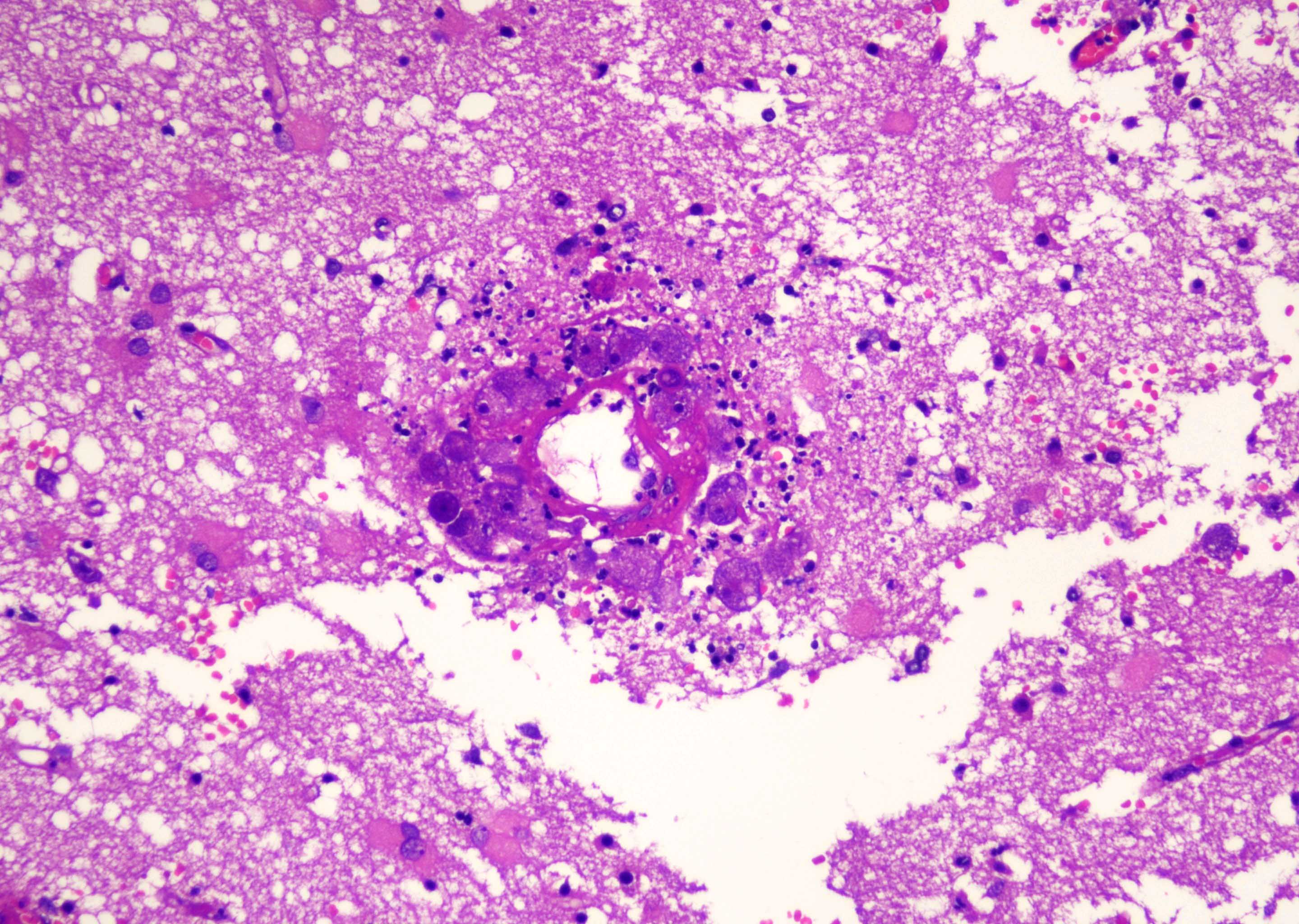
Necrotizing vasculitis
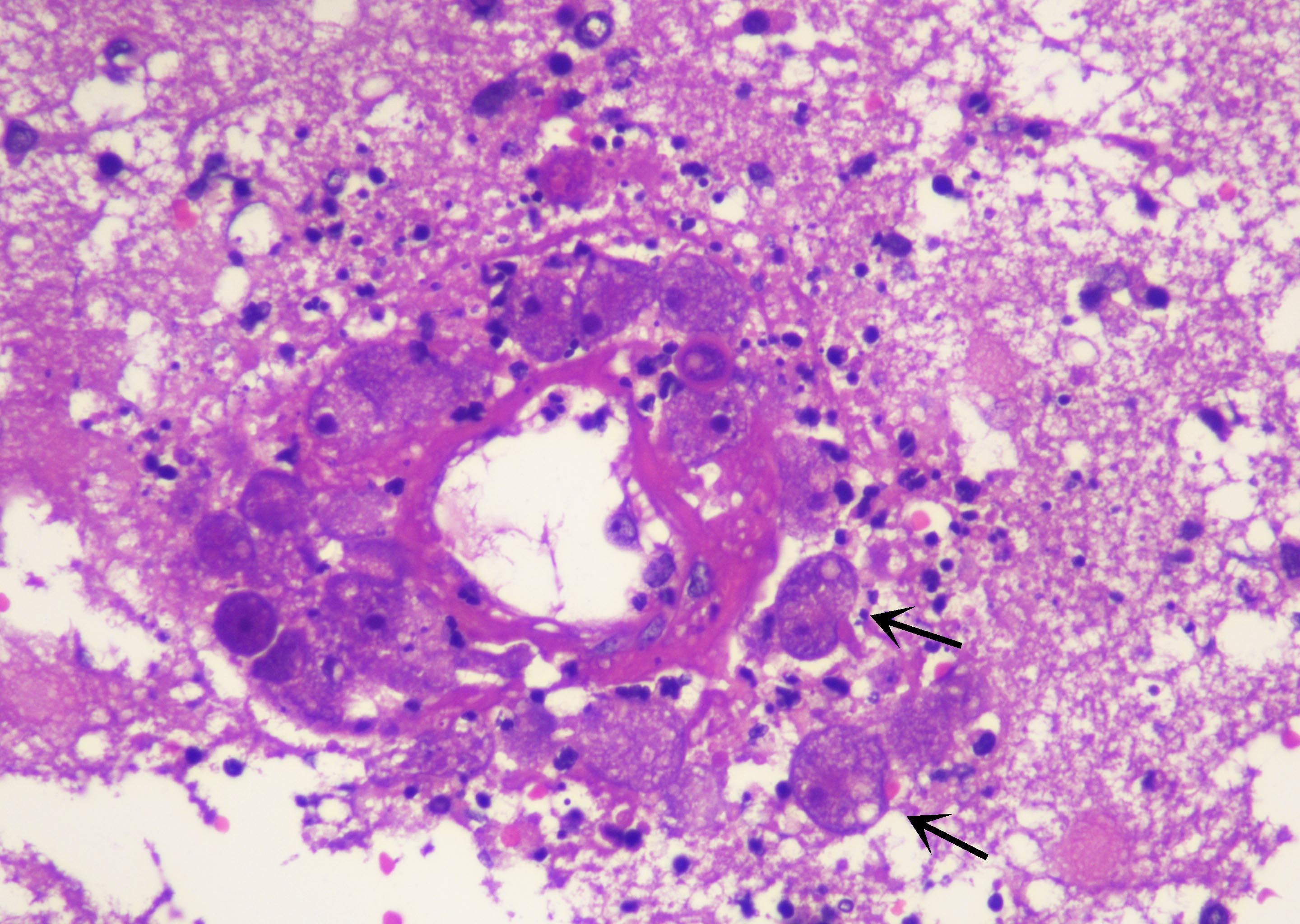
Obvious cytoplasmic vacuolations
Images hosted on other servers:

Trophozoites on CSF cytological examination
Lab detection of N. fowleri
Contributed by Mark Cohen, M.D.
White matter hyperintensity
Images hosted on other servers:

Fluid attenuated inversion recovery image

FLAIR sequence
Contributed by Mark Cohen, M.D.
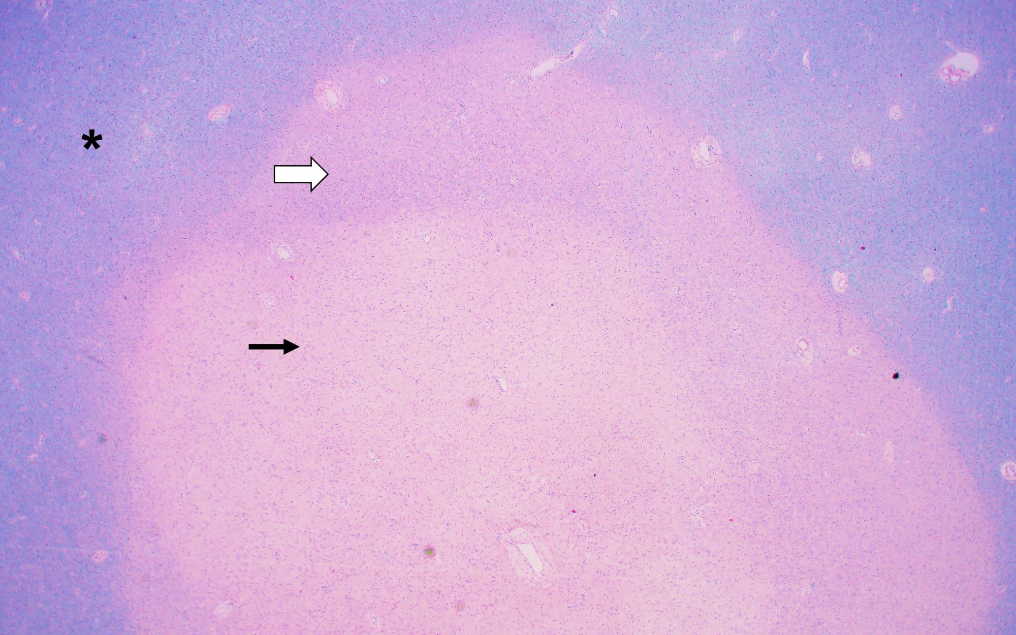
Degenerative change within the subcortical white matter
Contributed by Mark Cohen, M.D.
Extention into the overlying cerebral cortex
Perivascular inflammation
Cowdry B-like viral inclusions
Viral DNA

SV40
Images hosted on other servers:

Luxol fast blue staining

H&E, CD68, nuclear p53
Images hosted on other servers:

Frontal brain biopsy
Contributed by Kymberly A. Gyure, M.D.
Rabies

Images hosted on other servers:

Focal radiation necrosis

Differential diagnoses

Hemorrhagic radiation injury

Diffuse white matter injury

Diffuse white matter change (severe)

Concurrent focal and diffuse white matter injury

Diffuse necrotizing leukoencephalopathy


Radiation necrosis of pons

Abnormal enhancement in left occipital lobe

Irregular enhancement around surgical cavity

Pseudoprogression
Contributed by Palgun Nisarga, M.D.
Negative for tumor
Negative for tumor
Recurrent / residual tumor
Contributed by Palgun Nisarga, M.D.
Squash preparation (frozen)
Squash preparation (frozen)
Images hosted on other servers:

Cervical spinal cord MRI

Brain MRI
Contributed by Kymberly A. Gyure, M.D.

Subacute combined degeneration
Images hosted on other servers:

Bone marrow aspiration
Contributed by Benjamin Greenberg, M.D., James Luby, M.D. and Dennis K. Burns, M.D.
CT, TB meningitis
MRI, tuberculoma
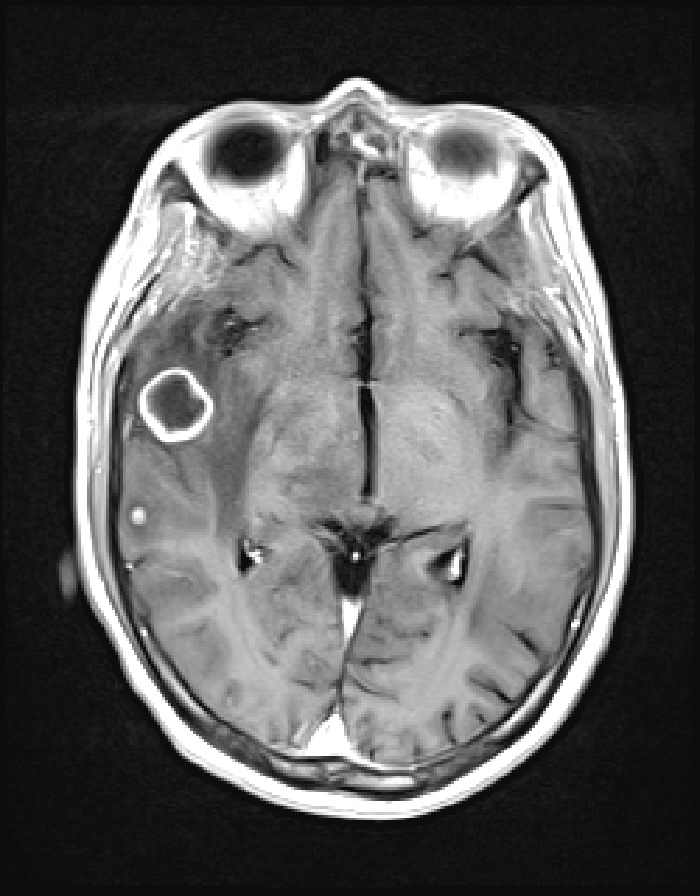
MRI, tuberculous abscess
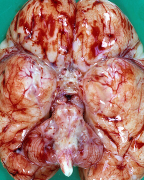
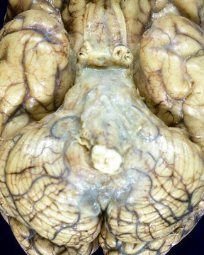
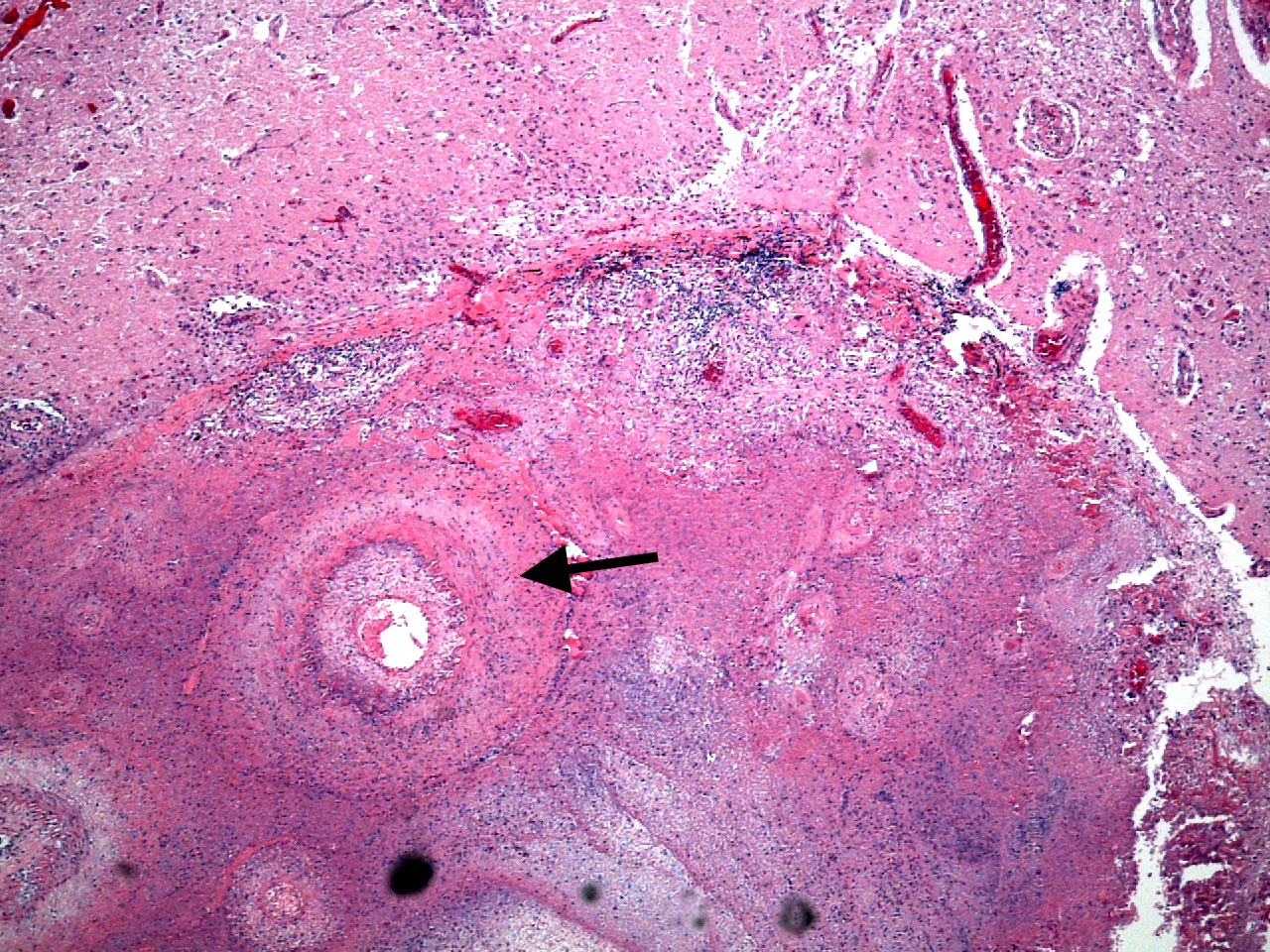
Contributed by Dennis K. Burns, M.D.
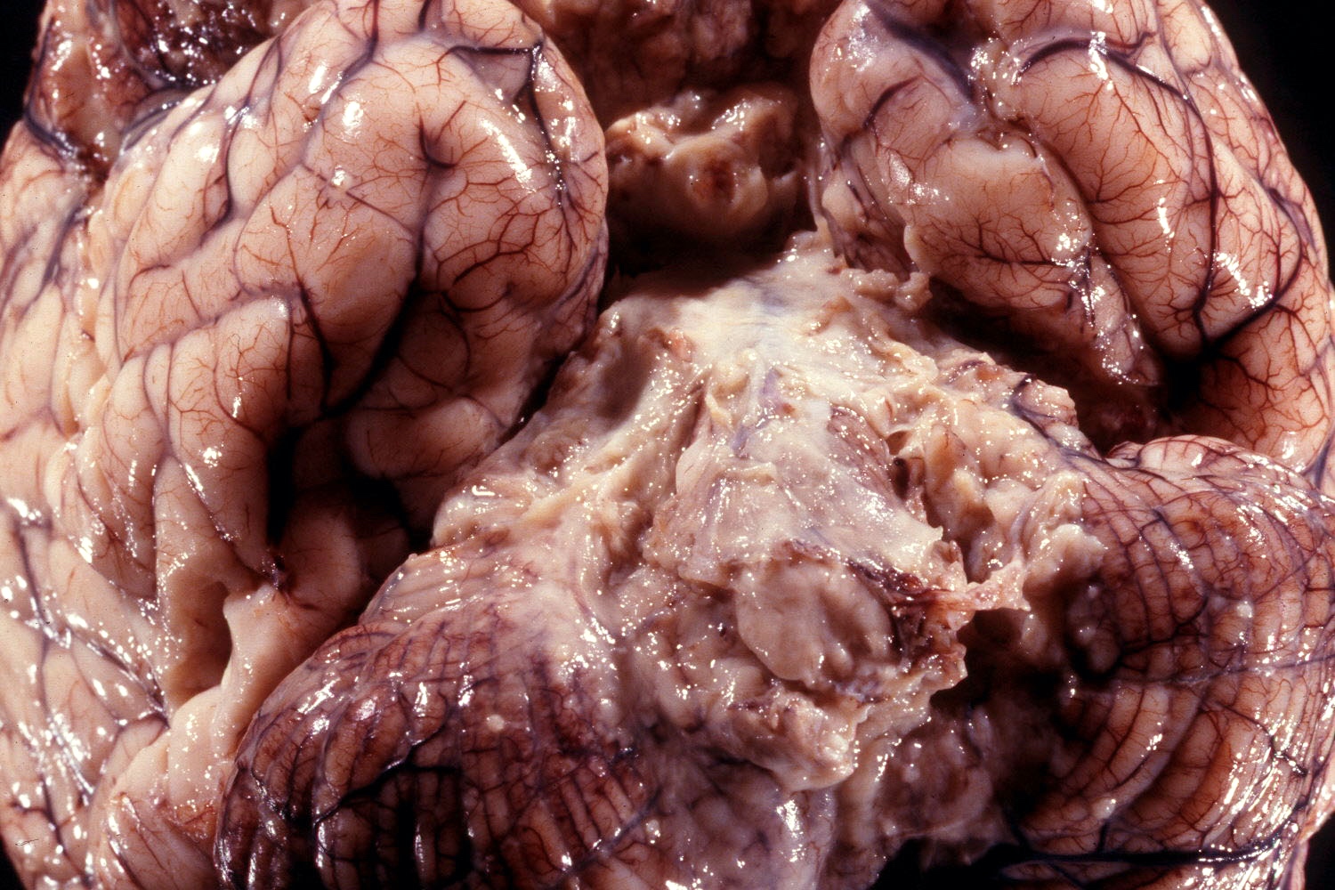
Tuberculous meningitis, ventral surface of brain
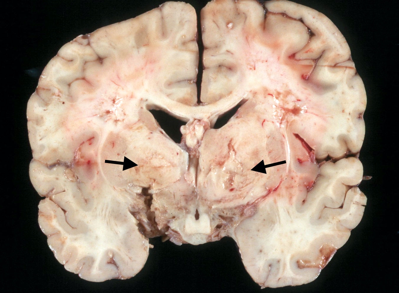
Tuberculous meningitis, coronal section
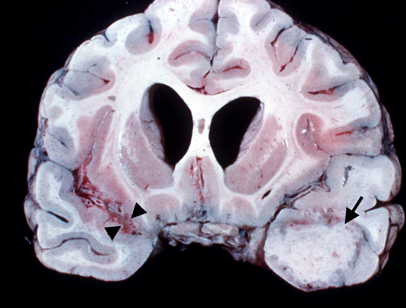
Tuberculoma, cerebral hemispheres
Contributed by Dennis K. Burns, M.D.
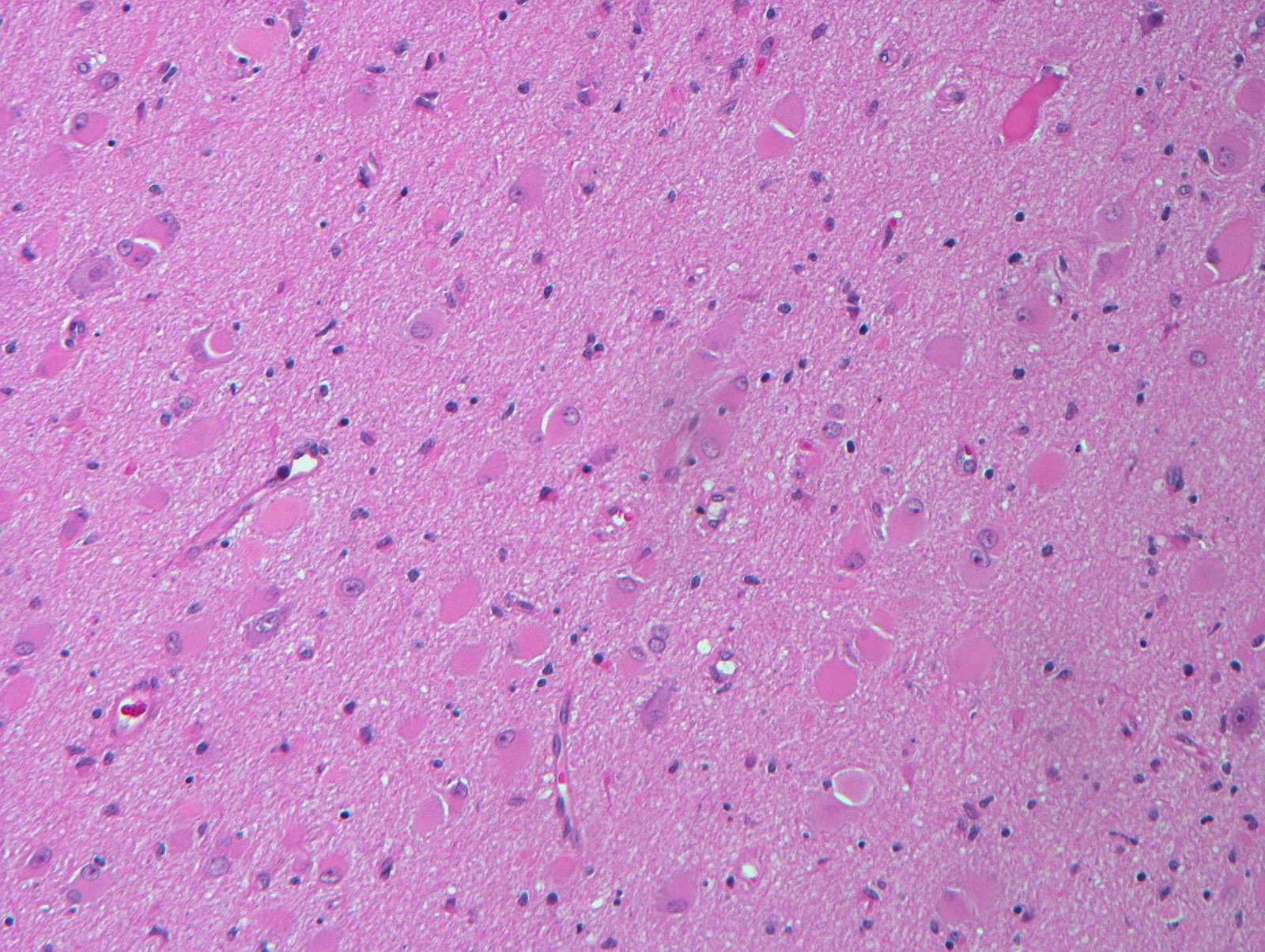
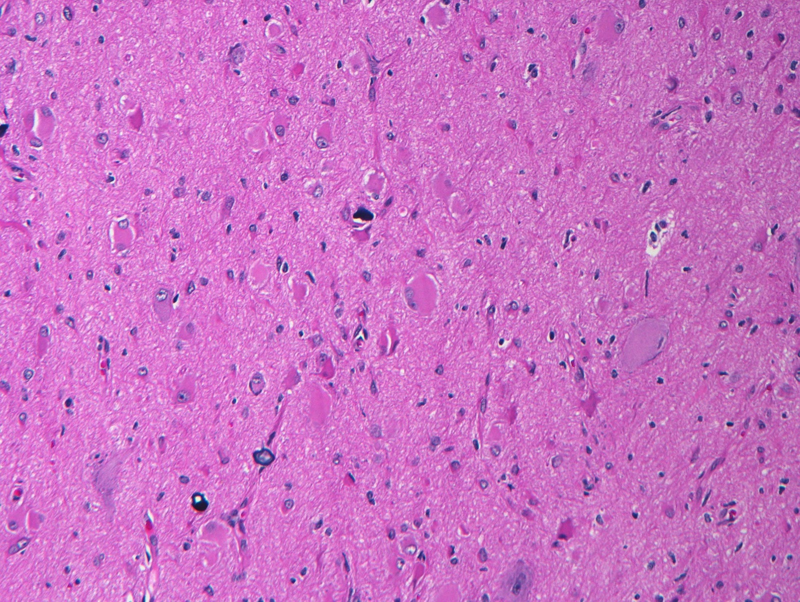
Paraffin section, tuberculous meningitis
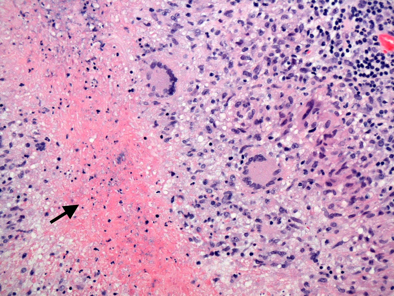
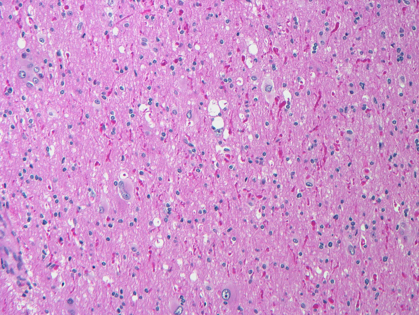
Paraffin section, tuberculoma
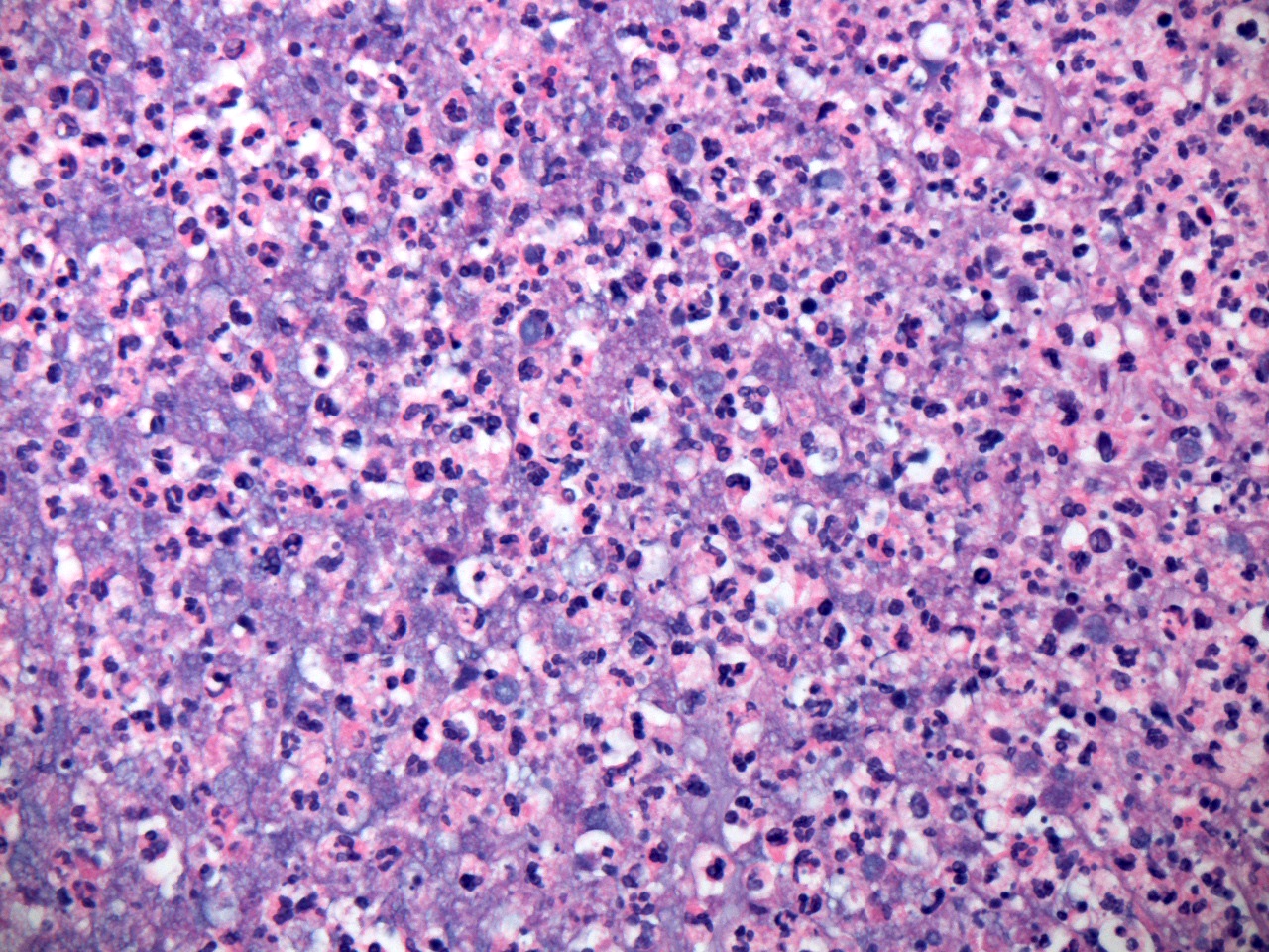
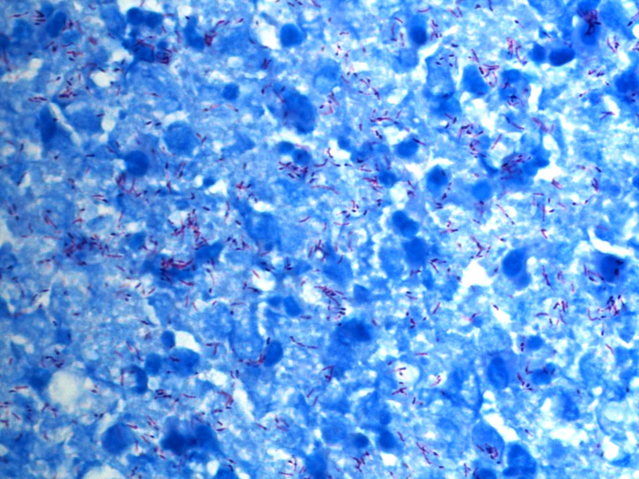
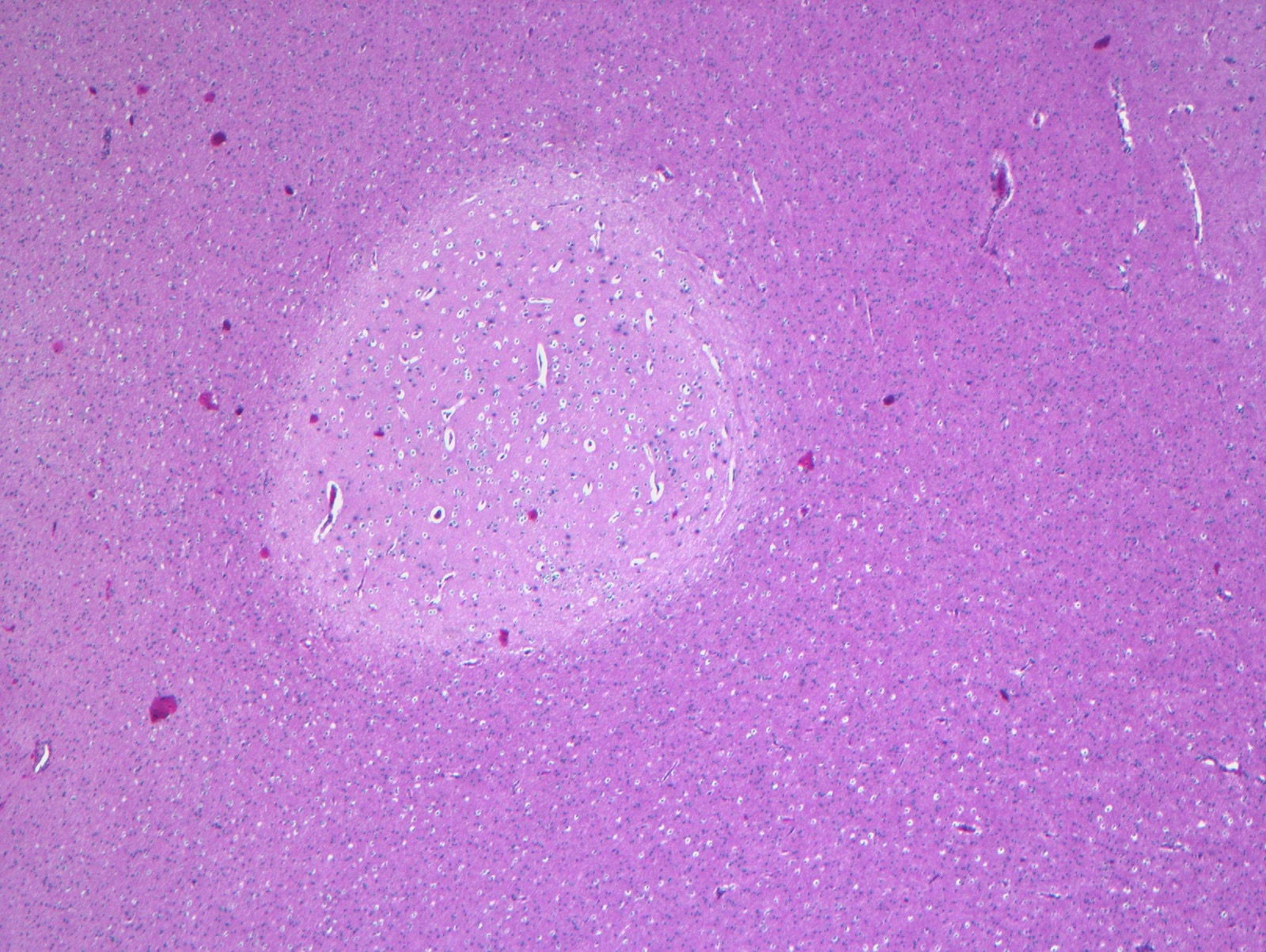
Paraffin section, tuberculous abscess
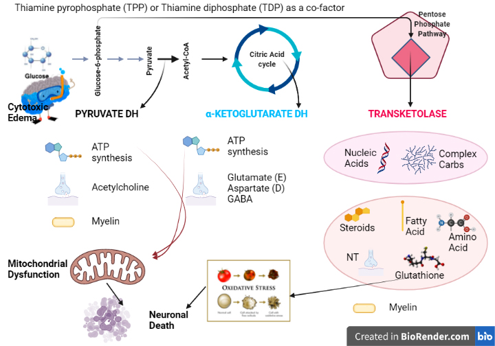


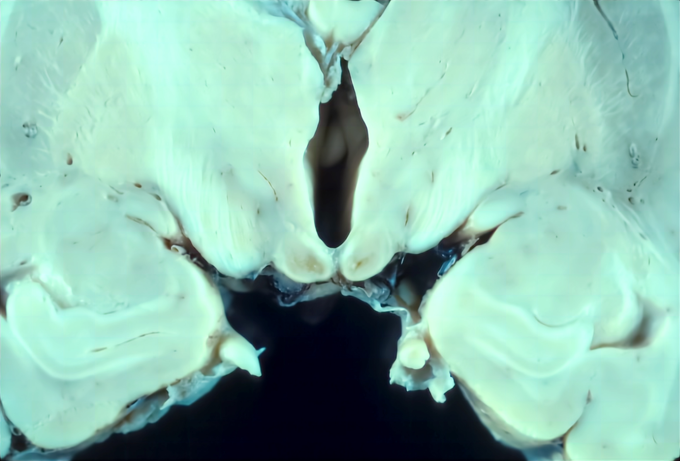
Contributed by Kymberly A. Gyure, M.D.
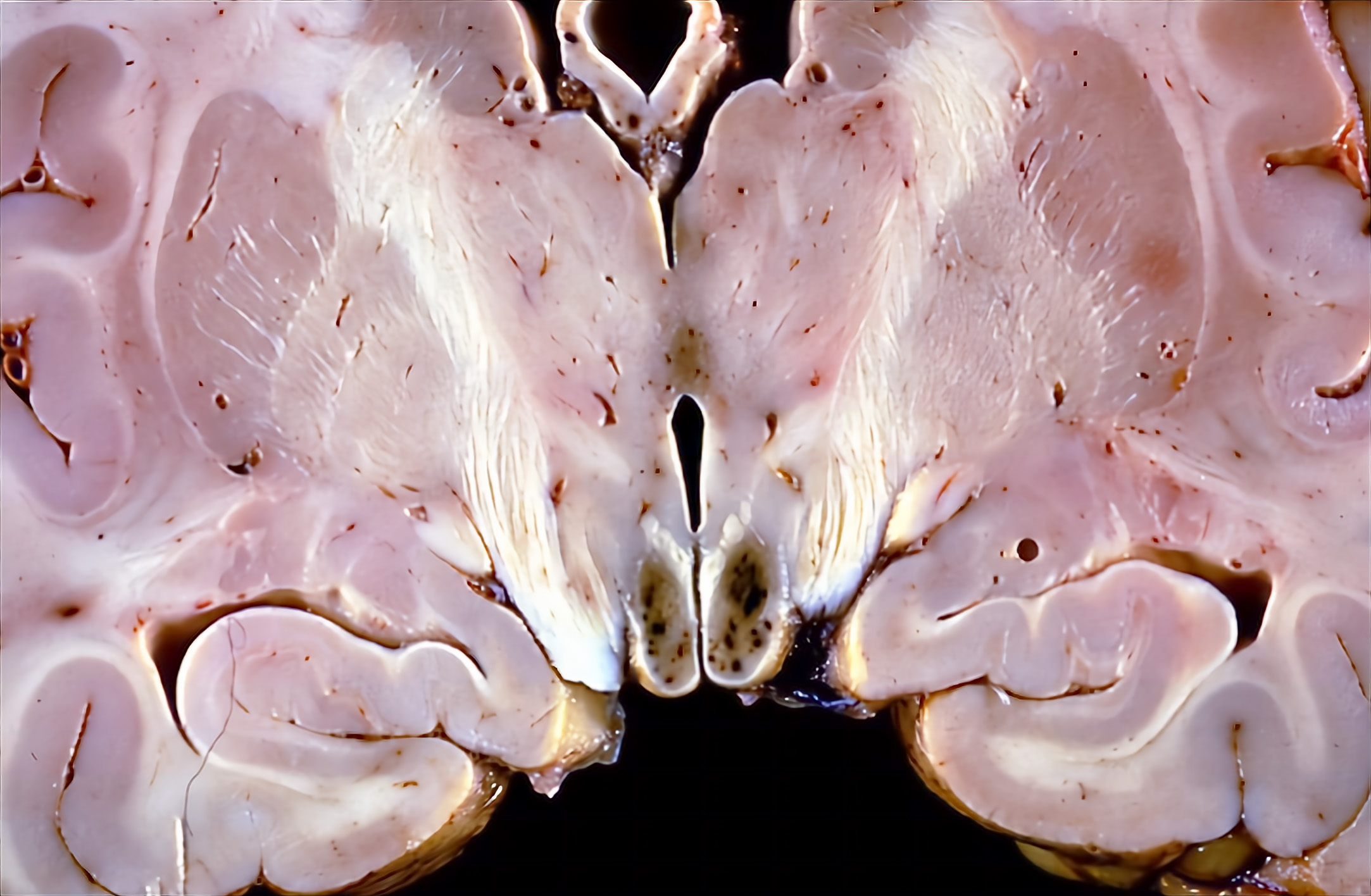
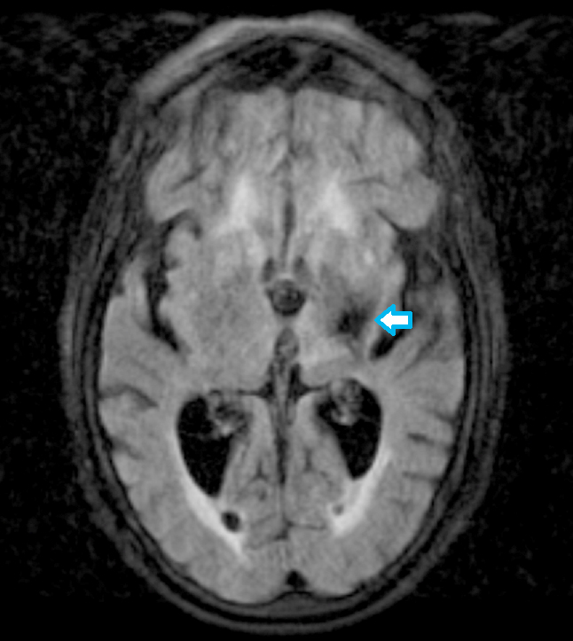
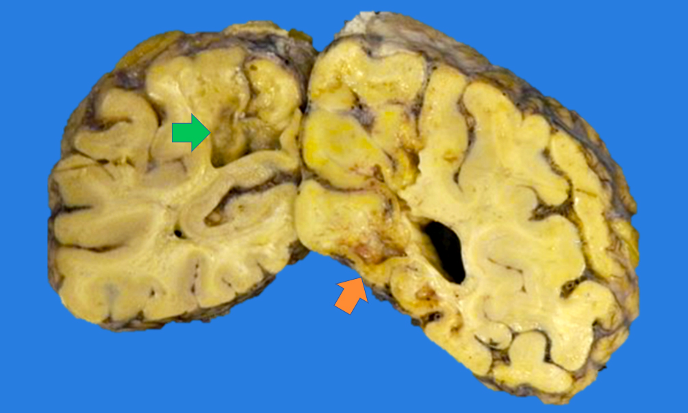
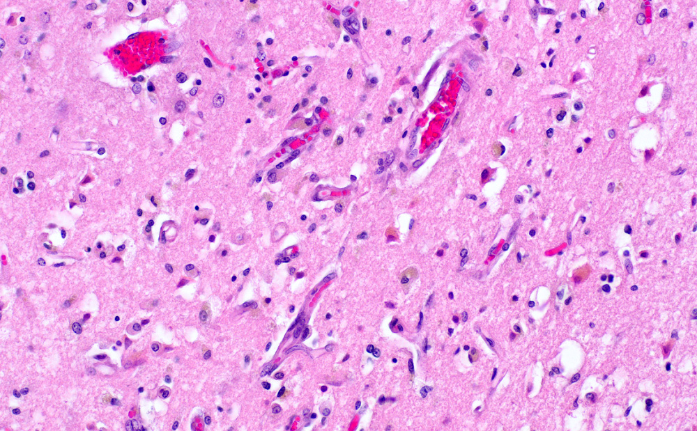
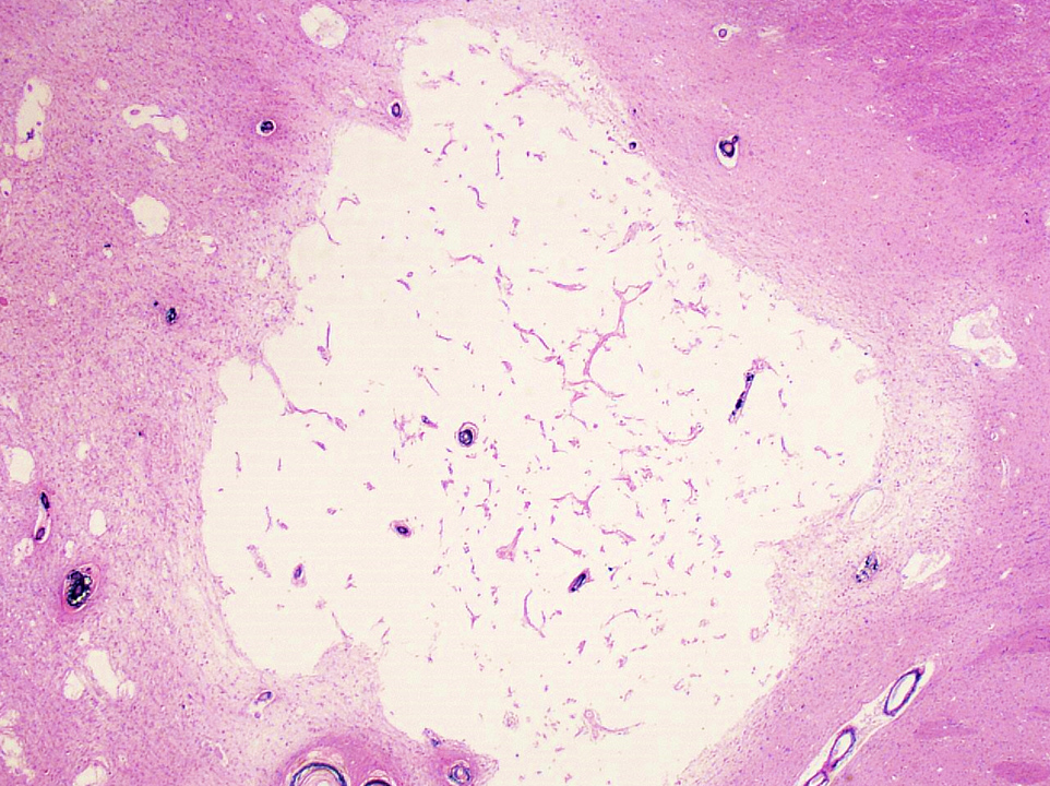
Mammillary bodies - acute Wernicke encephalopathy
Mammillary bodies - chronic Wernicke encephalopathy
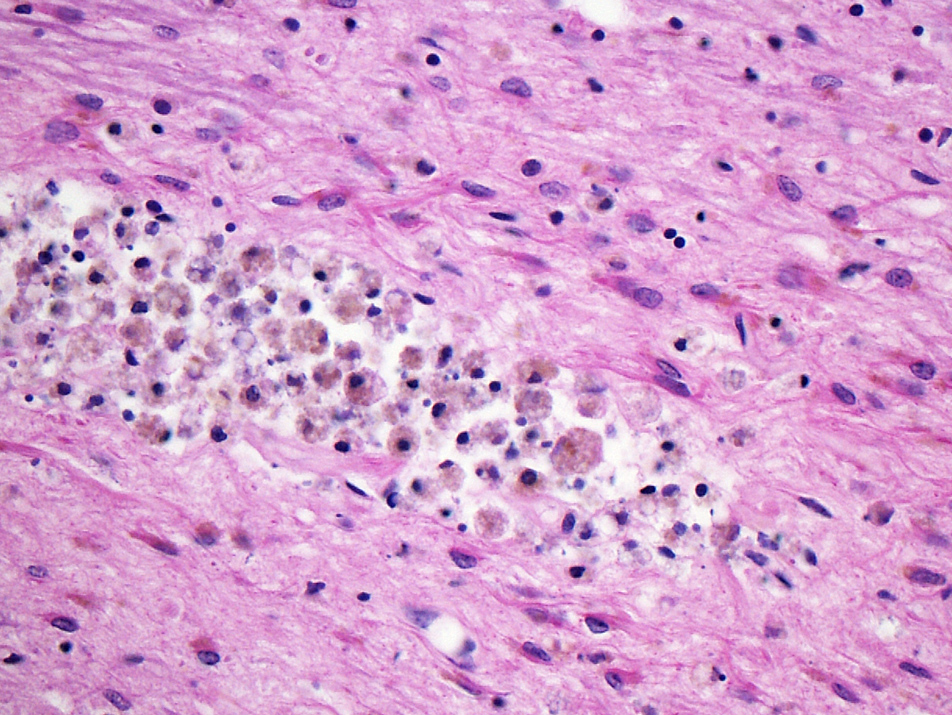
Thalamus - acute Wernicke encephalopathy
Contributed by Kymberly A. Gyure, M.D.
Acute Wernicke encephalopathy
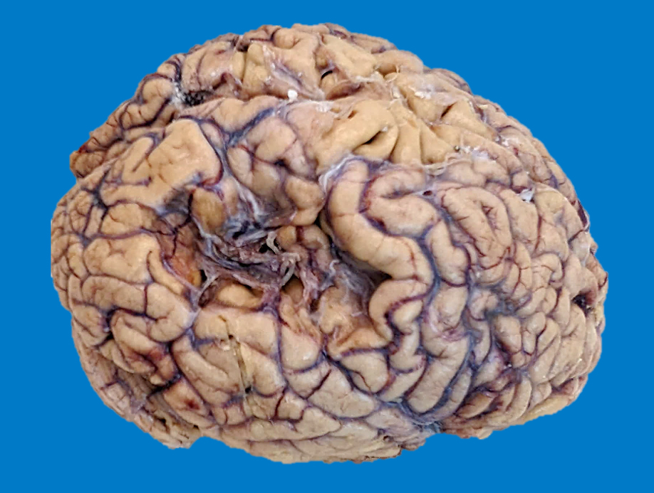
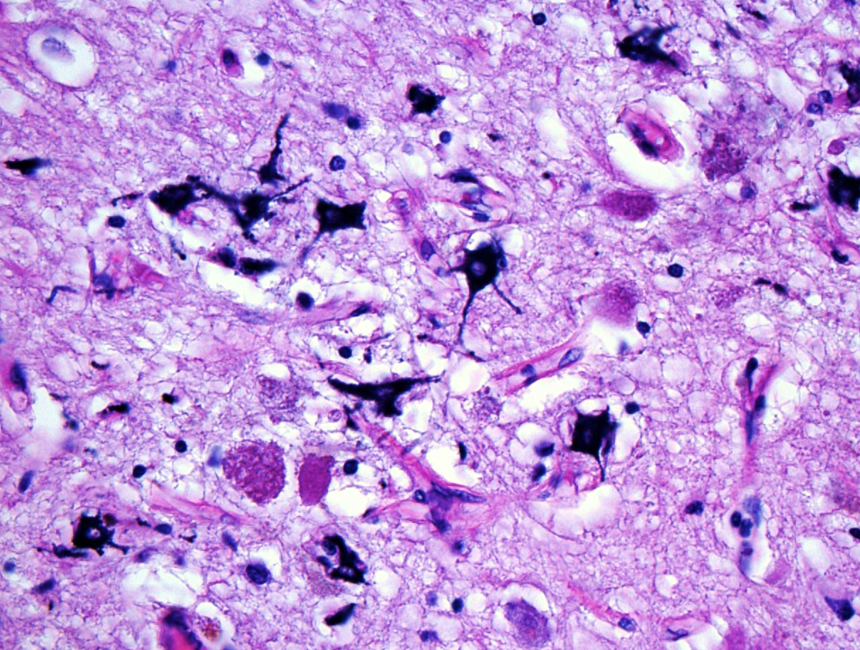
Cerebellar vermis - chronic Wernicke encephalopathy
Images hosted on other servers:

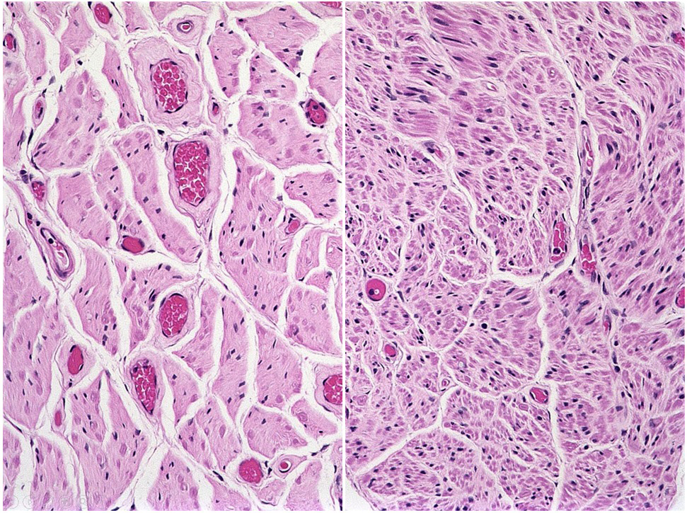
Impacts of spinal and bulbar muscular atrophy
Images hosted on other servers:

Tongue atrophy

Asymmetric facial muscle atrophy
Images hosted on other servers:

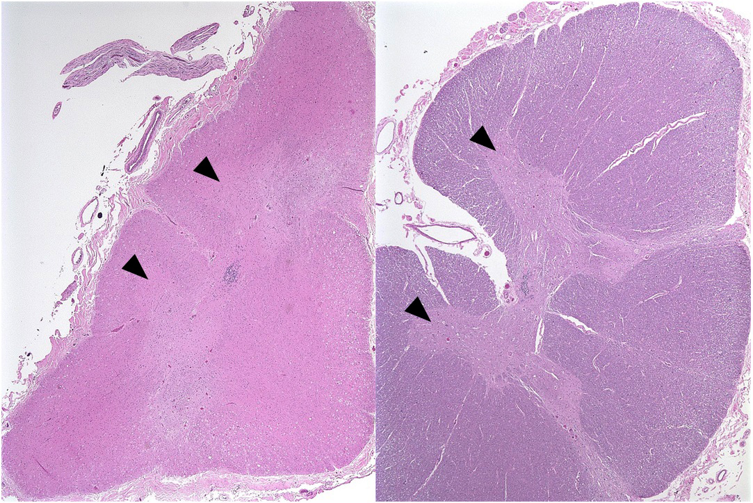
Muscle biopsy

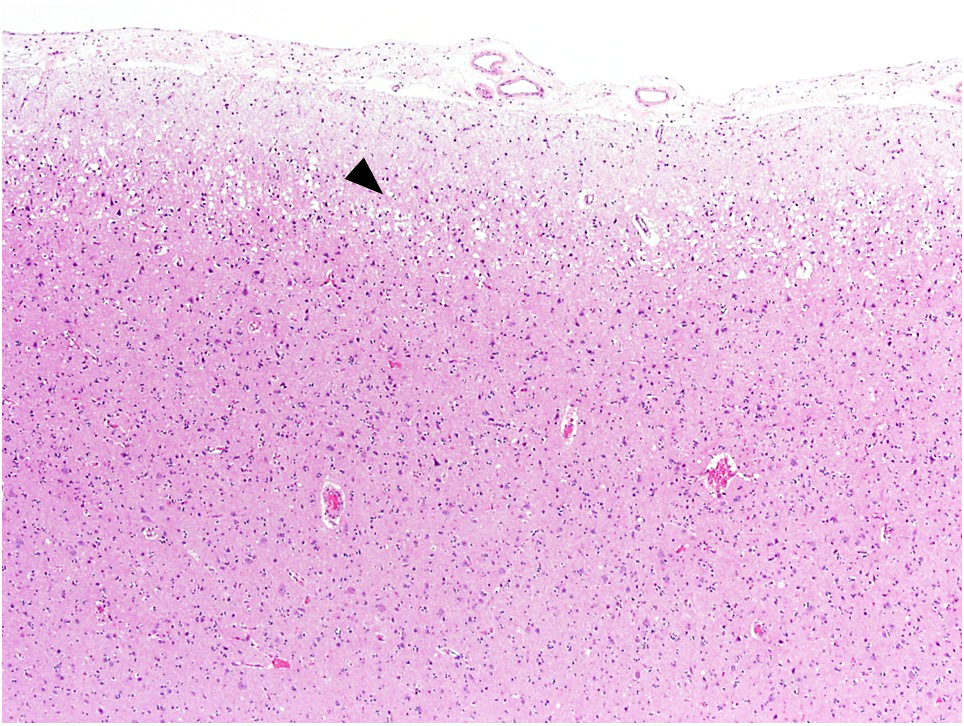
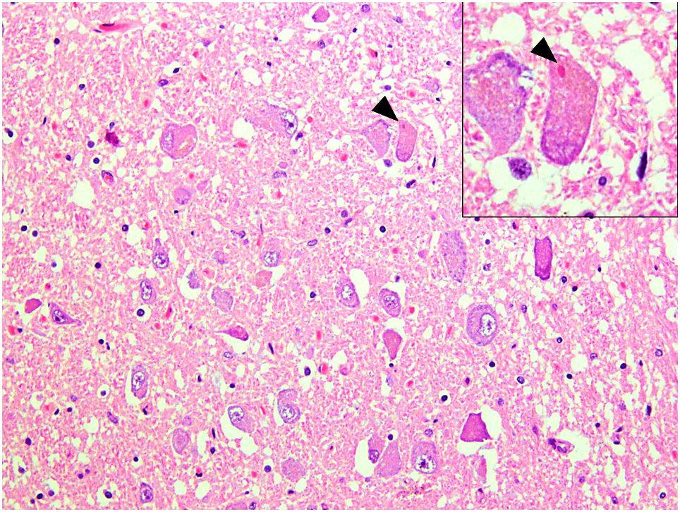
Pathology of Kennedy disease
Hand tremor, tongue fasciculation
Barbato: 2016
Bault : 2015
Braak: 2015
Gray: 2018
Kleinschmidt-DeMasters: 2022
Kovacs: 2015
Love: 2015
Perry: 2017
Rub: 2015
Find related Pathology books: neuropathology, muscle and peripheral nerve nontumor
