

Images hosted on other servers:

Acalculous choleystitis imaging
Images hosted on other servers:

Inflamed gallbladder with exudate
Images hosted on other servers:
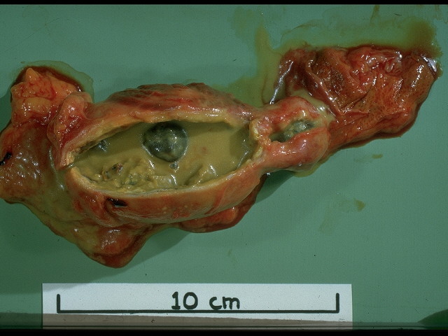
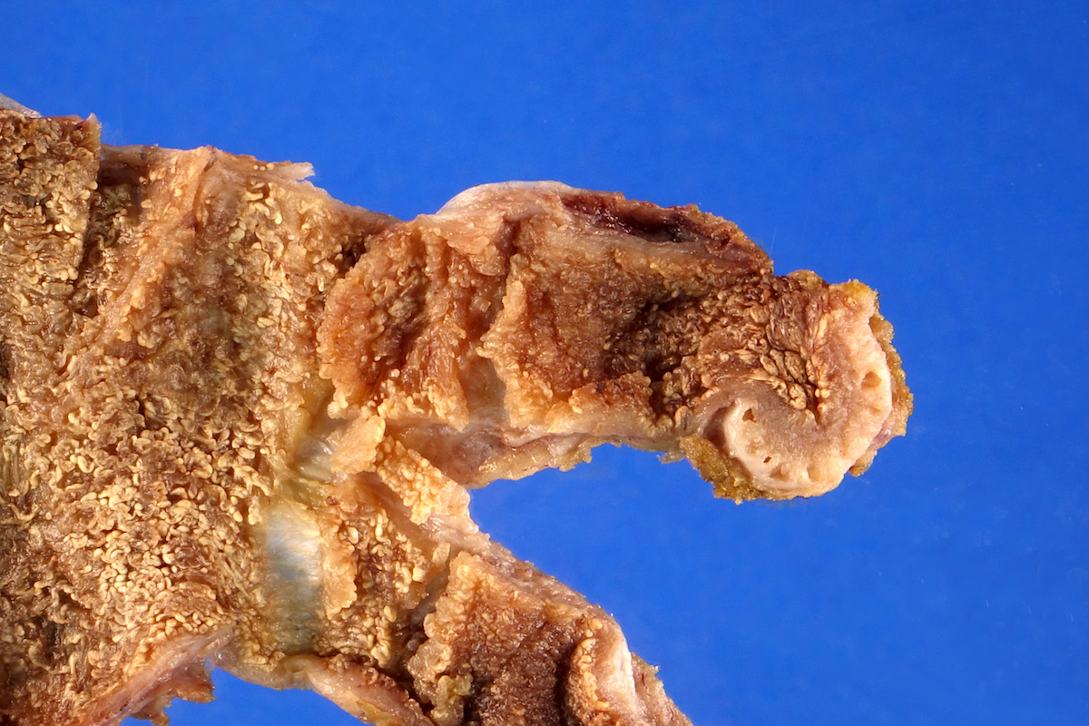
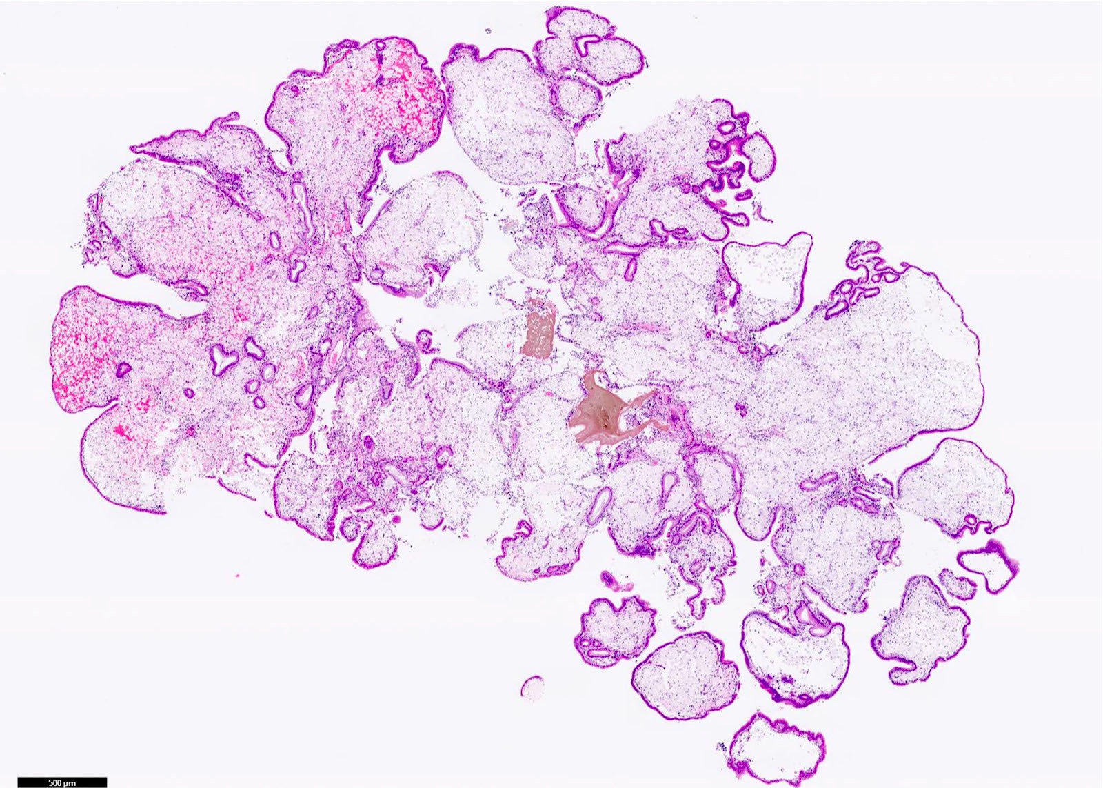
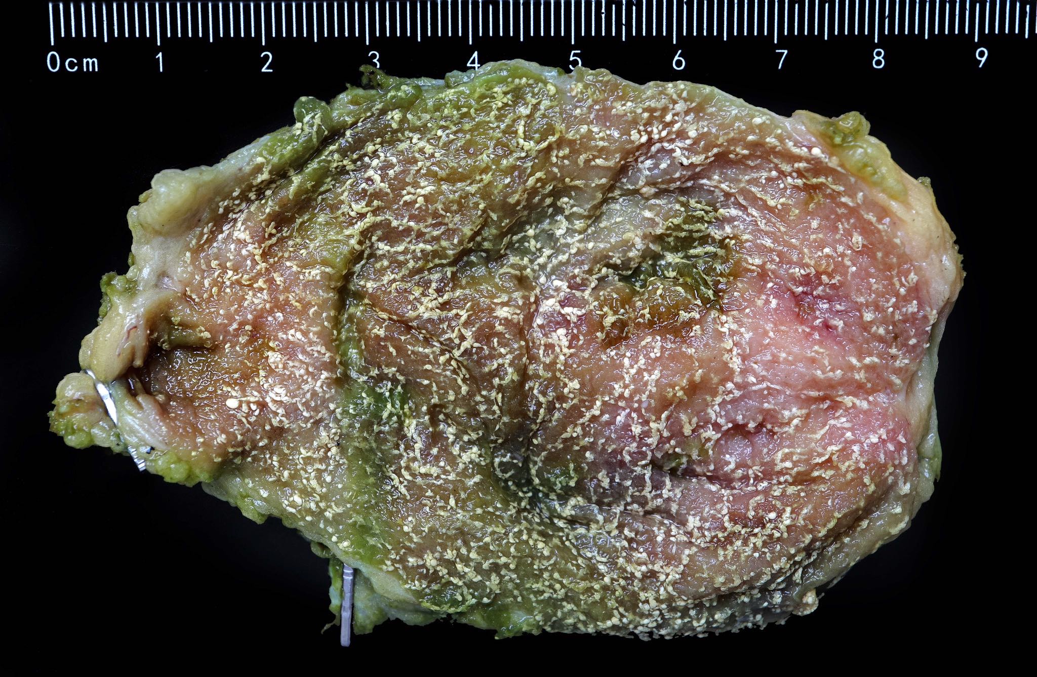
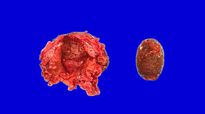
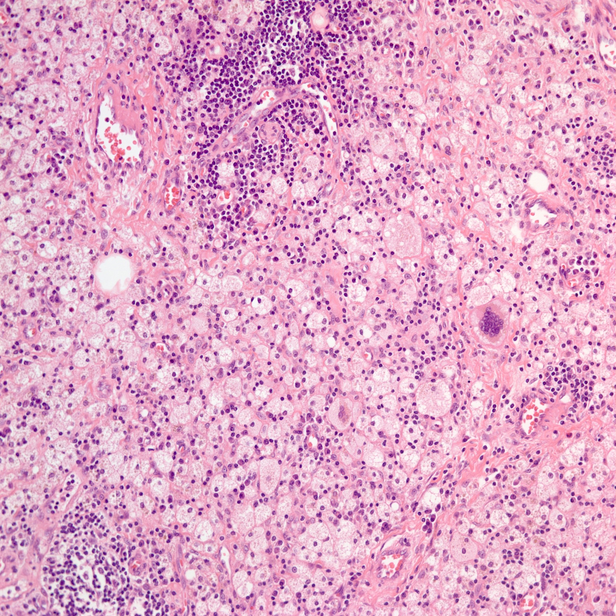
Acute (with
empyema) and
chronic cholecystitis
with gallstone
Contributed by Aaron Huber, D.O.
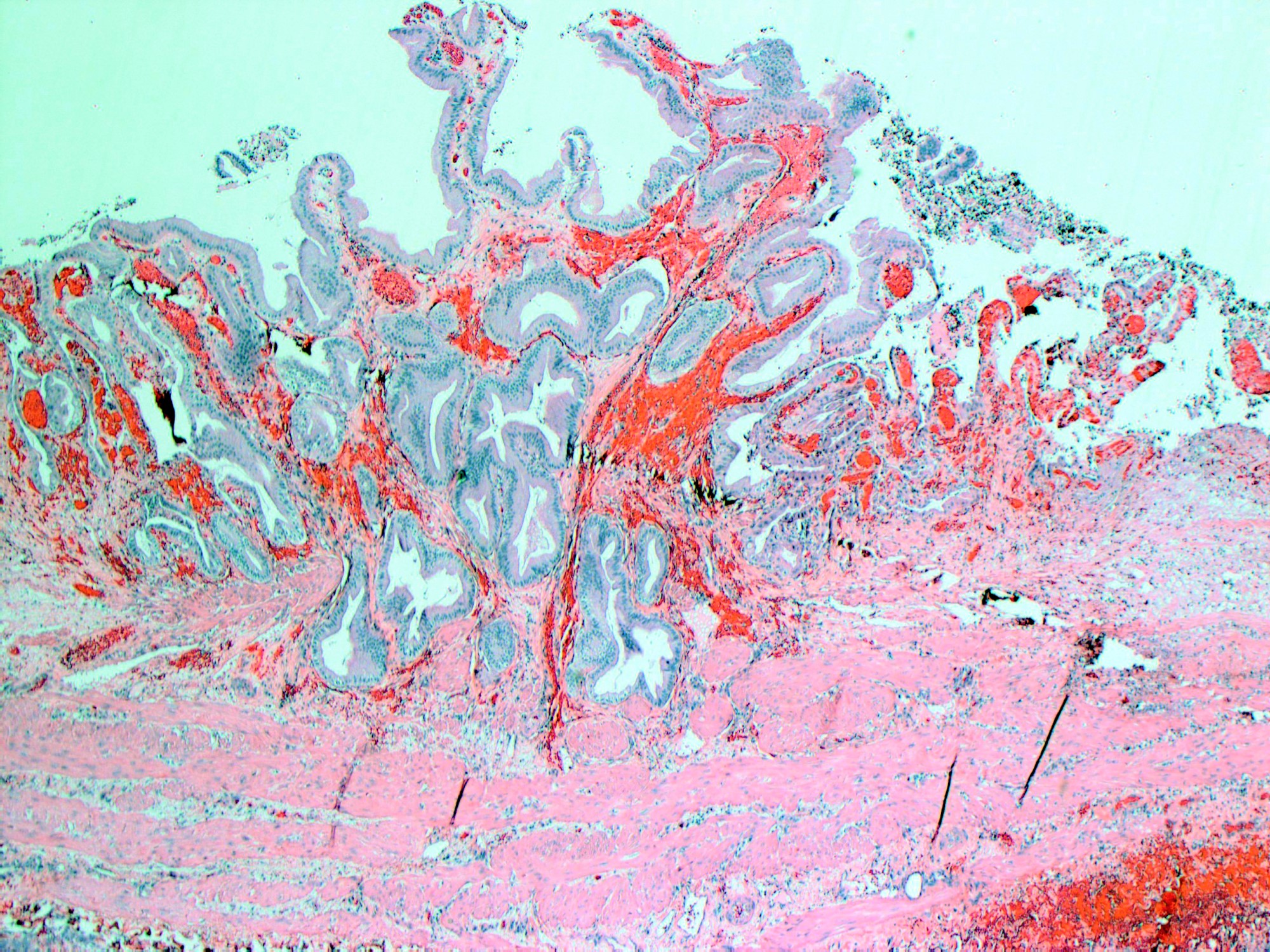
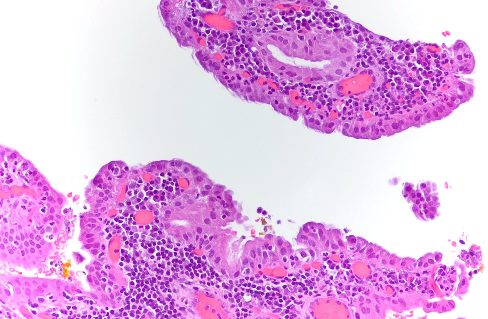
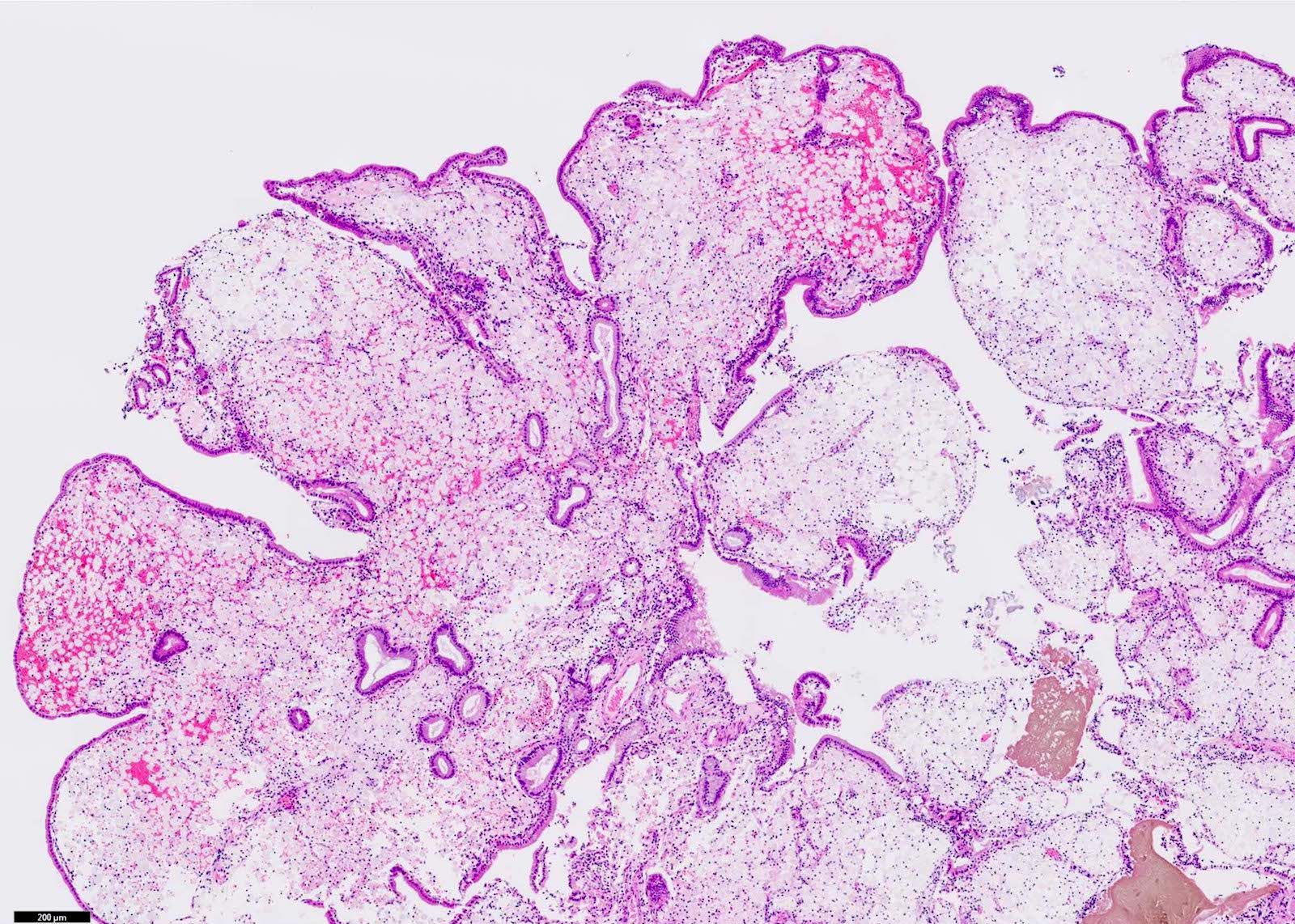
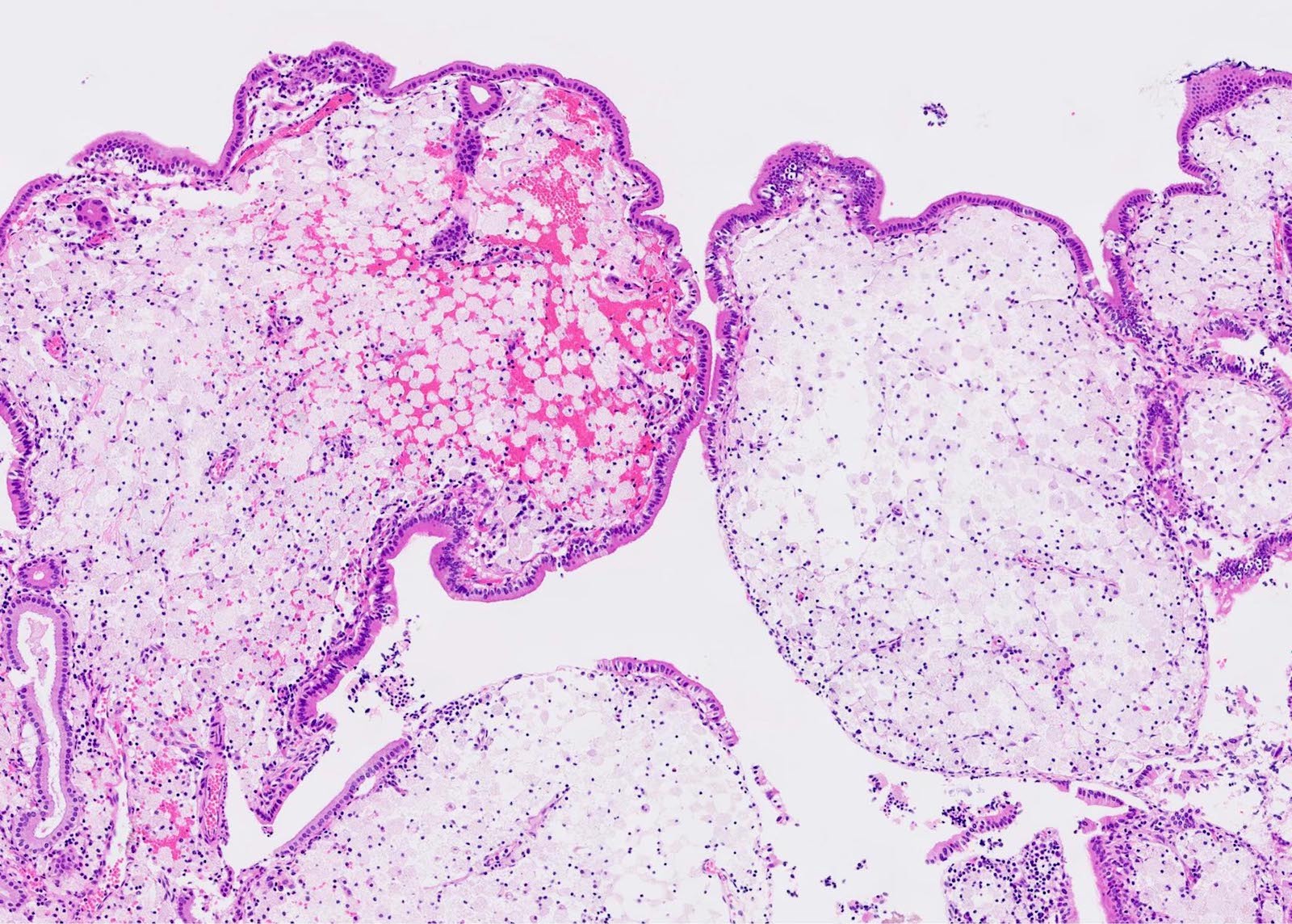
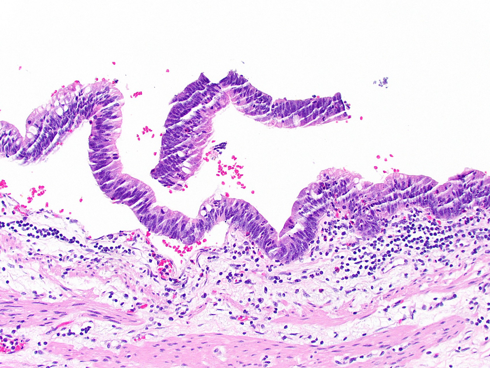
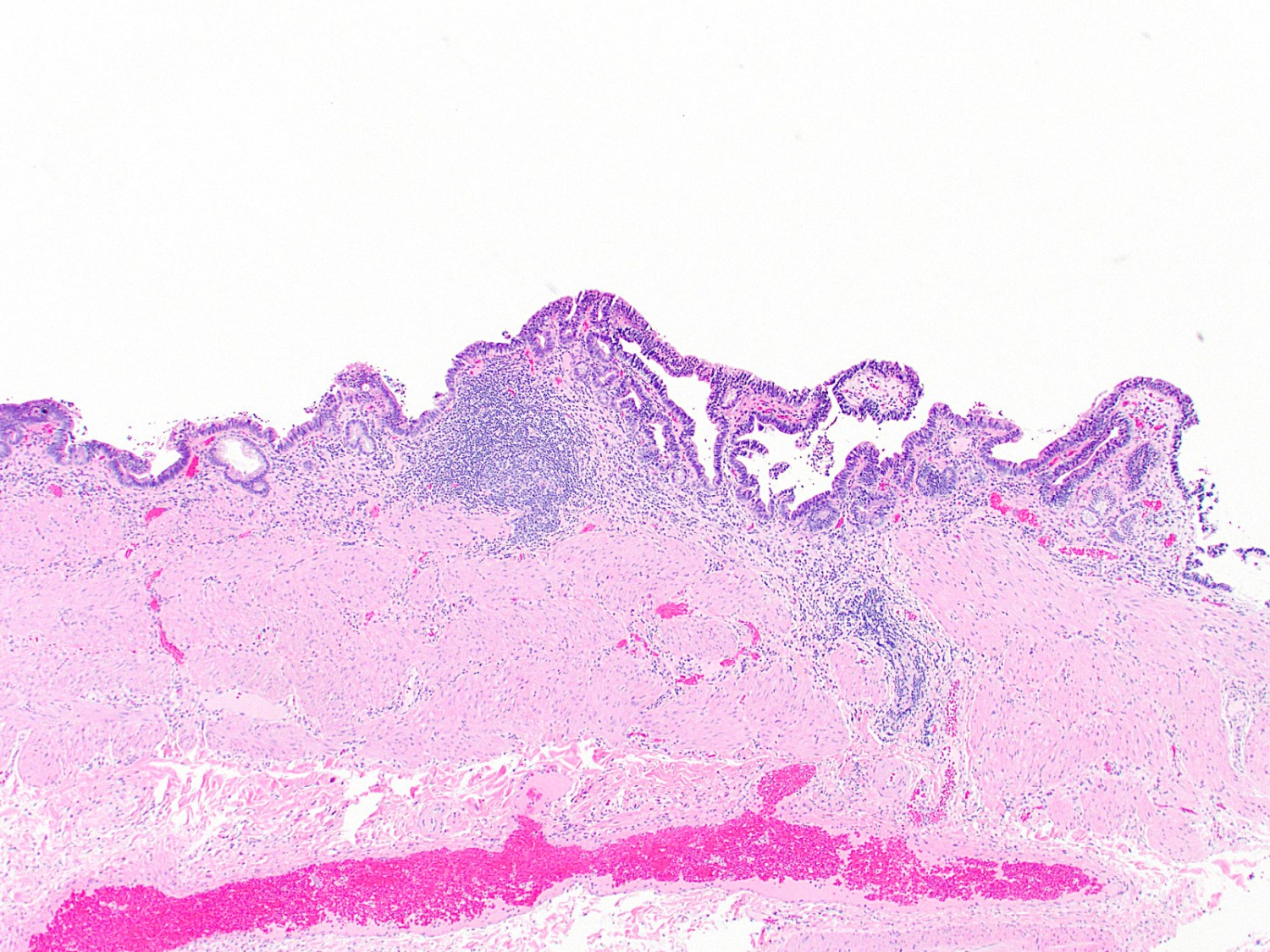
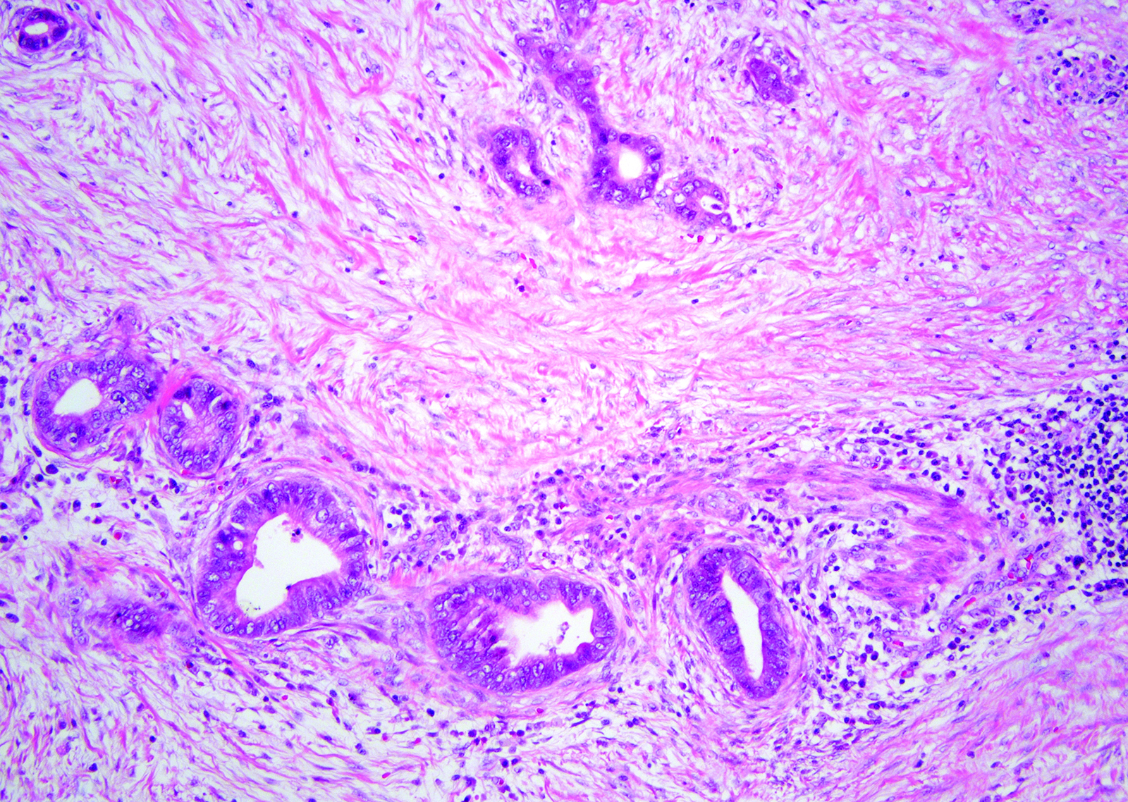
Hemorrhagic mucosa
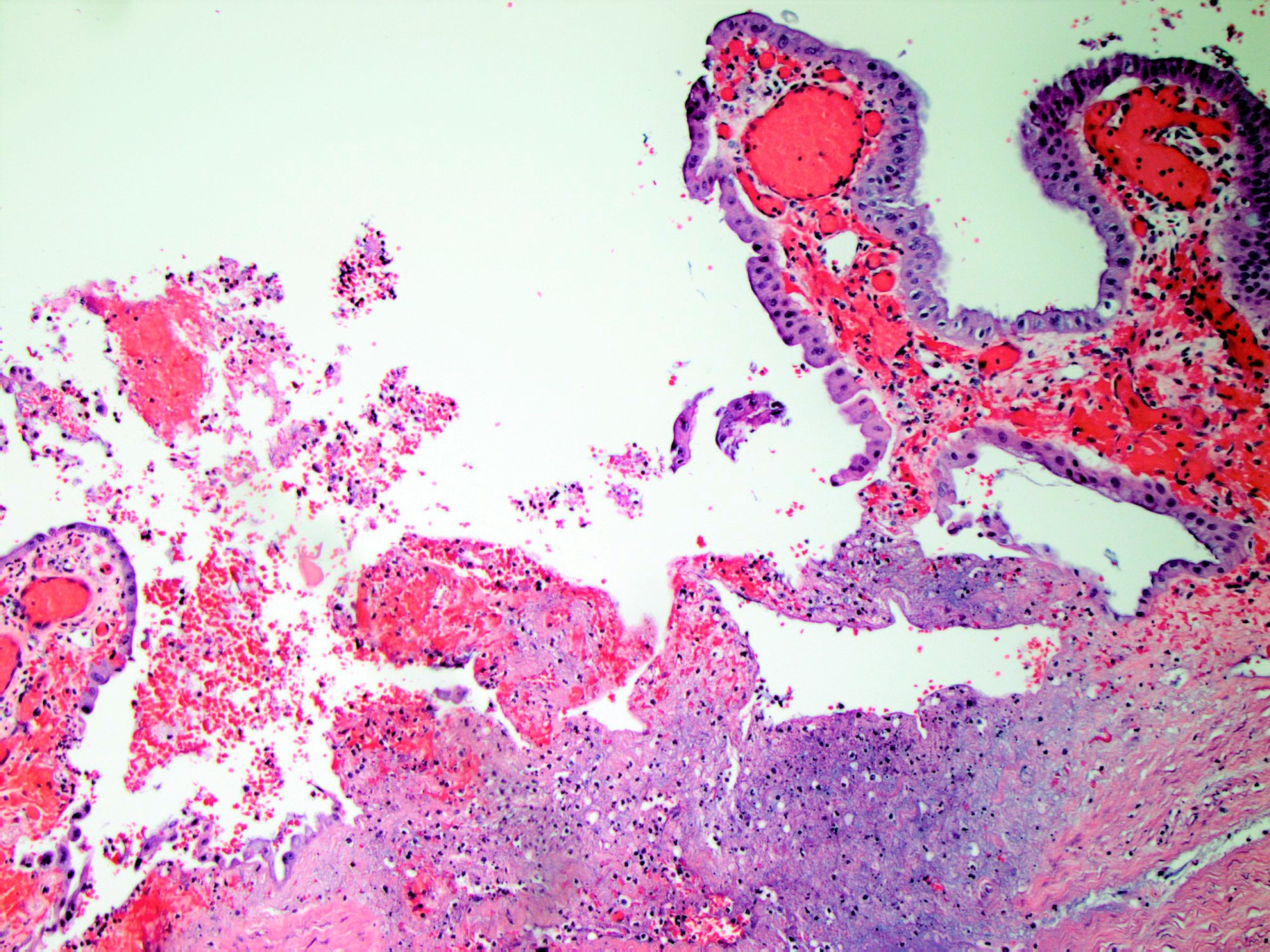
Acute inflammation and erosion
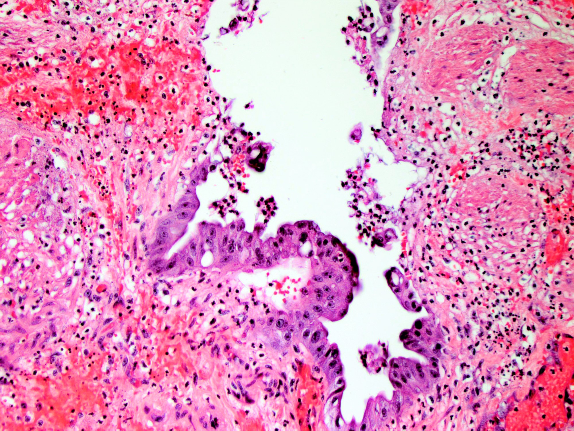
Inflamed and reactive epithelium
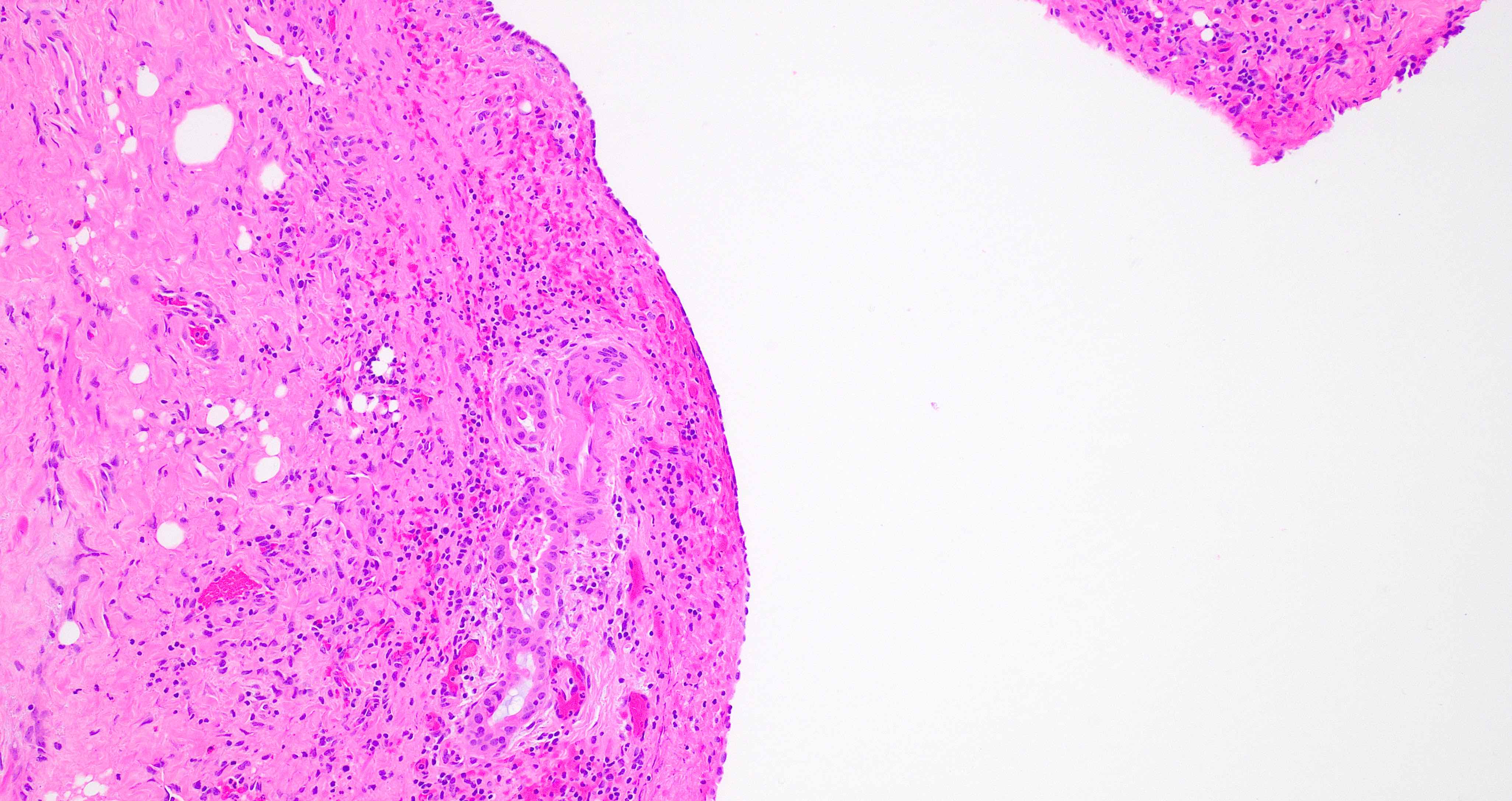
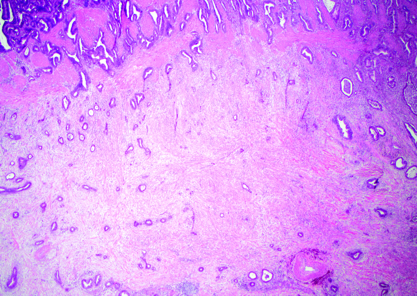
Overview of acute cholecystitis
Imaging findings in cholelithiasis and acute cholecystitis
Images hosted on other servers:


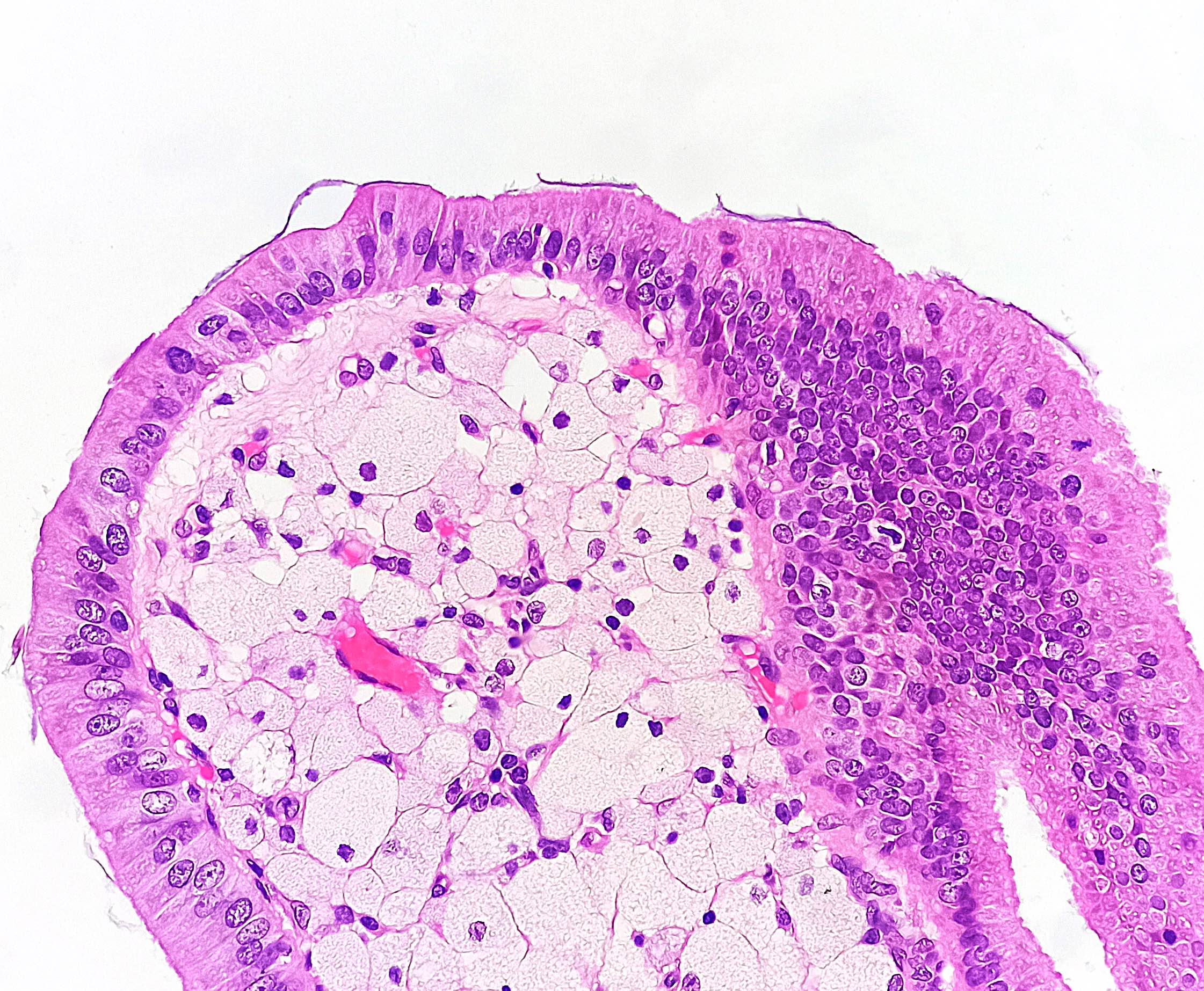
Ultrasound images of gallbladder adenomyoma
Contributed by Alan A. George, D.O. and Monica T. Garcia-Buitrago, M.D.
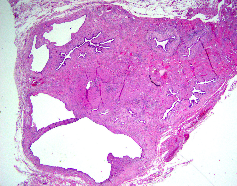
Localized adenomyoma
Images hosted on other servers:

Focal adenomyoma
Contributed by Monica T. Garcia-Buitrago, M.D.

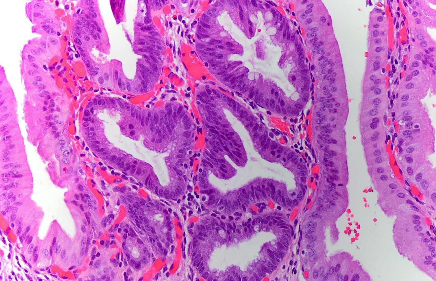
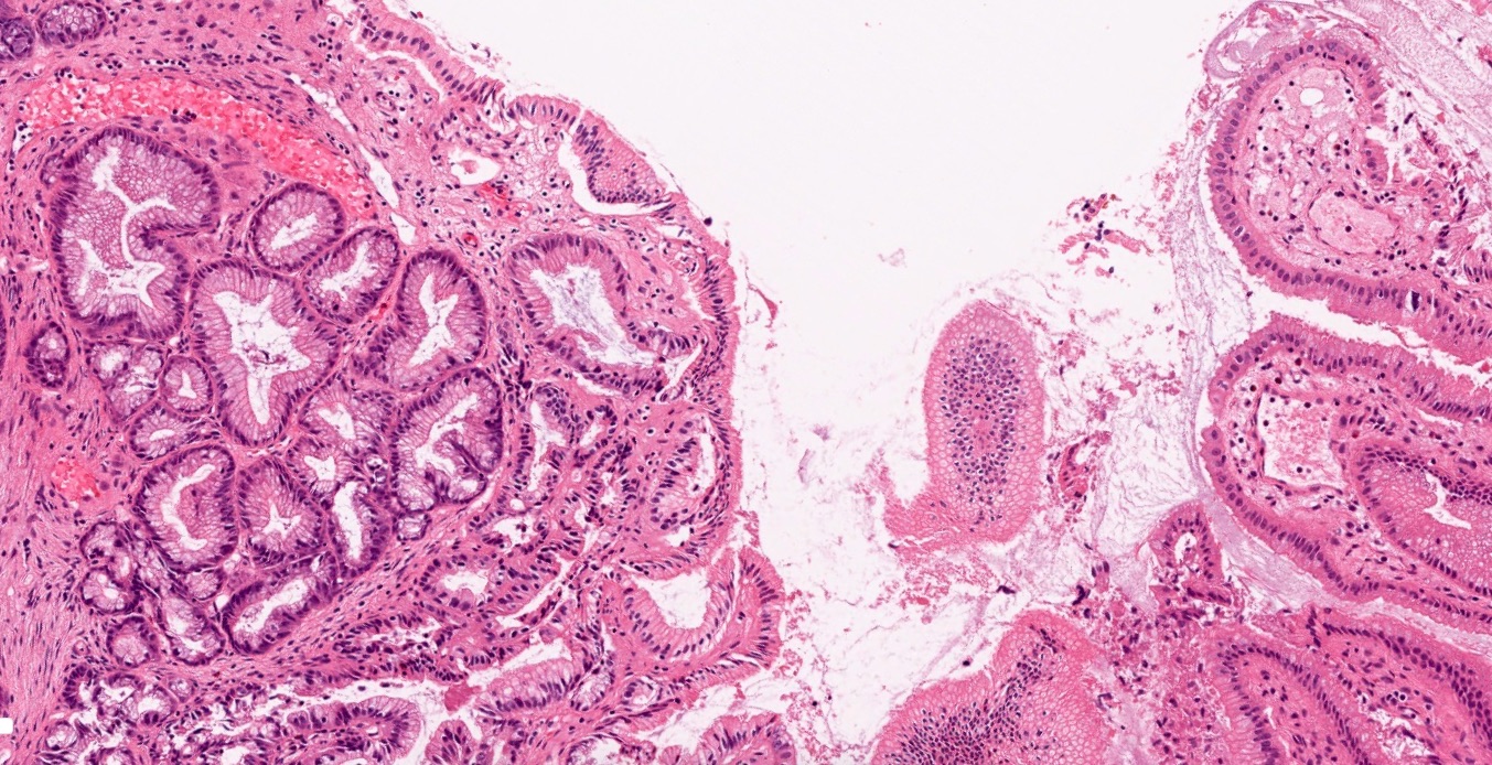
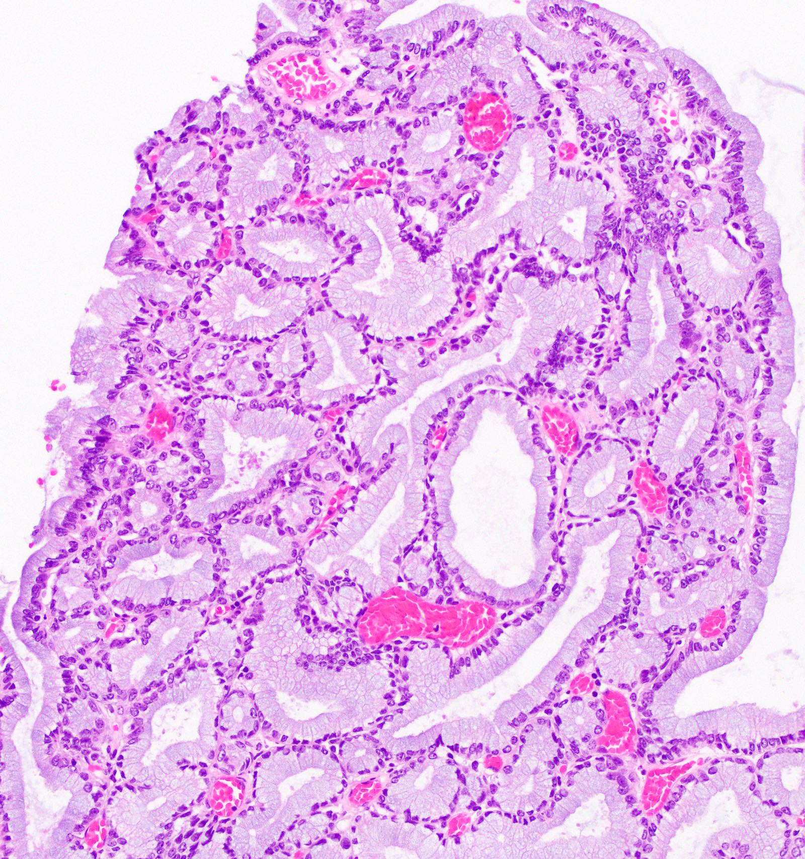
Cystically dilated biliary glands
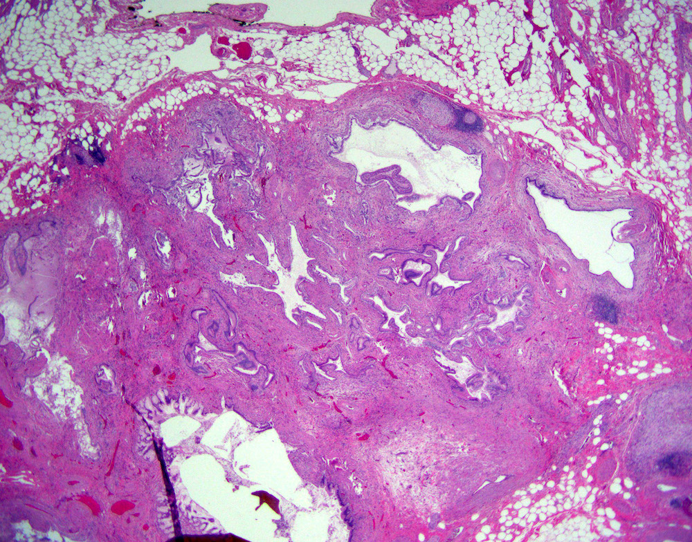
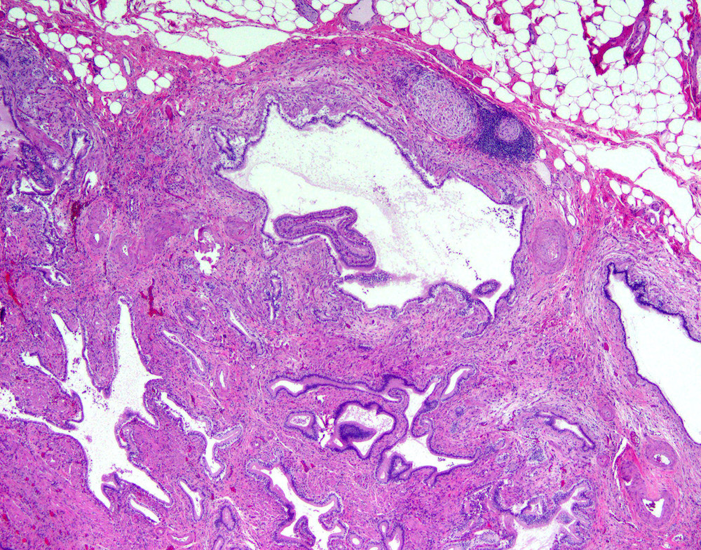
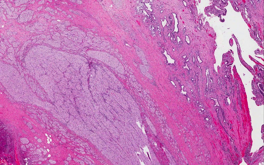
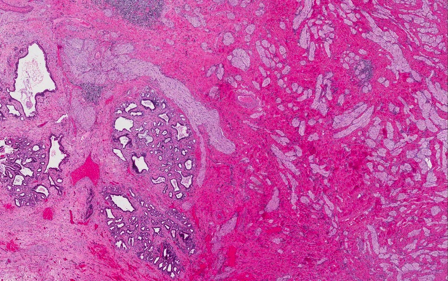
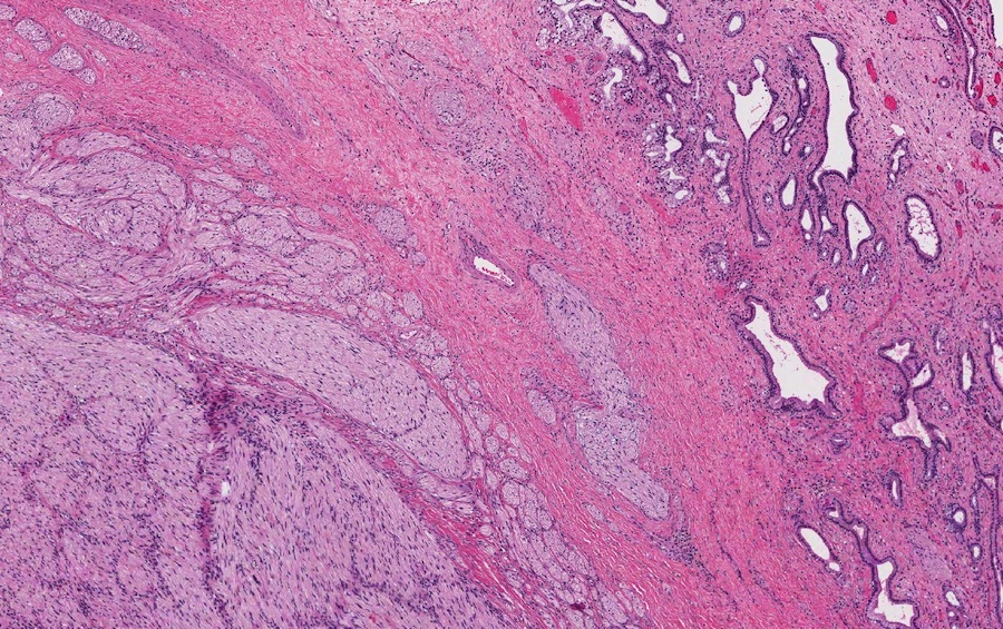
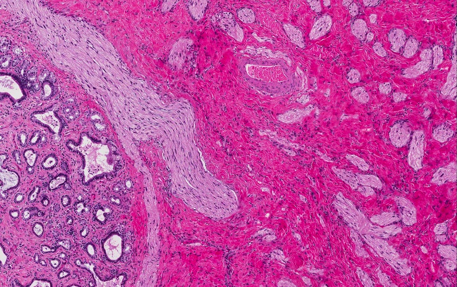
Adenomyomatous hyperplasia / adenomyomatous nodule
Dilated biliary gland, surrounding smooth muscle

Smooth muscle hyperplasia surrounding biliary glands
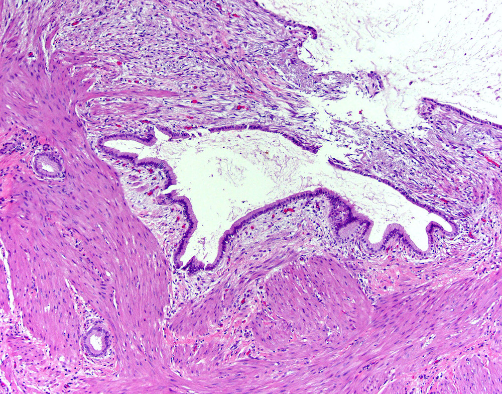
Thickened smooth muscle surrounding biliary gland
Cystically dilated biliary glands, adjacent nerve
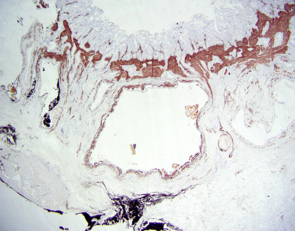
Smooth muscle surrounding cystically dilated biliary glands
Images hosted on other servers:

Inferior surface of the liver

Posterior and inferior surfaces of the liver

Gallbladder and bile ducts laid open
Contributed by Naziheh Assarzadegan, M.D.
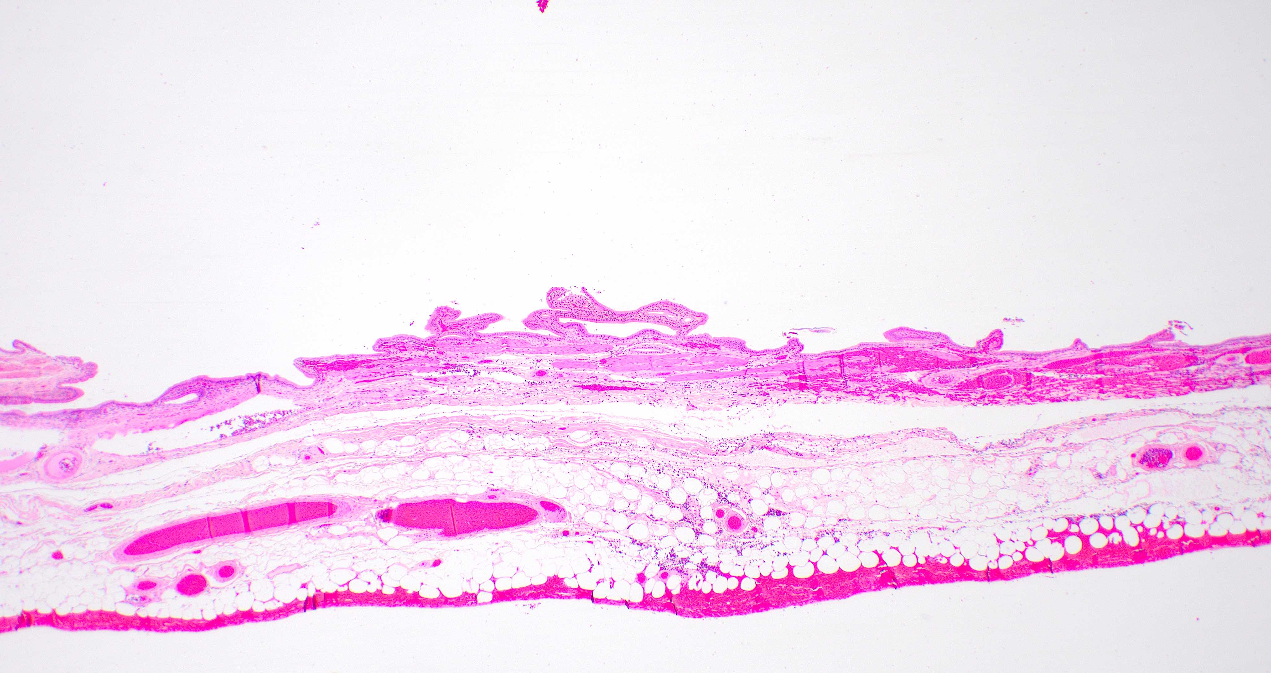
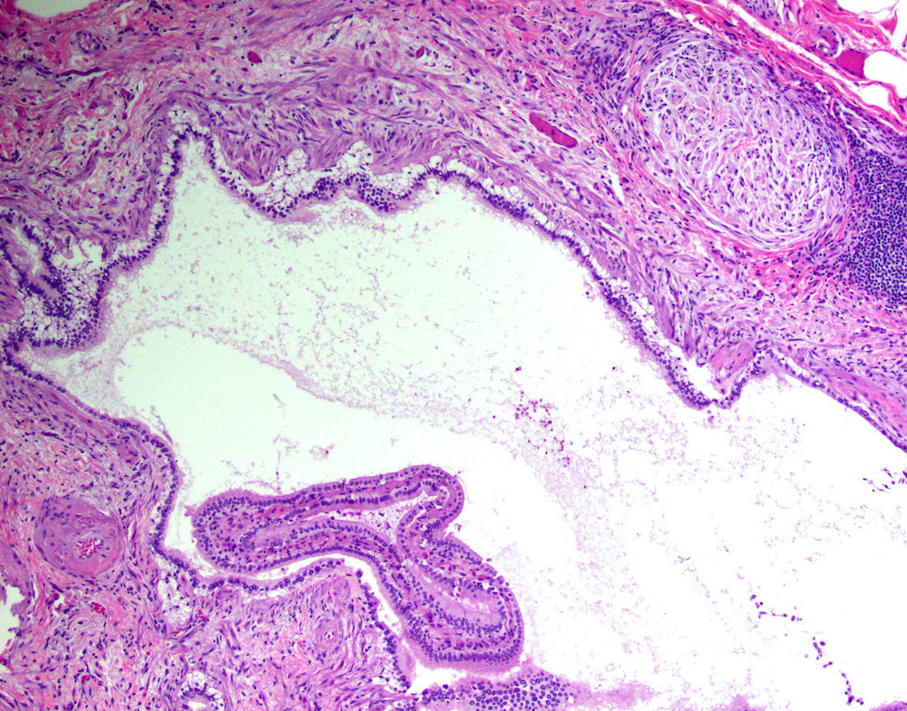
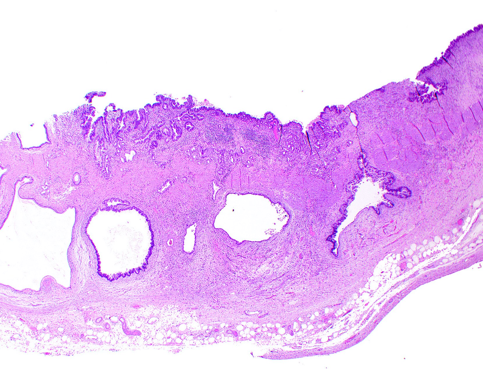
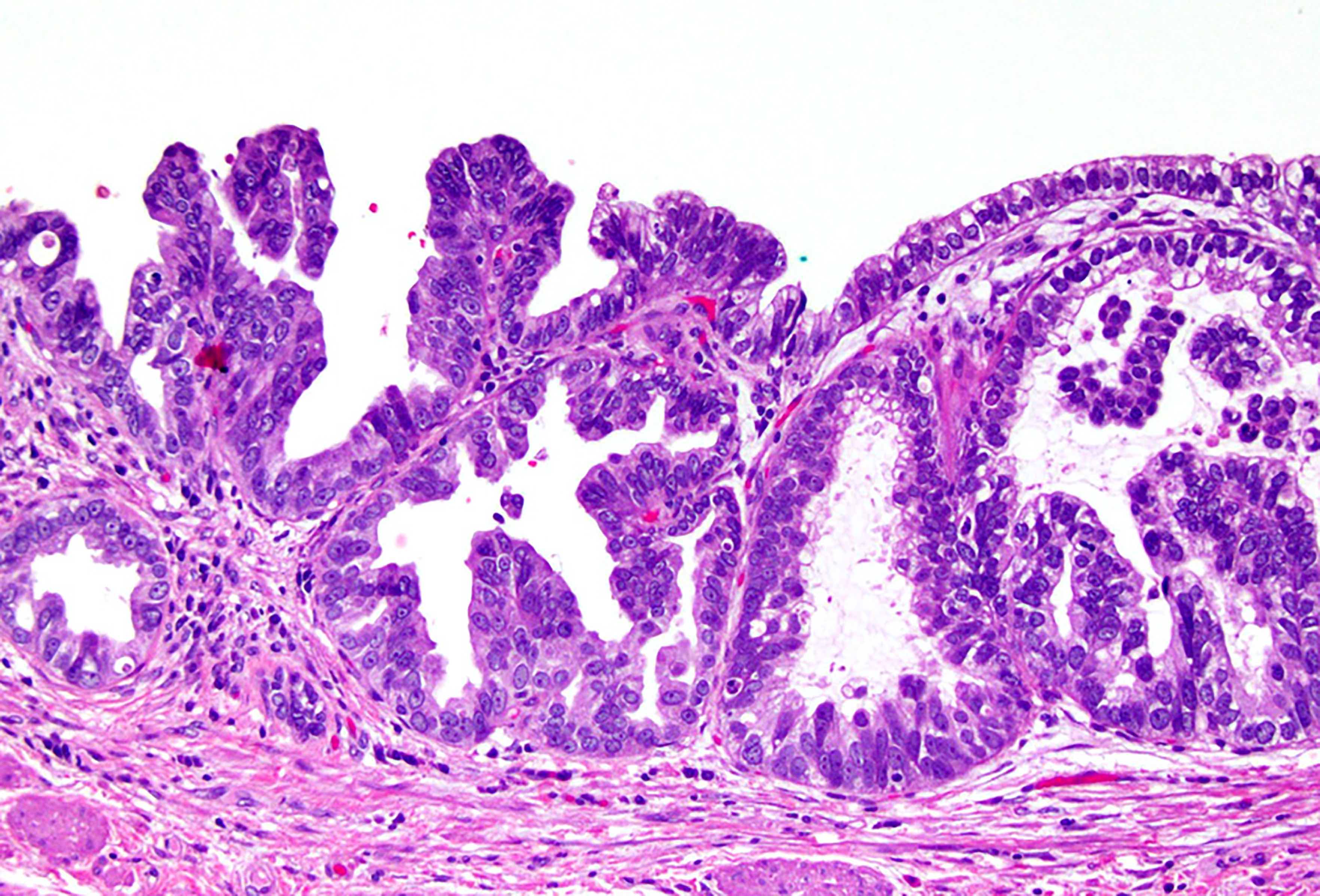
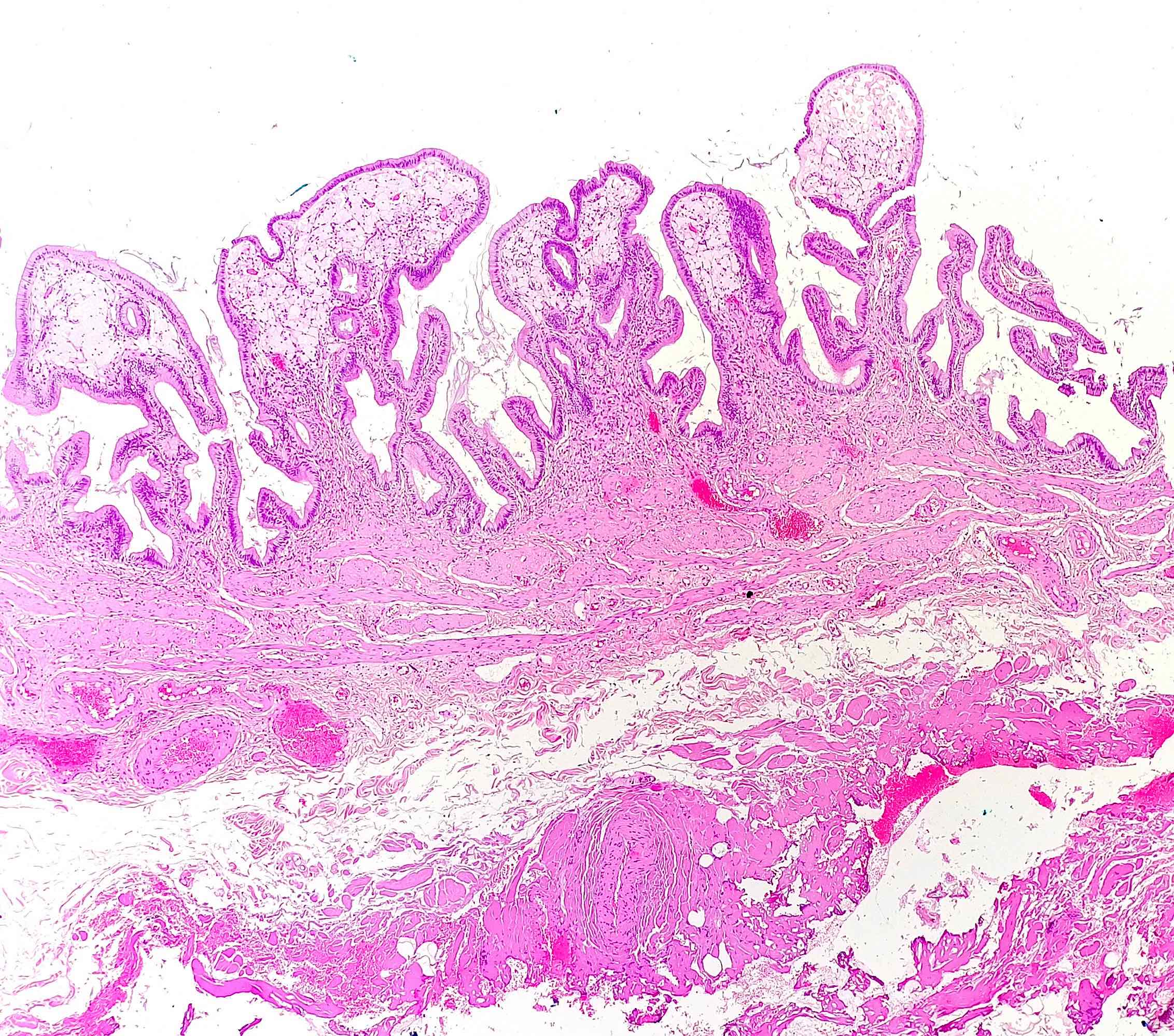
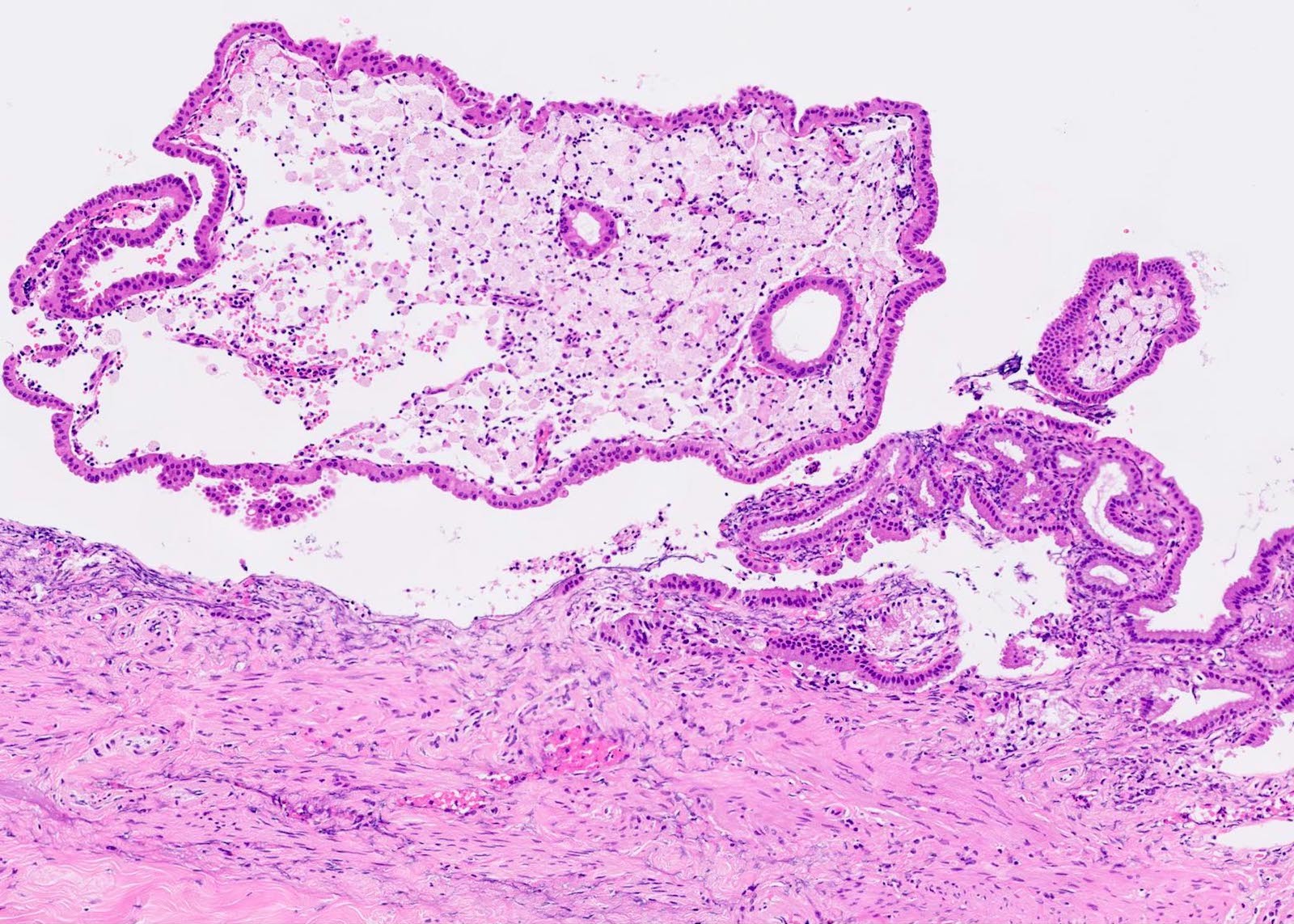
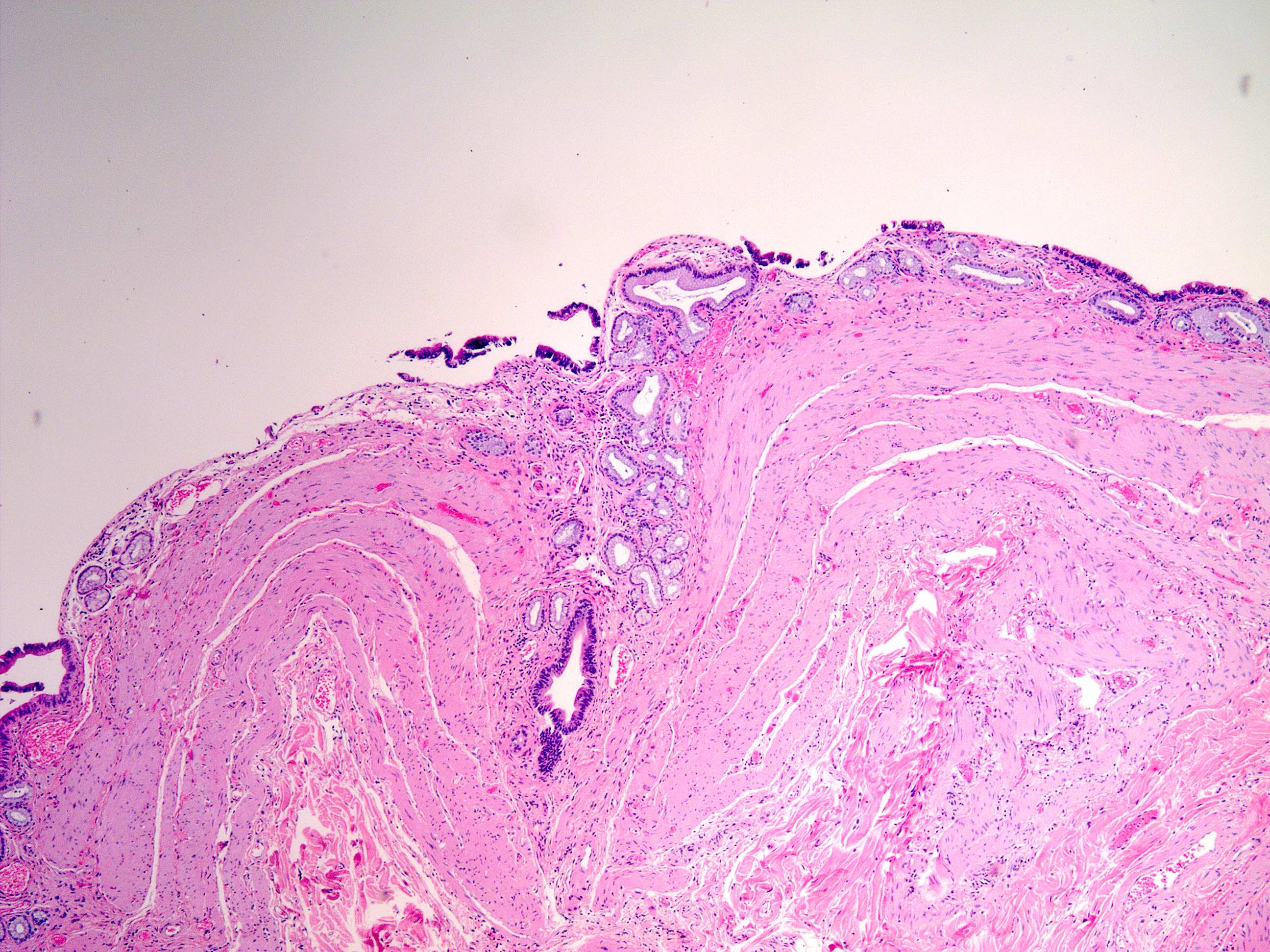
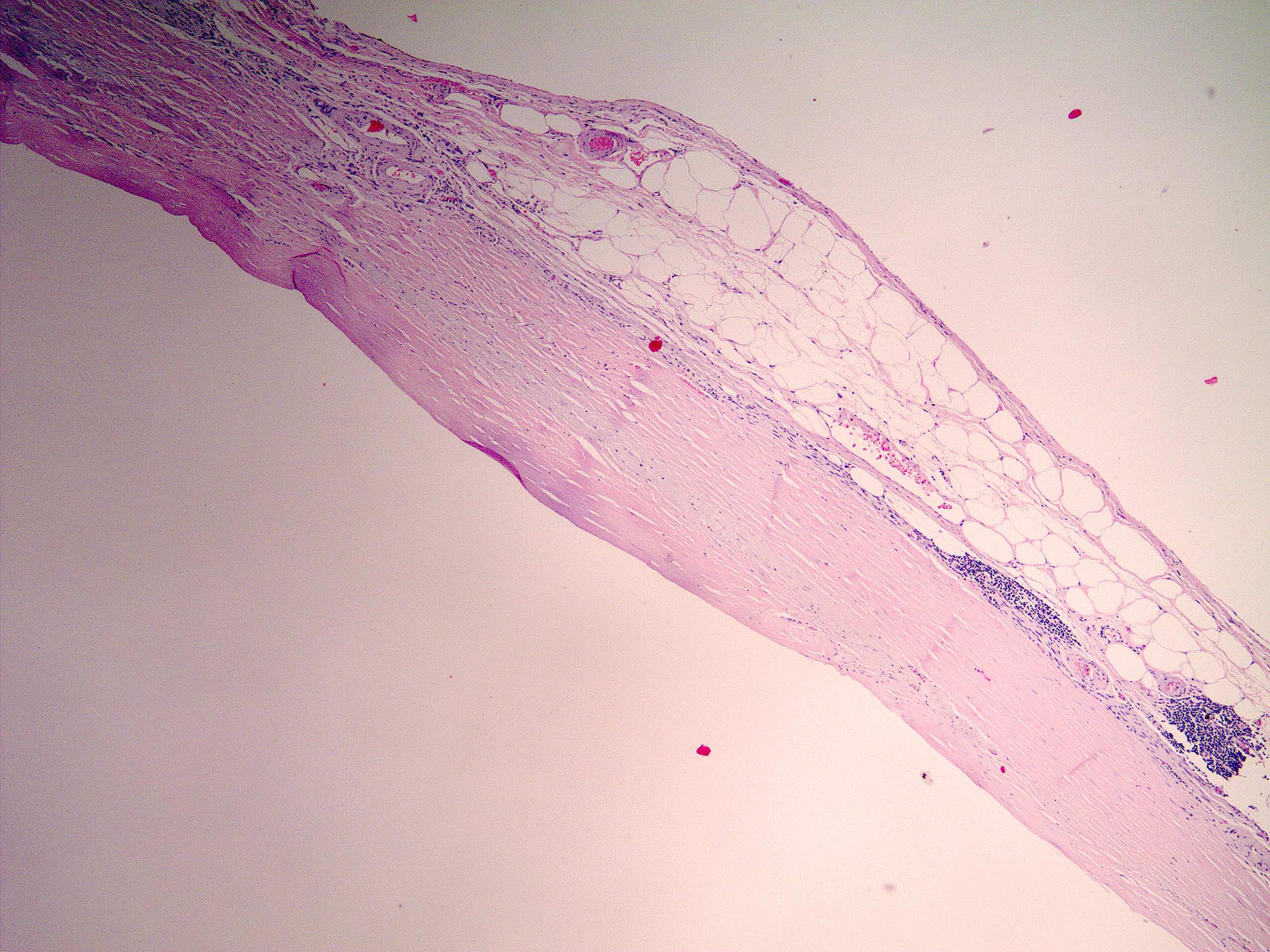
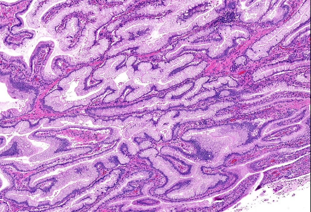
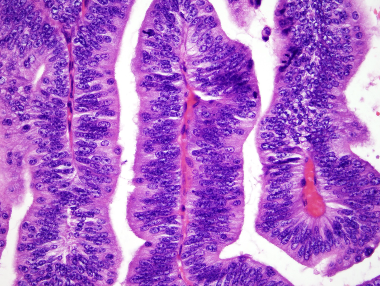
Normal layers
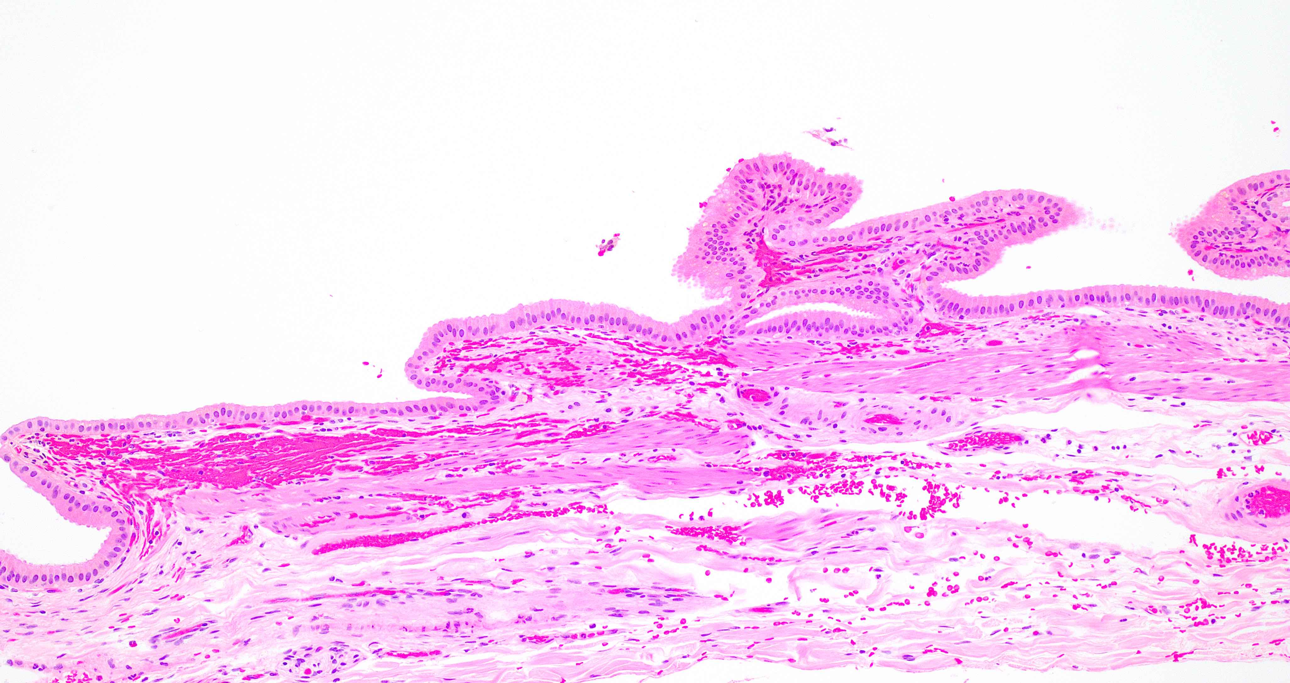
Mucosa
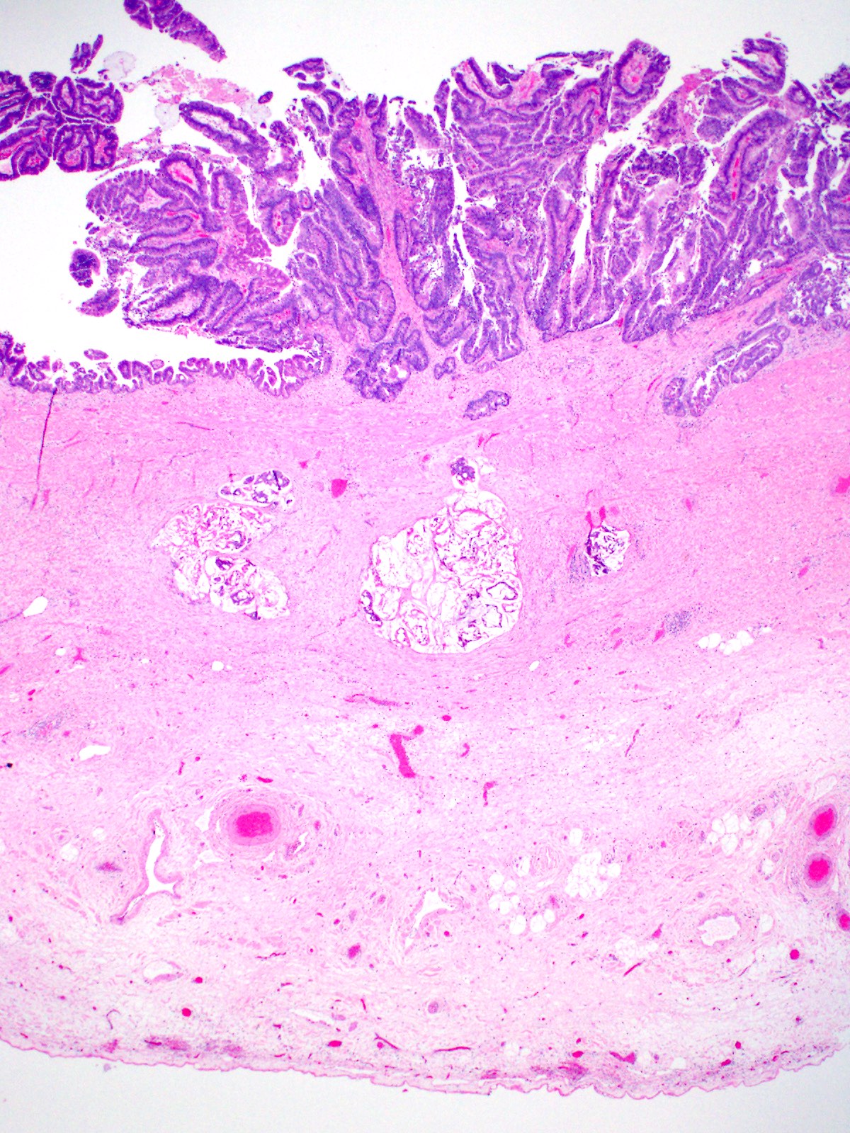
Luschka ducts
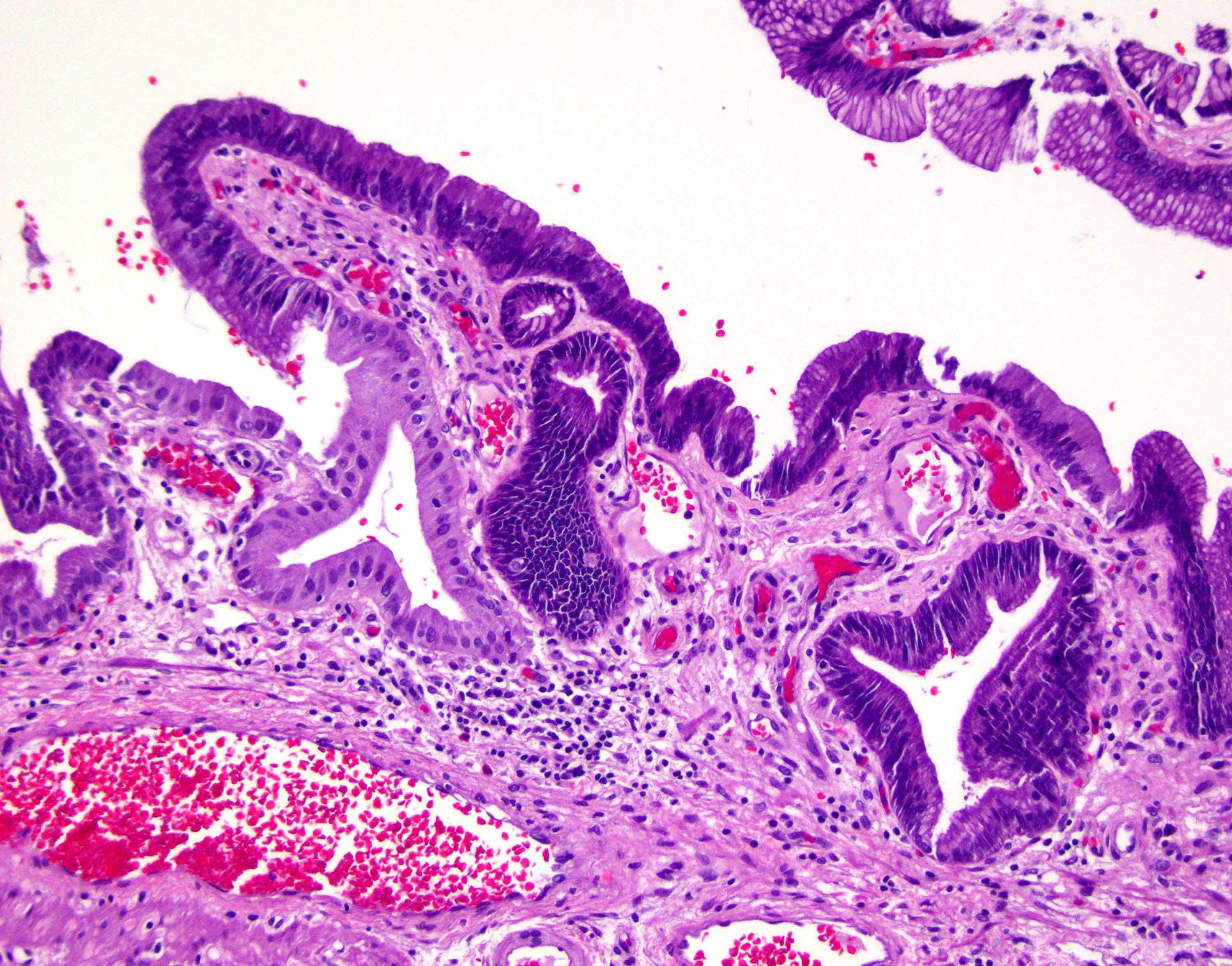
Contributed by Jian-Hua Qiao, M.D.
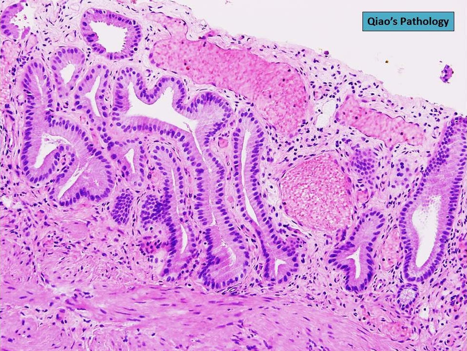
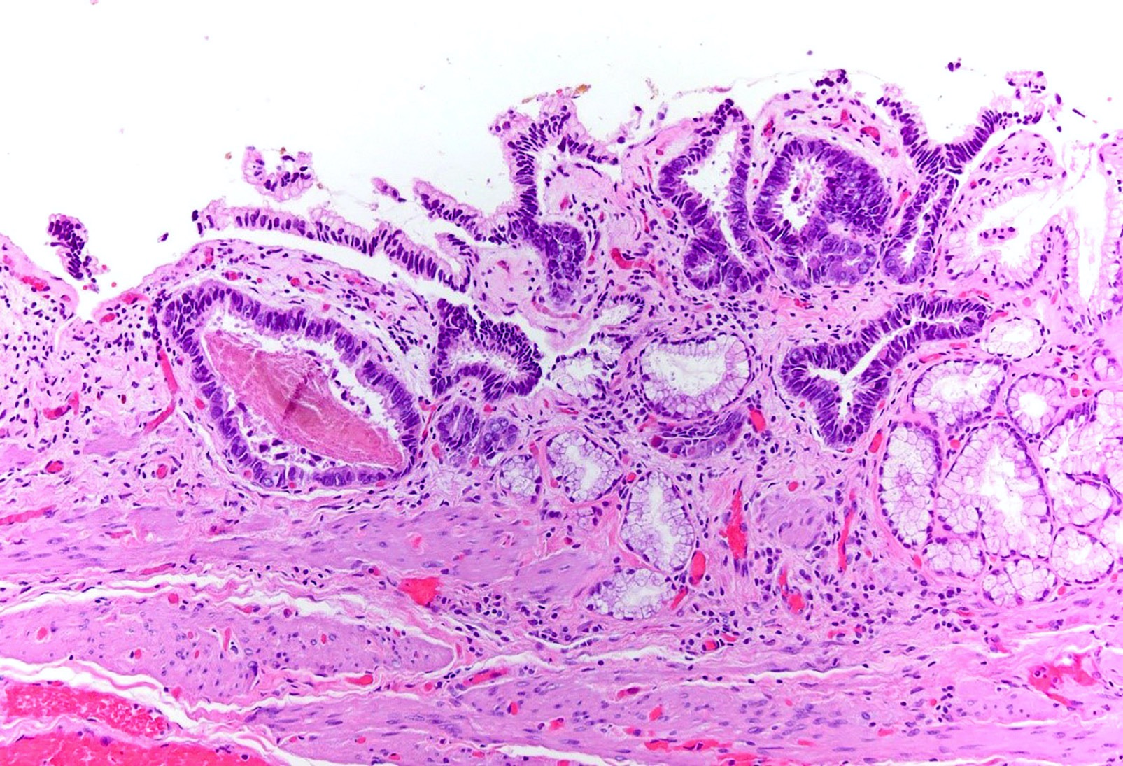
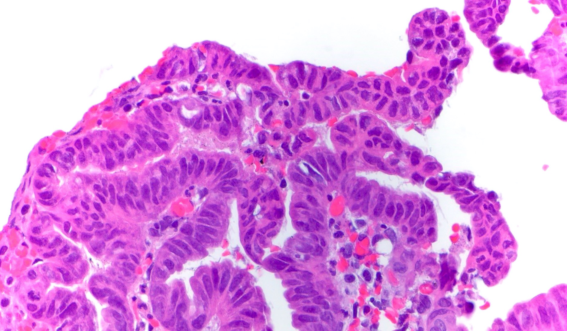
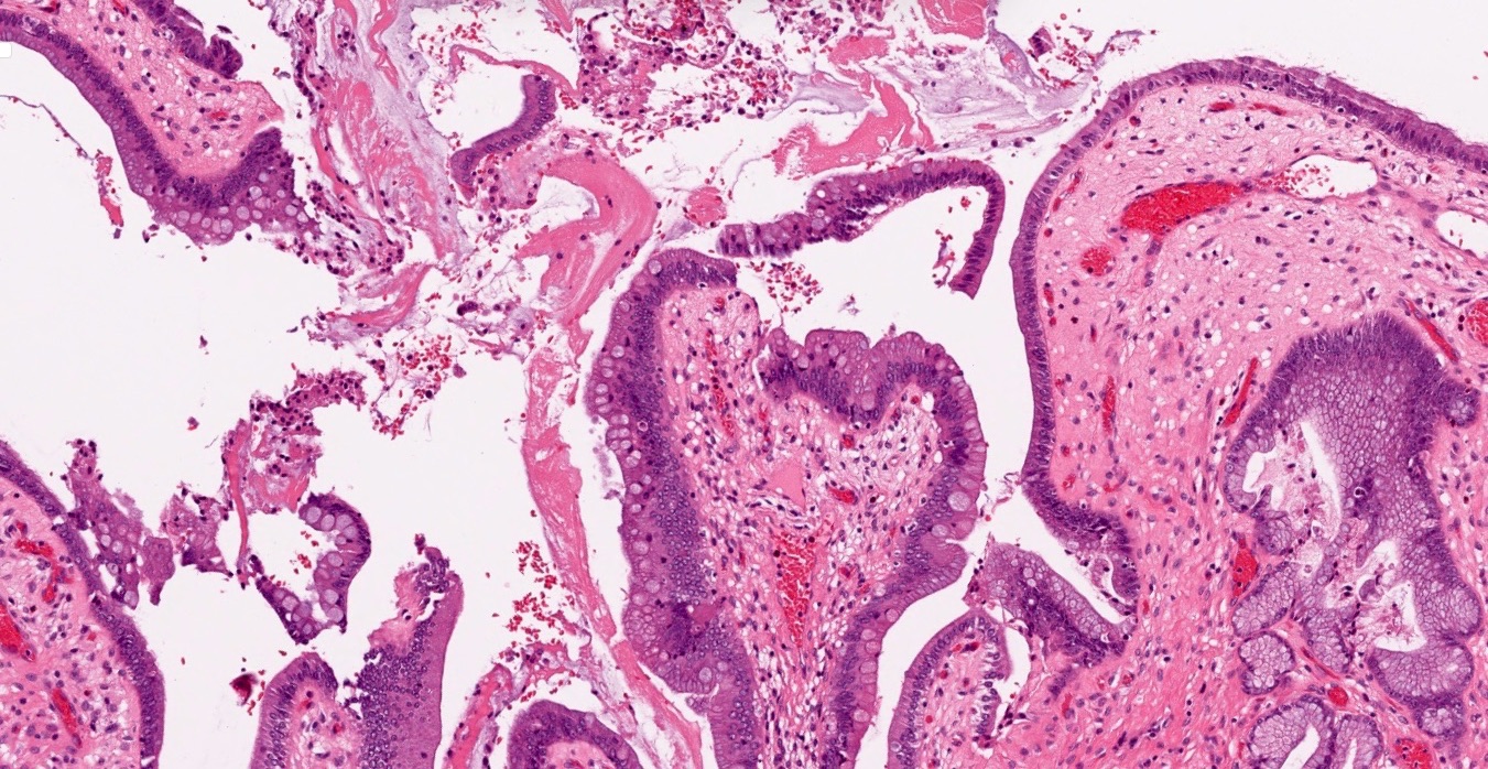
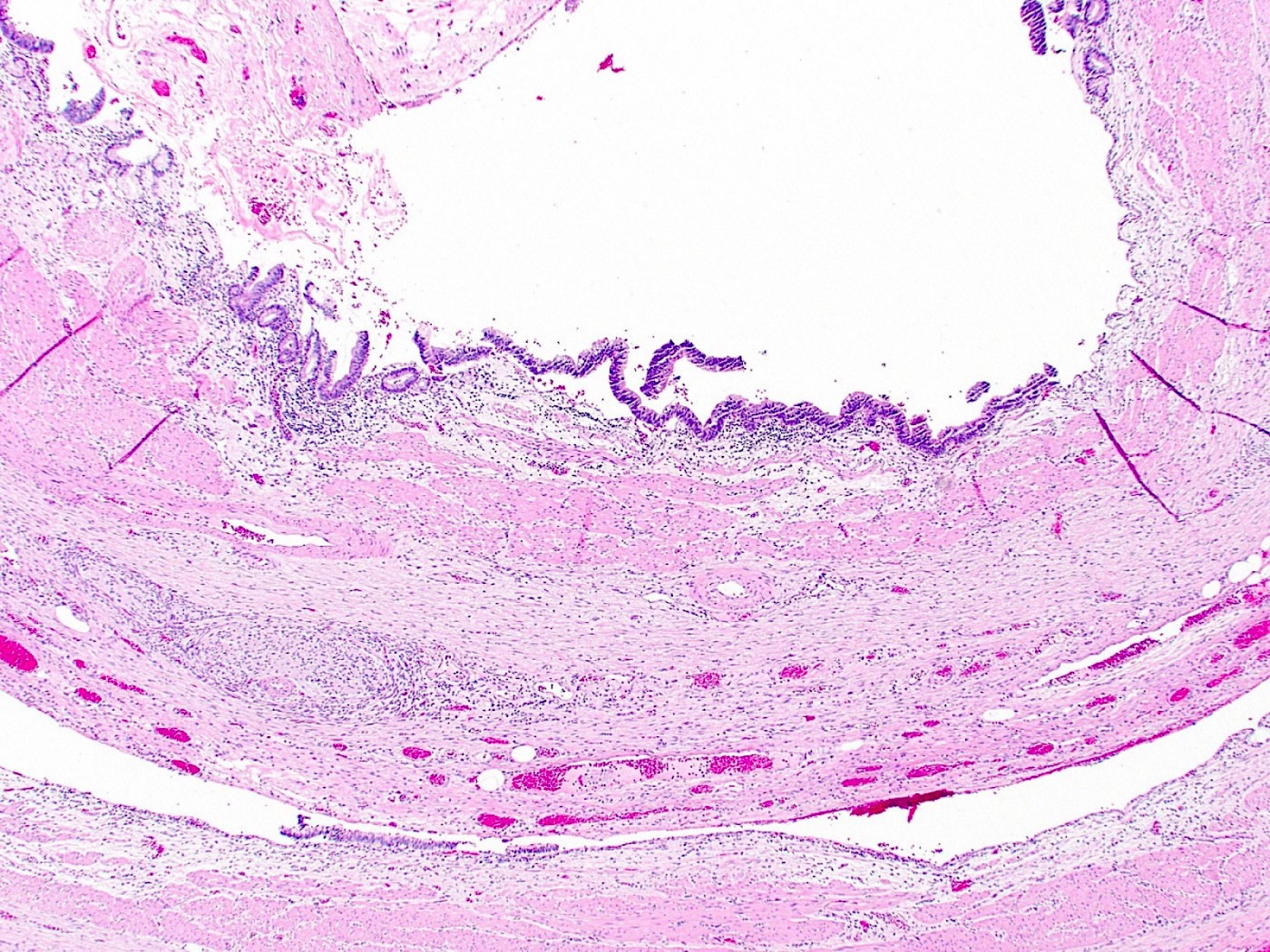
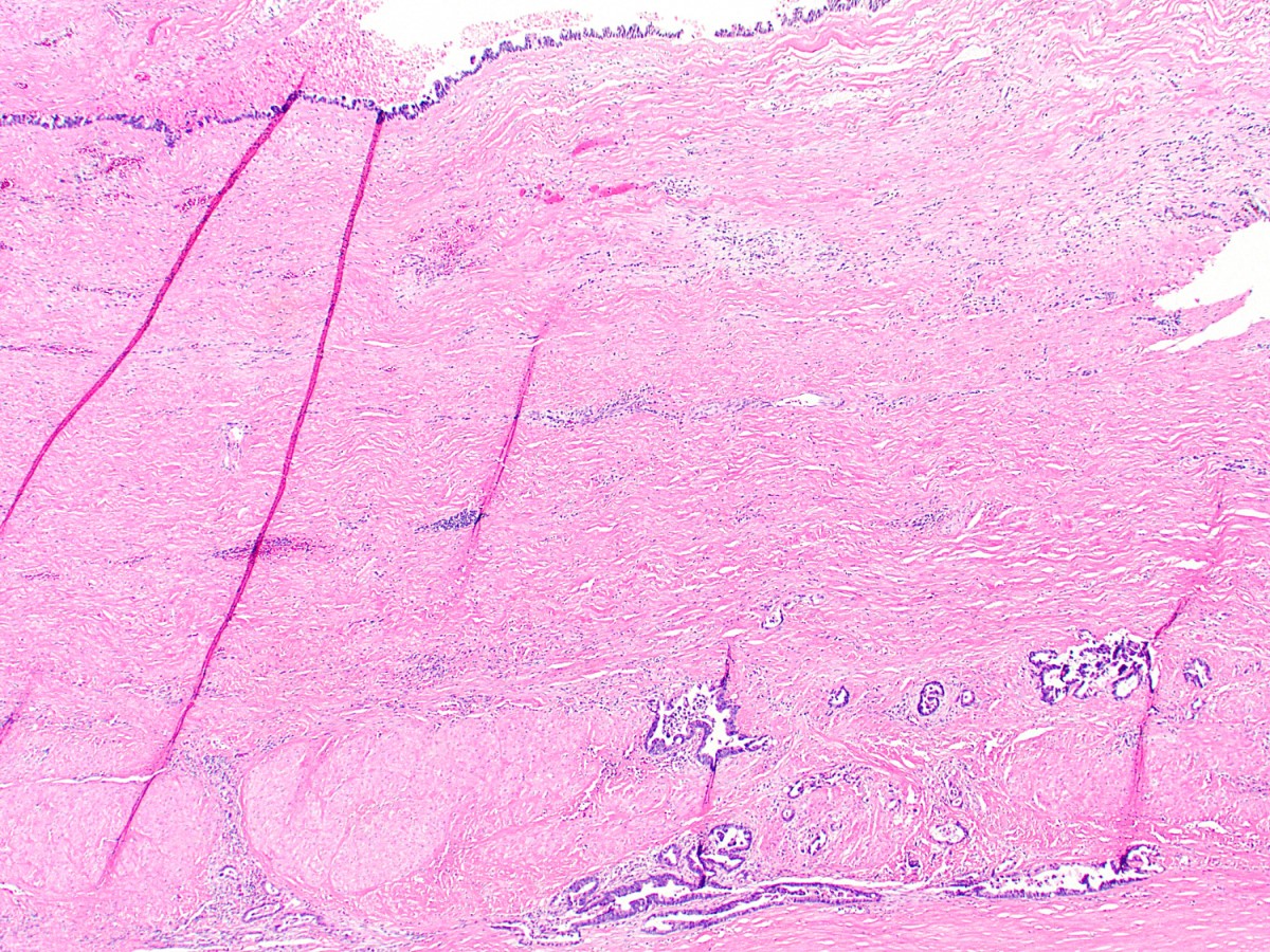
Surface denudation / ulceration

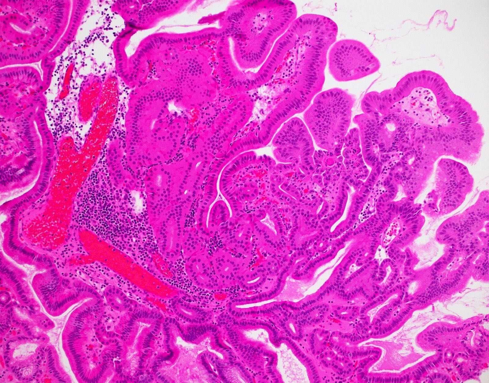
Multiple dilated vessels in lamina propria
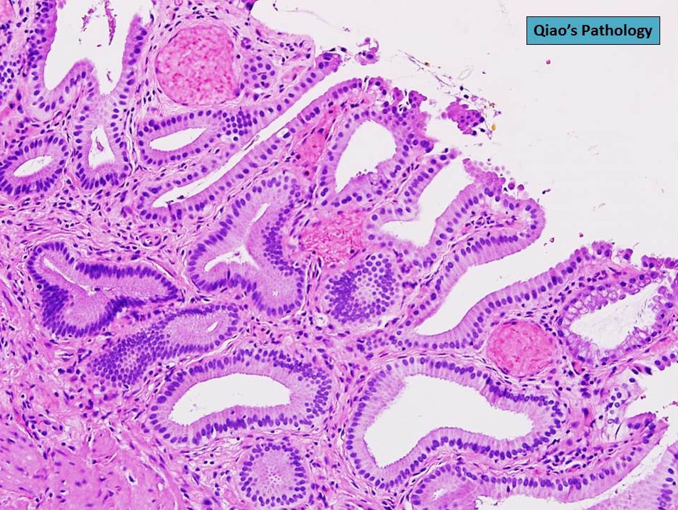
Widened mucosal folds

Disorganized veins and arteries
Contributed by Monica T. Garcia-Buitrago, M.D.
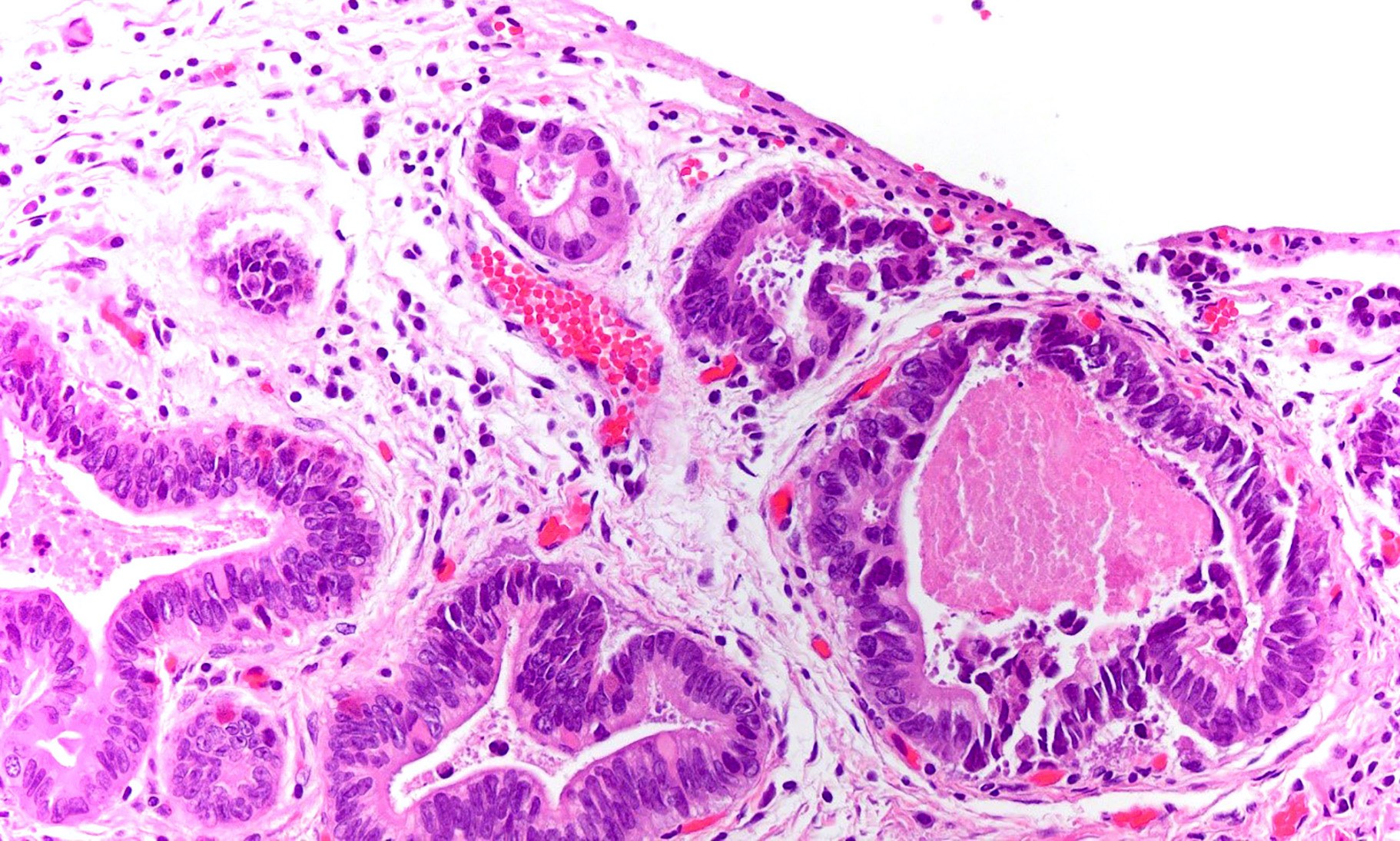
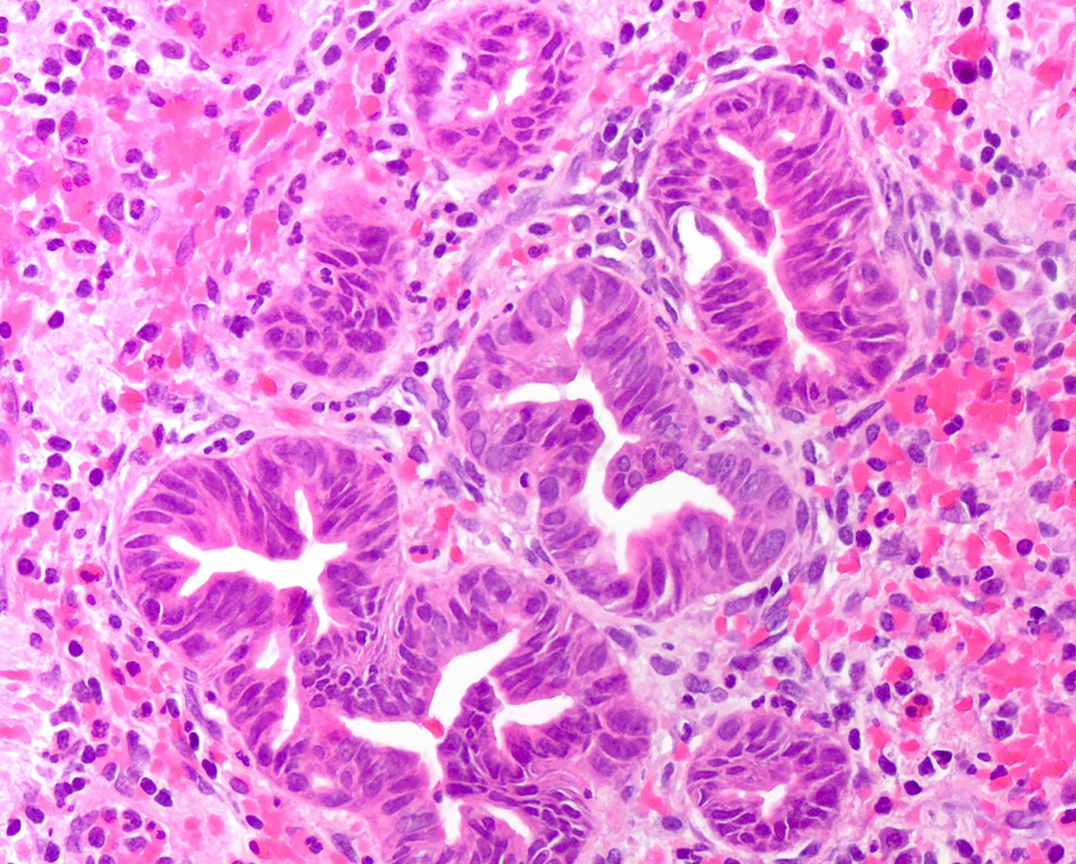
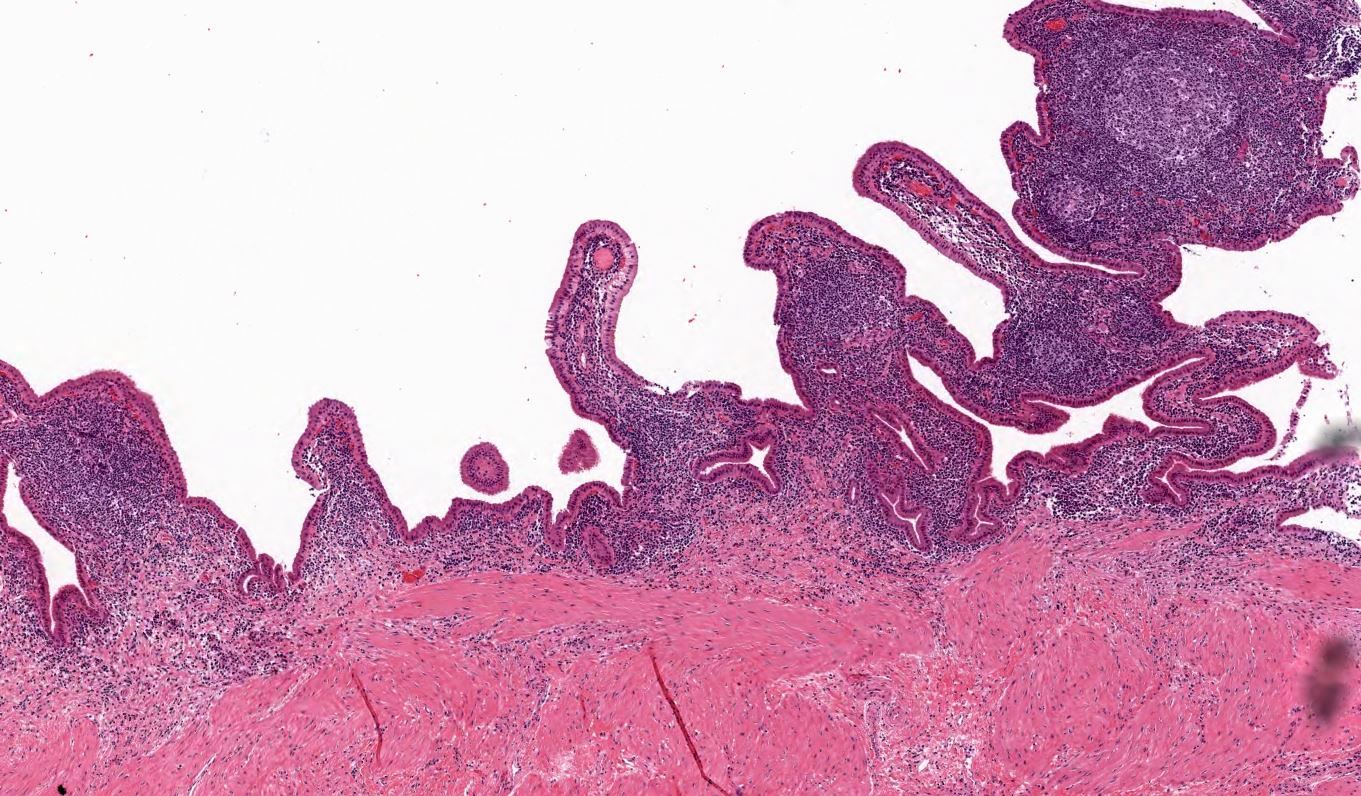
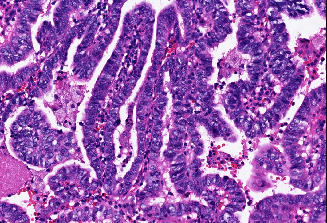
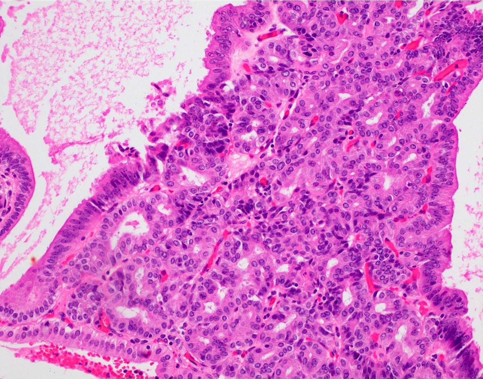
High grade BilIN
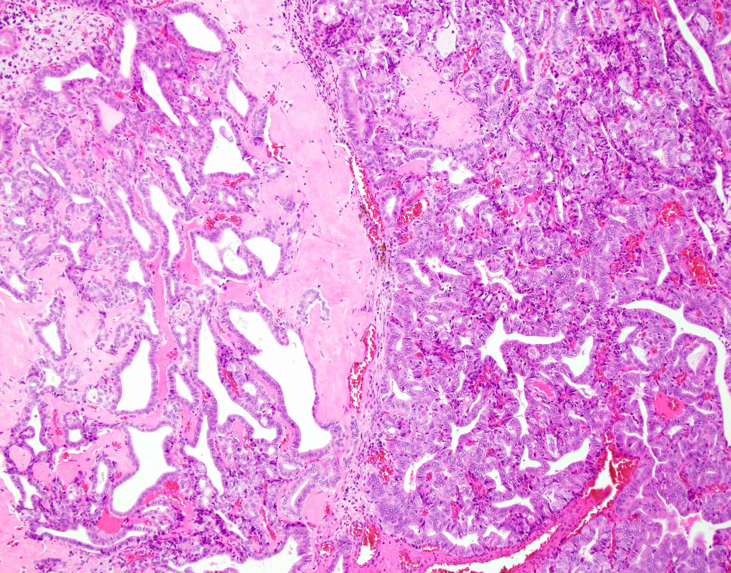
Contributed by Satyapal Chahar, M.D. and Monica T. Garcia-Buitrago, M.D.
Low grade dysplasia
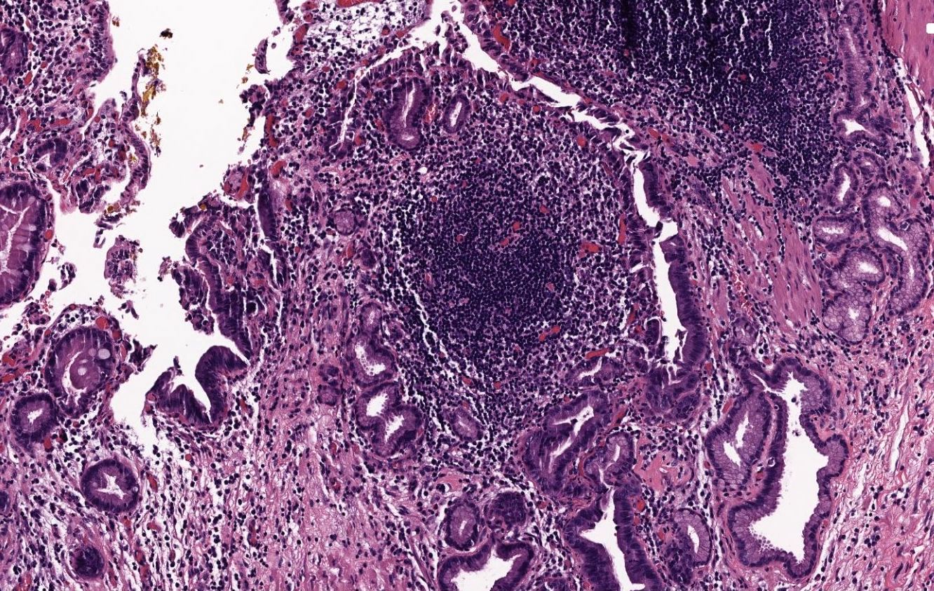
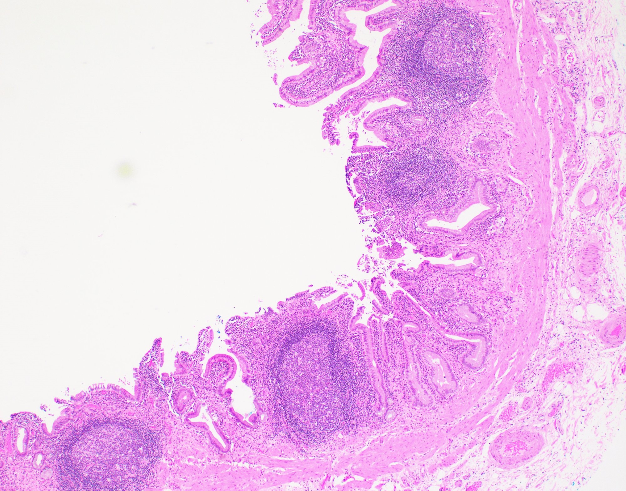
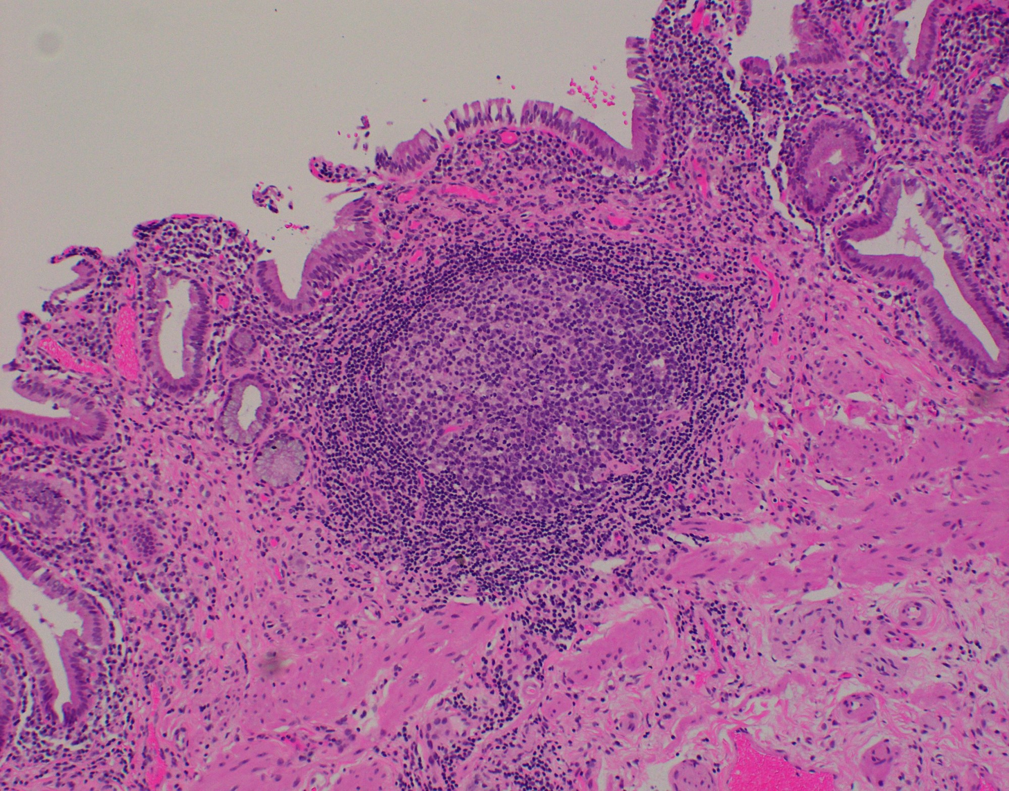
Acute inflammation
Rokitansky-Aschoff sinus involvement
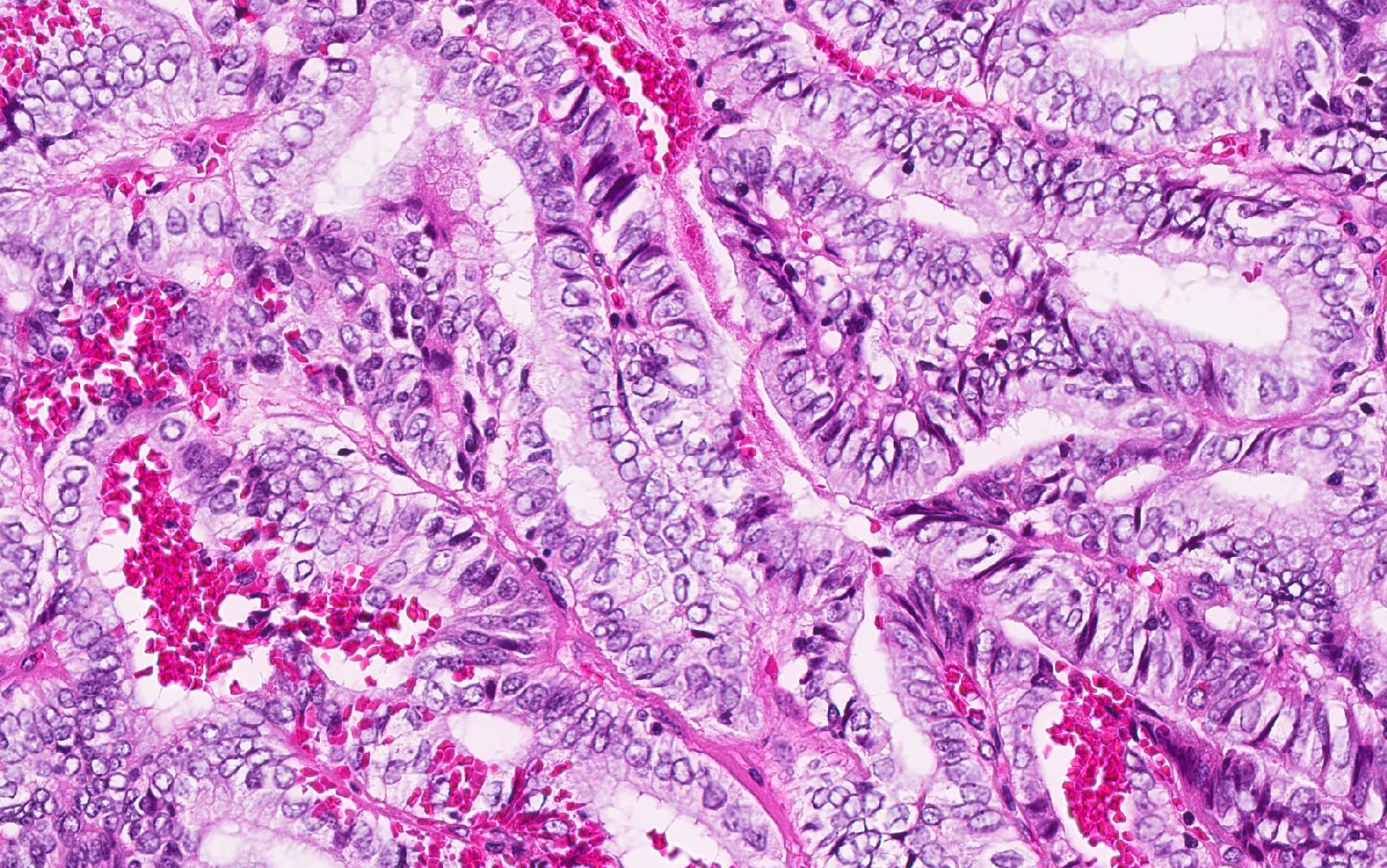
High grade dysplasia
High grade dysplasia
High grade dysplasia
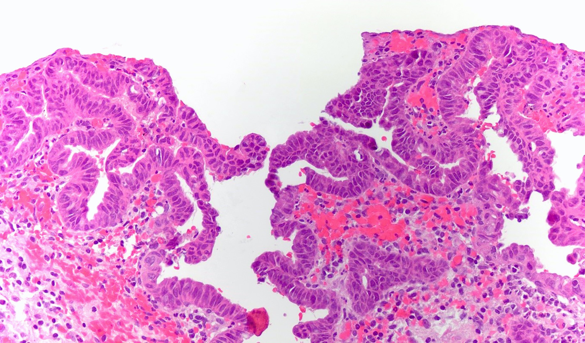
High grade dysplasia
Images hosted on other servers:

Bismuth-Corlette classification
Images hosted on other servers:

Imaging of Klatskin tumor

Imaging of
perihilar
cholangiocarcinoma


ERCP, mass forming extrahepatic cholangiocarcinoma

Ultrasound,
mass forming
extrahepatic
cholangiocarcinoma
Contributed by Raul S. Gonzalez, M.D.

Perihilar
cholangiocarcinoma
Intrapancreatic cholangiocarcinoma

Cystic duct
cholangiocarcinoma
Contributed by Raul S. Gonzalez, M.D. and Andrey Bychkov, M.D., Ph.D.
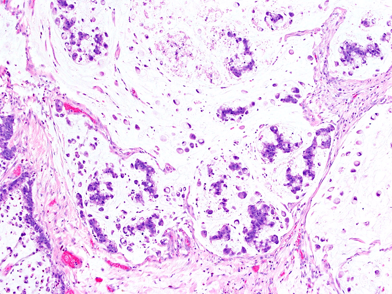
Arising from IPNB
Infiltrative glands

Periampullary origin

Small dispersed glands

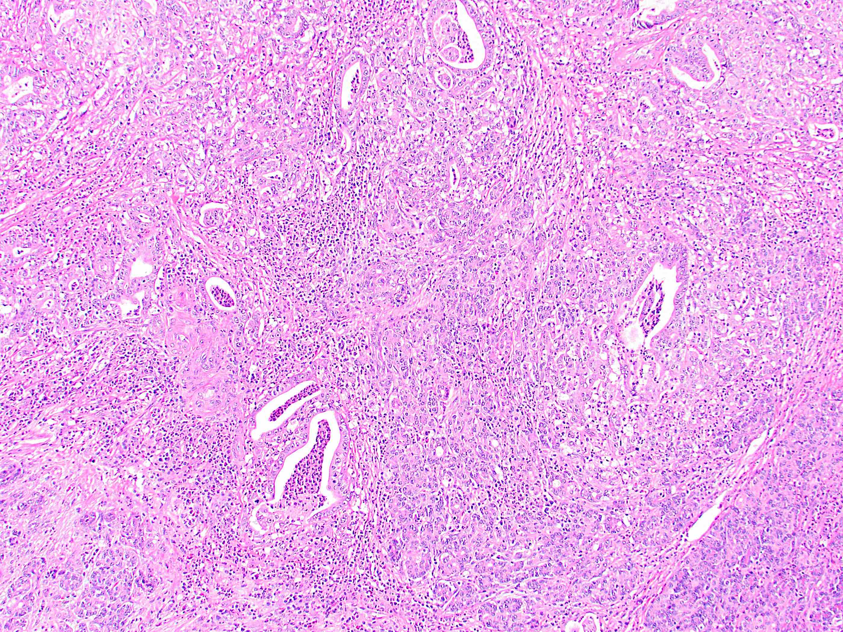
Adenocarcinoma


Common bile duct
Contributed by Jian-Hua Qiao, M.D.
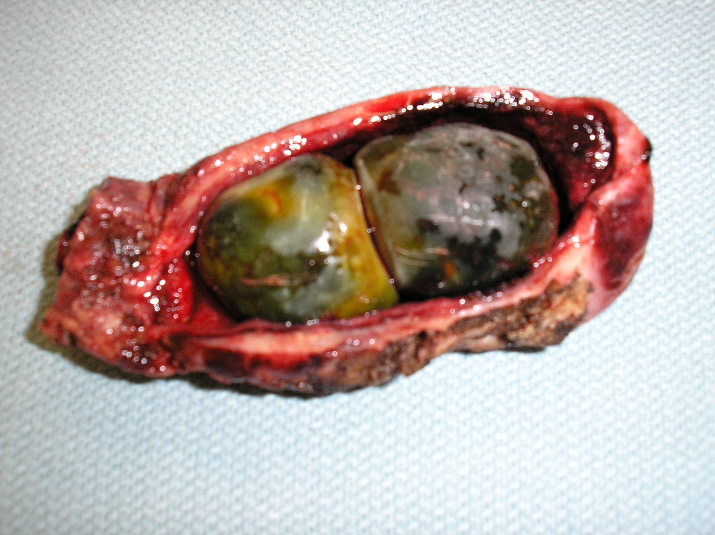
2 pigment gallstones
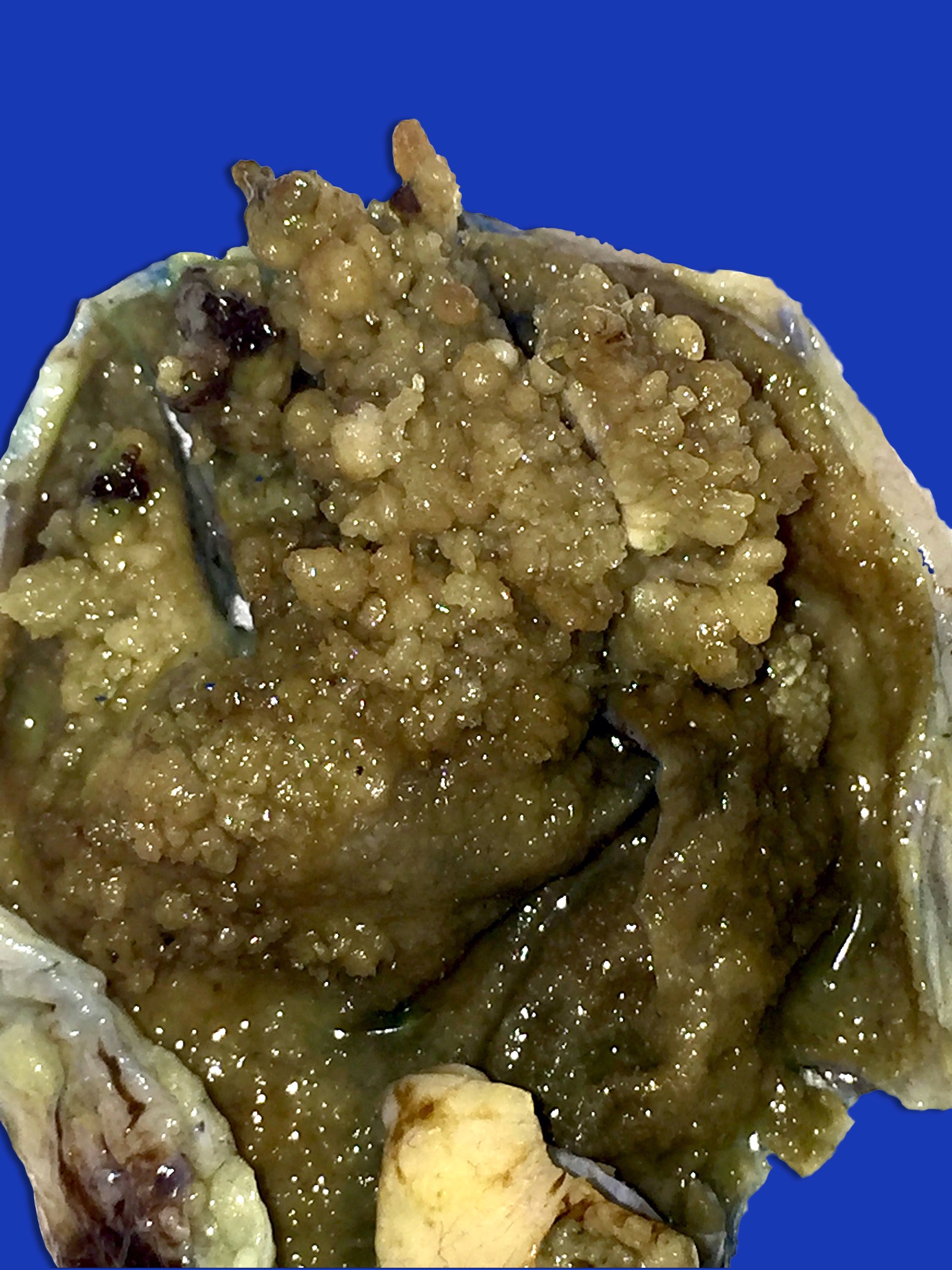
Images hosted on other servers:

Hyperechoic homogeneous pedunculated polyps
Images hosted on other servers:

Multiple yellow polyps
Contributed by Raul S. Gonzalez, M.D., Andrey Bychkov, M.D., Ph.D. and Jijgee Munkhdelger, M.D., Ph.D.
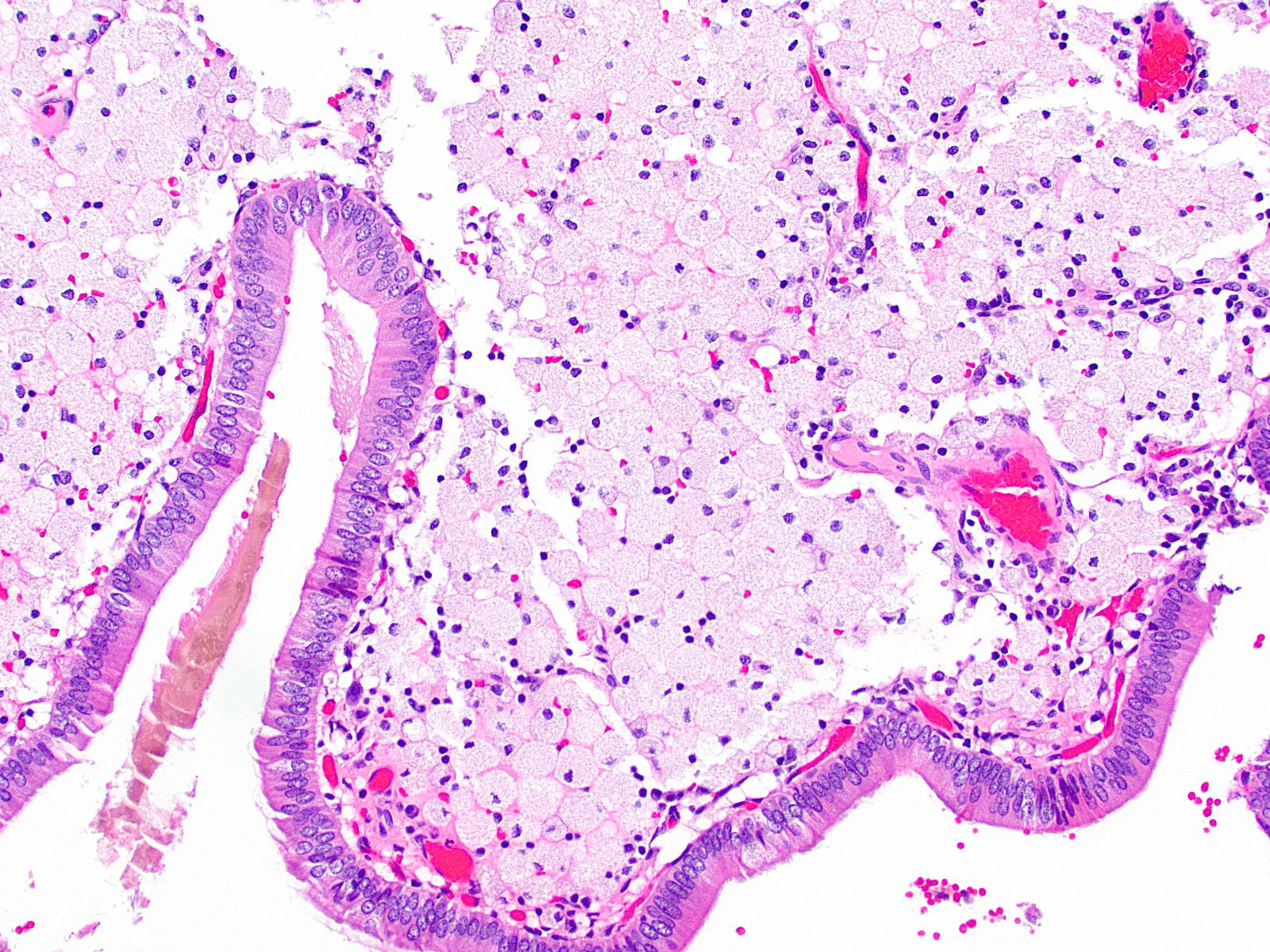
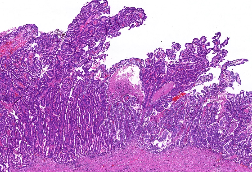
Lipid laden macrophages
Normal lining epithelium
Cauliflower-like architecture
Lipid laden macrophages
Foamy lipid laden macrophages
Polypoid lesion
Stromal macrophages
Lipid laden macrophages
Images hosted on other servers:

Reverberation (comet tail artifact)

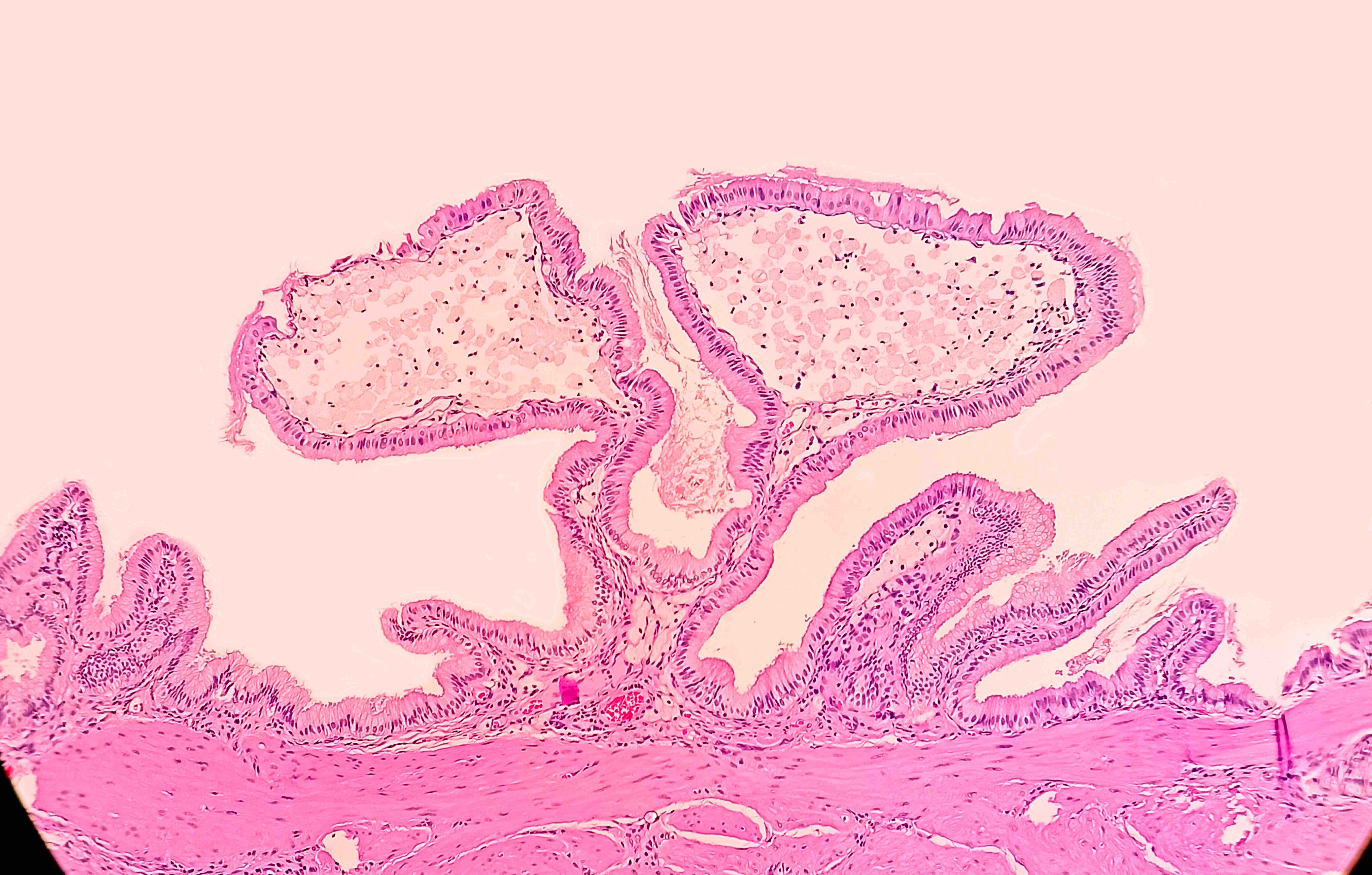
Cholesterolosis

Cholesterol polyp
Contributed by Faris Alshammas, M.D. and @Andrew_Fltv on Twitter
Numerous mucosal yellow specks
Cholesterolosis
Contributed by Reem Hamasha, M.D., Faris Alshammas, M.D., Andrey Bychkov, M.D., Ph.D. and Jijgee Munkhdelger, M.D., Ph.D.
Hypertrophy of gallbladder villi
Hypertrophy of a gallbladder villus
Foamy lipid laden macrophages

Foamy macrophages in lamina propria

3 small foci of foamy macrophages
Mucosal projection
Contributed by Kelsey E. McHugh, M.D.
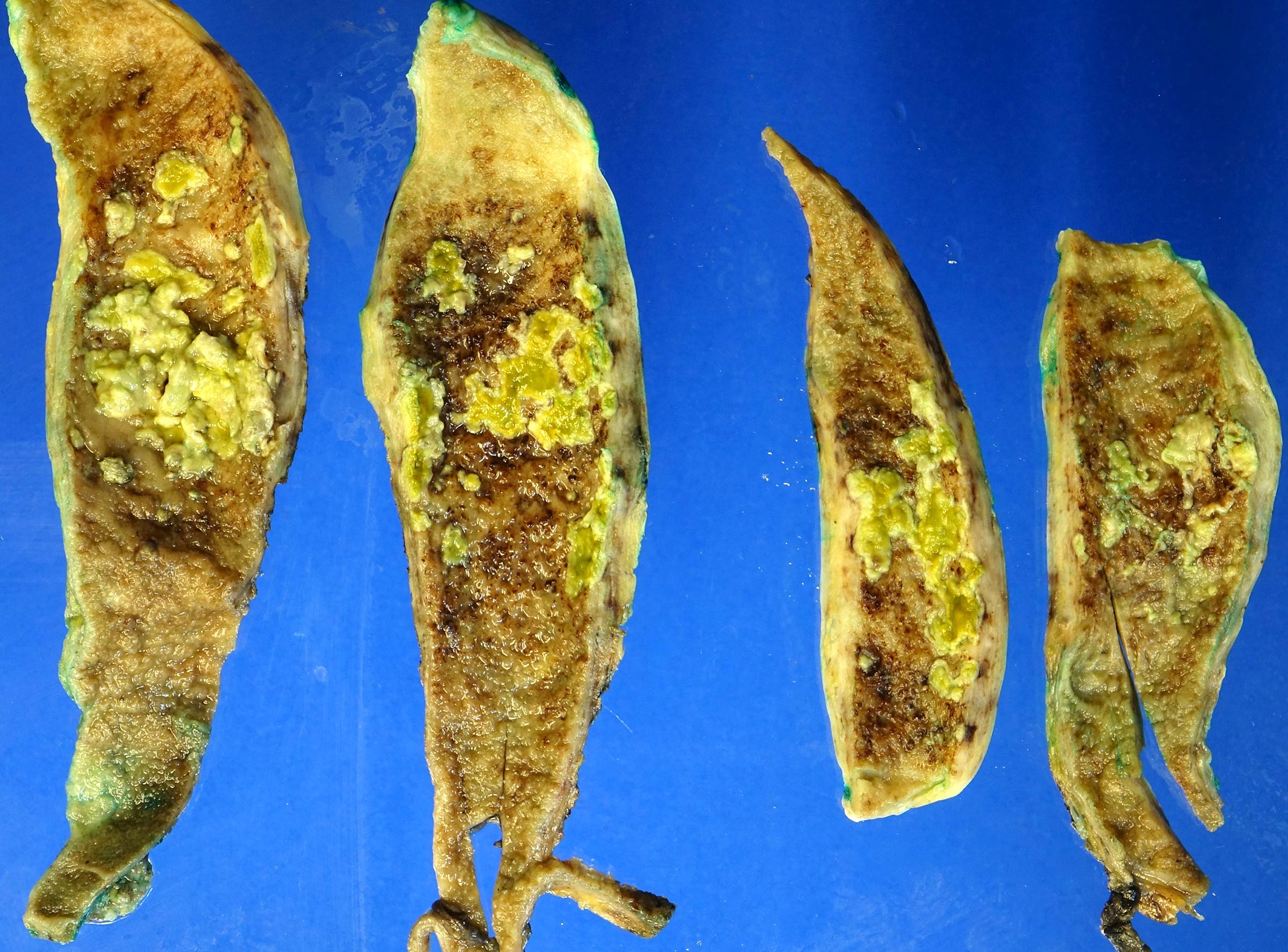
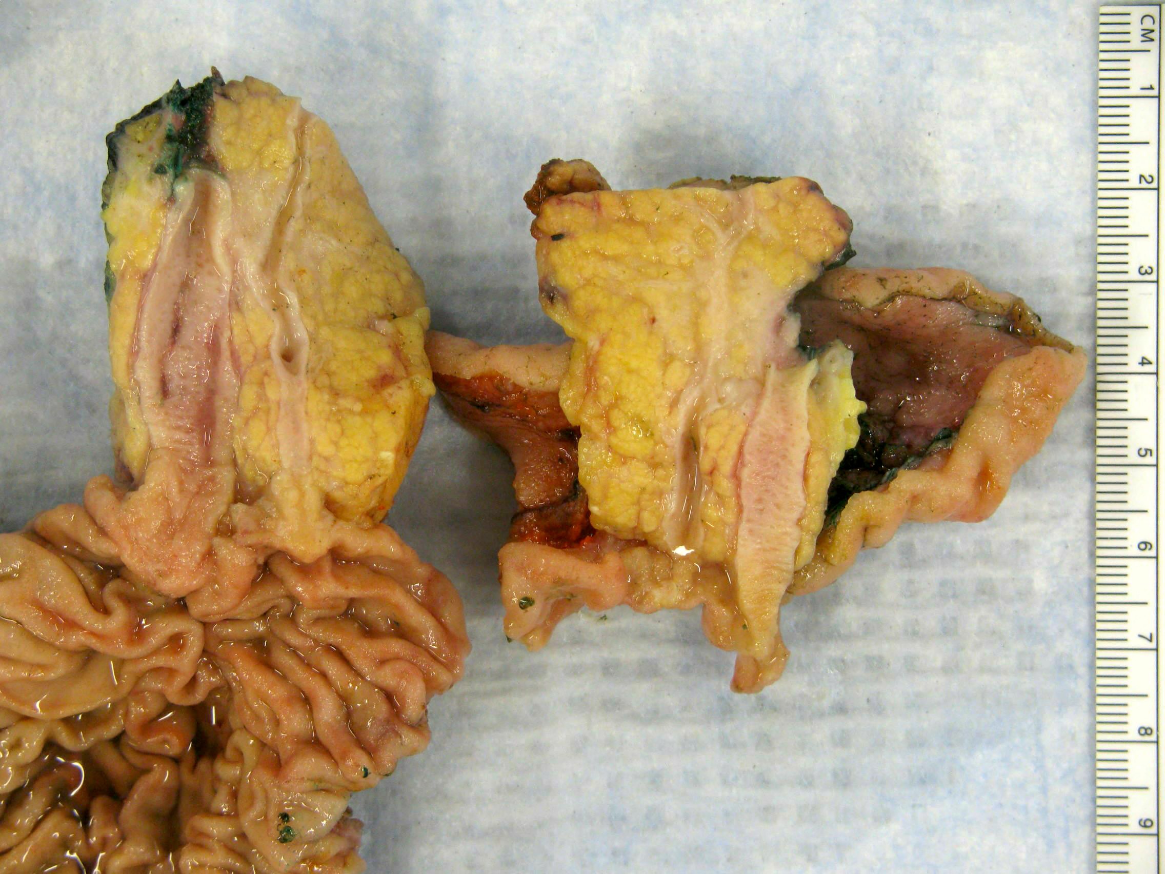
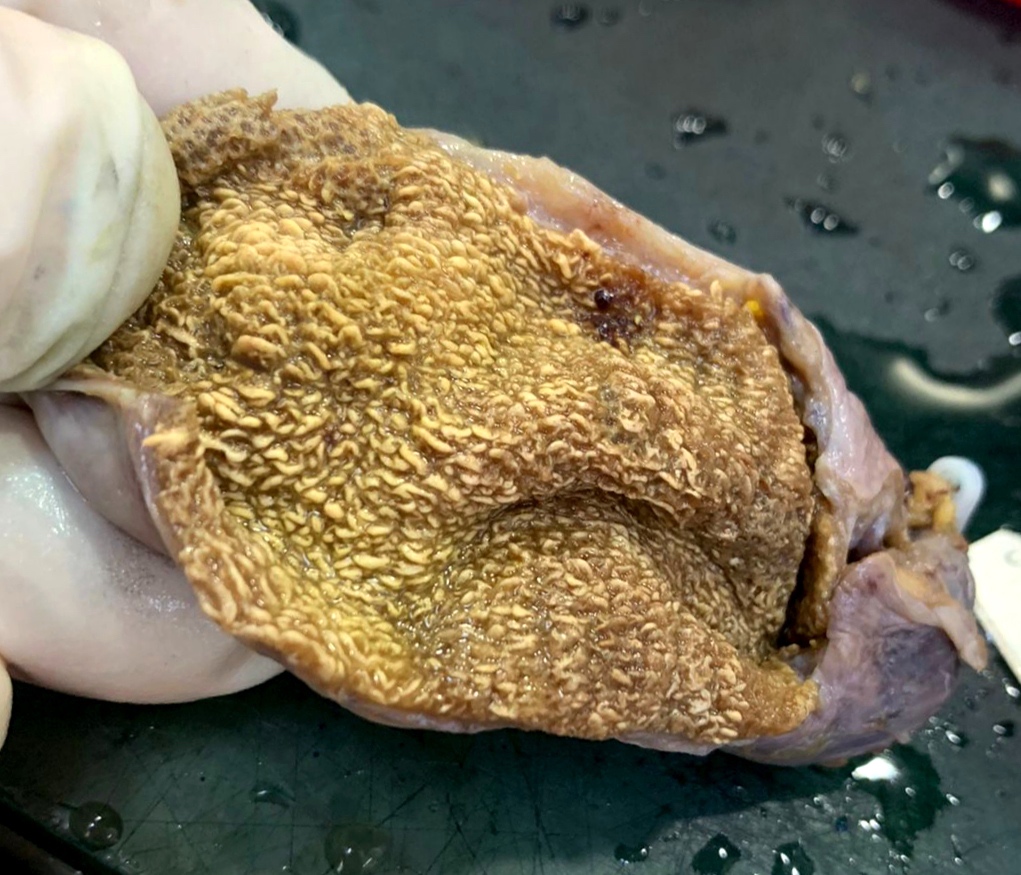
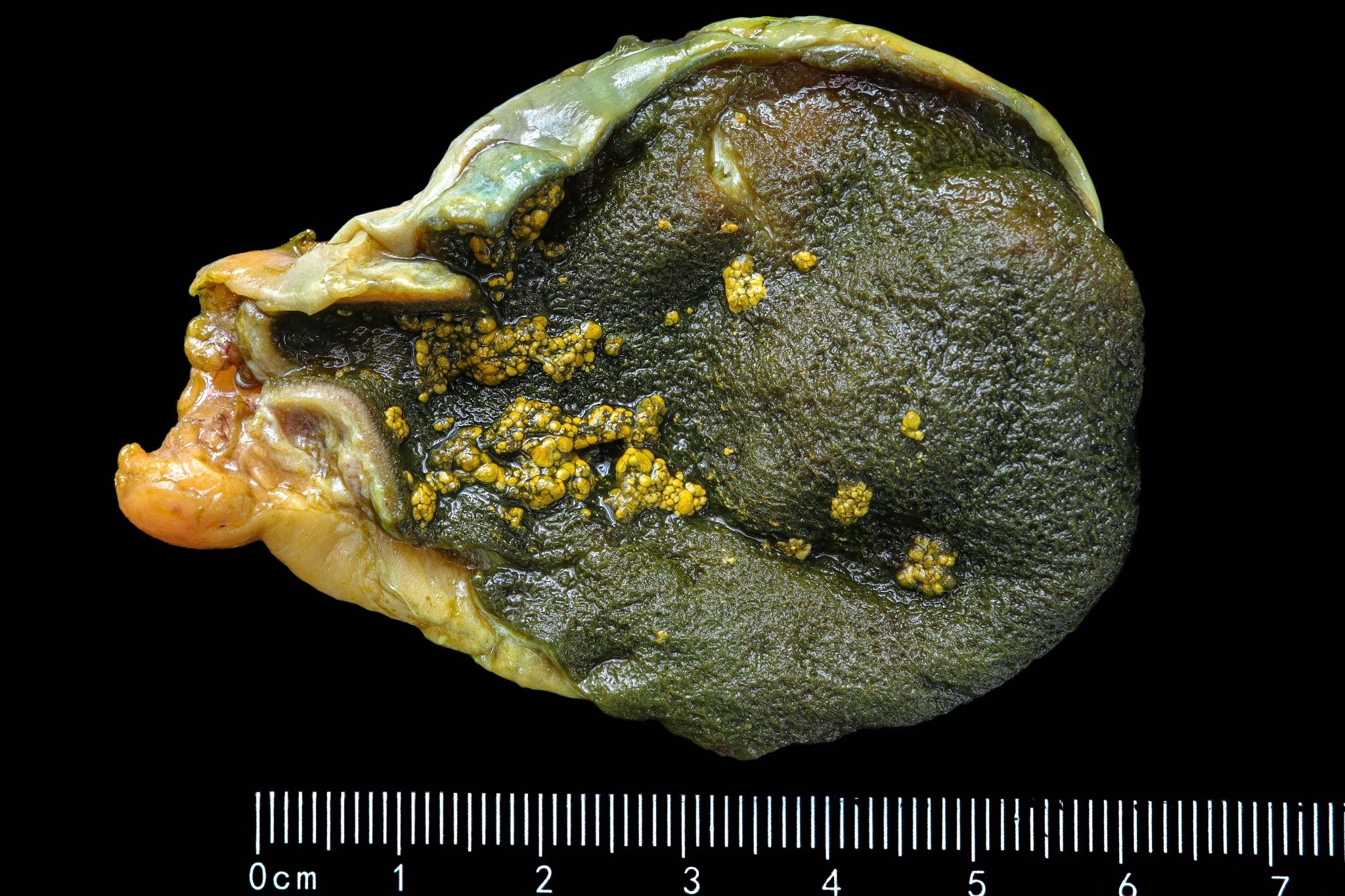
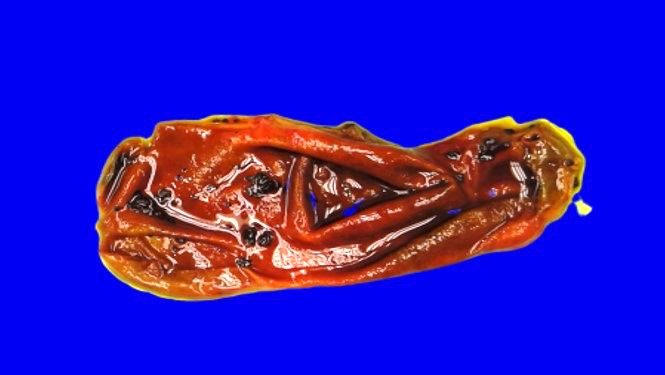
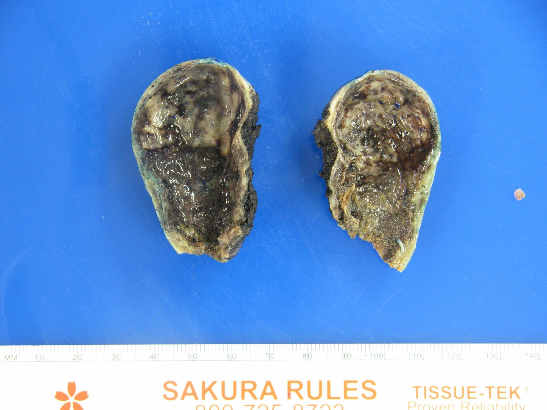
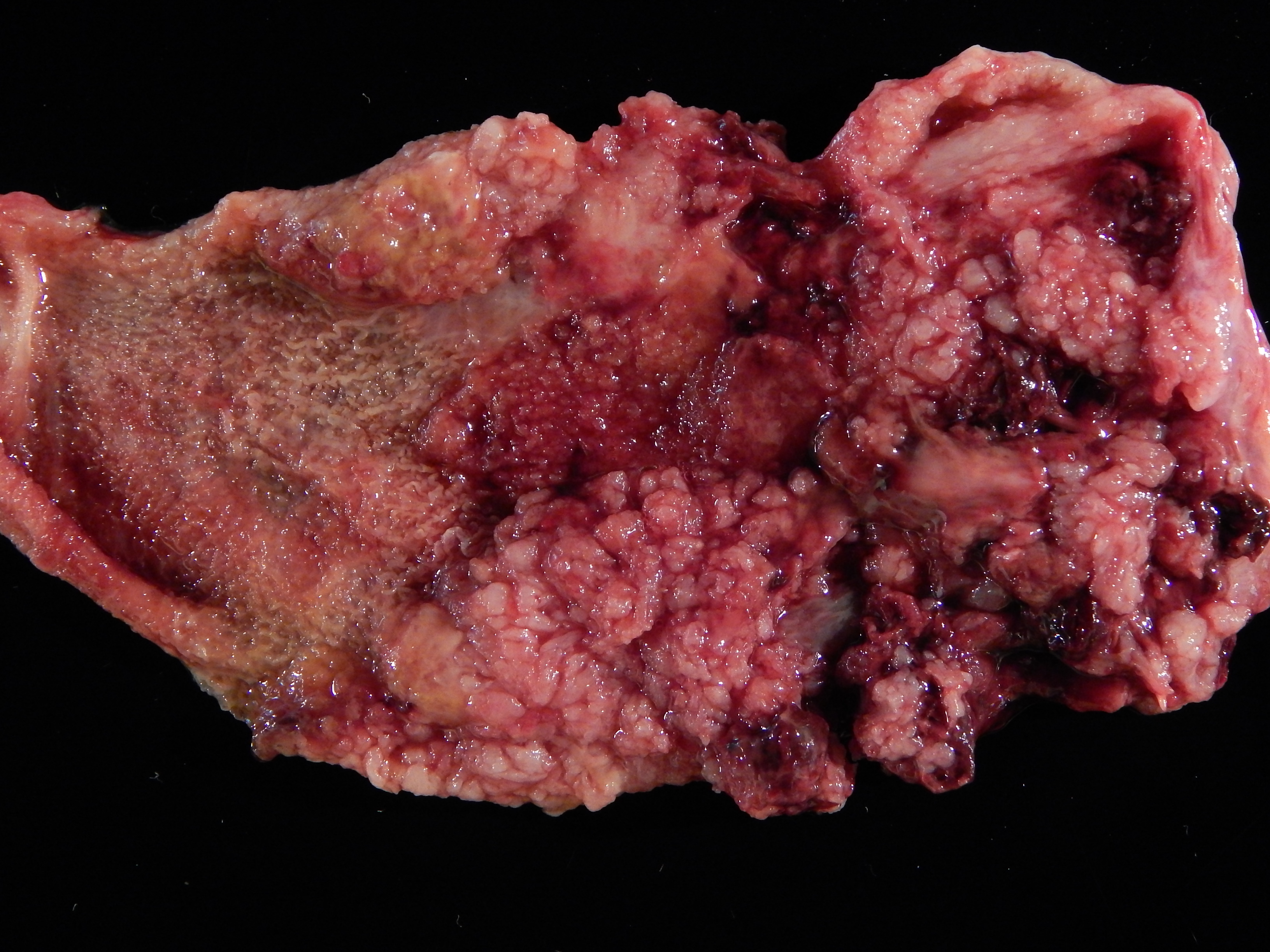
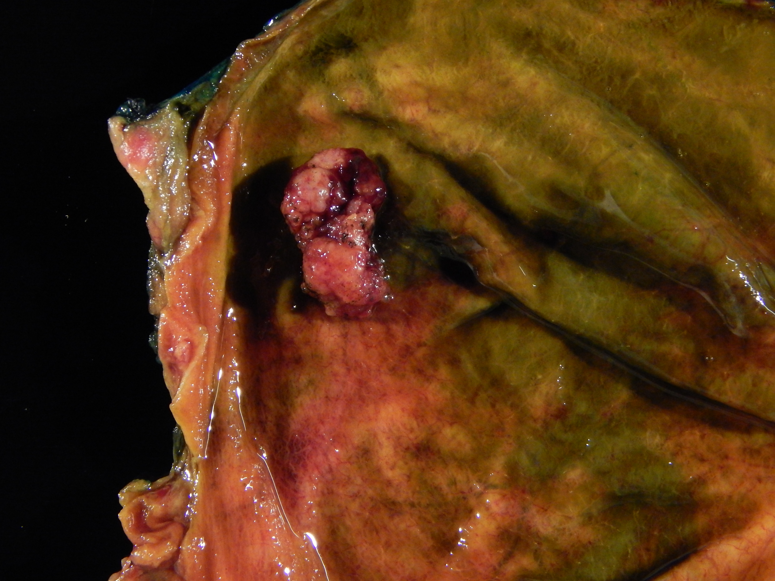
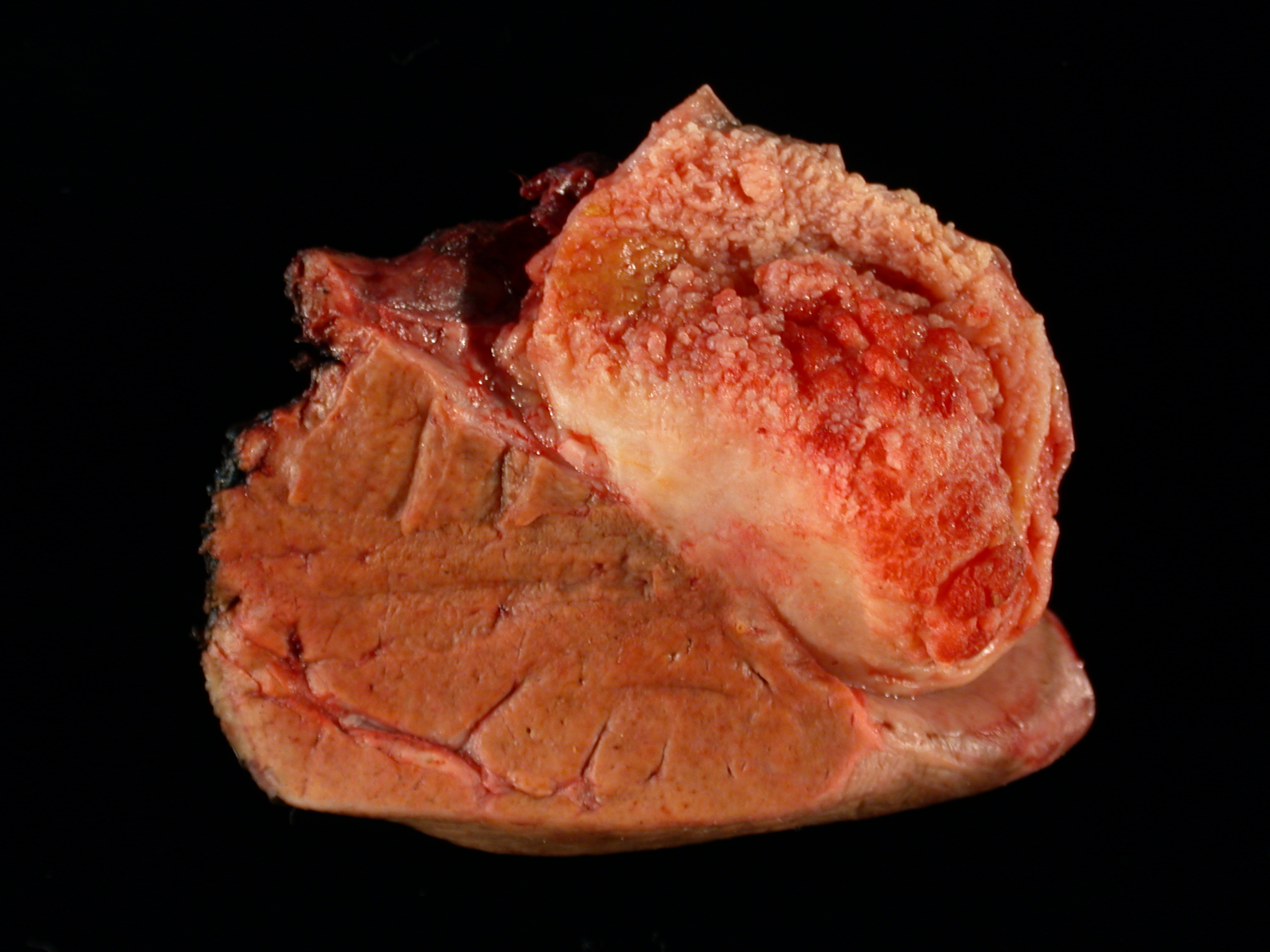
Distended gallbladder with stones

Contracted gallbladder with stones
Thickened gallbladder wall with stones
Contributed by Kelsey E. McHugh, M.D.
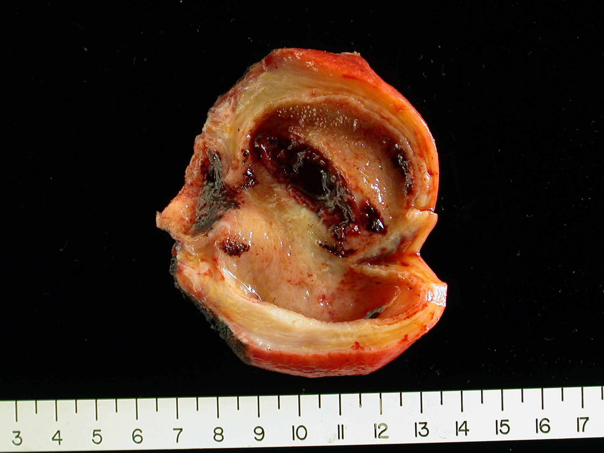
Contracted gallbladder with stones
Distended gallbladder with stones
Contributed by Kelsey E. McHugh, M.D.
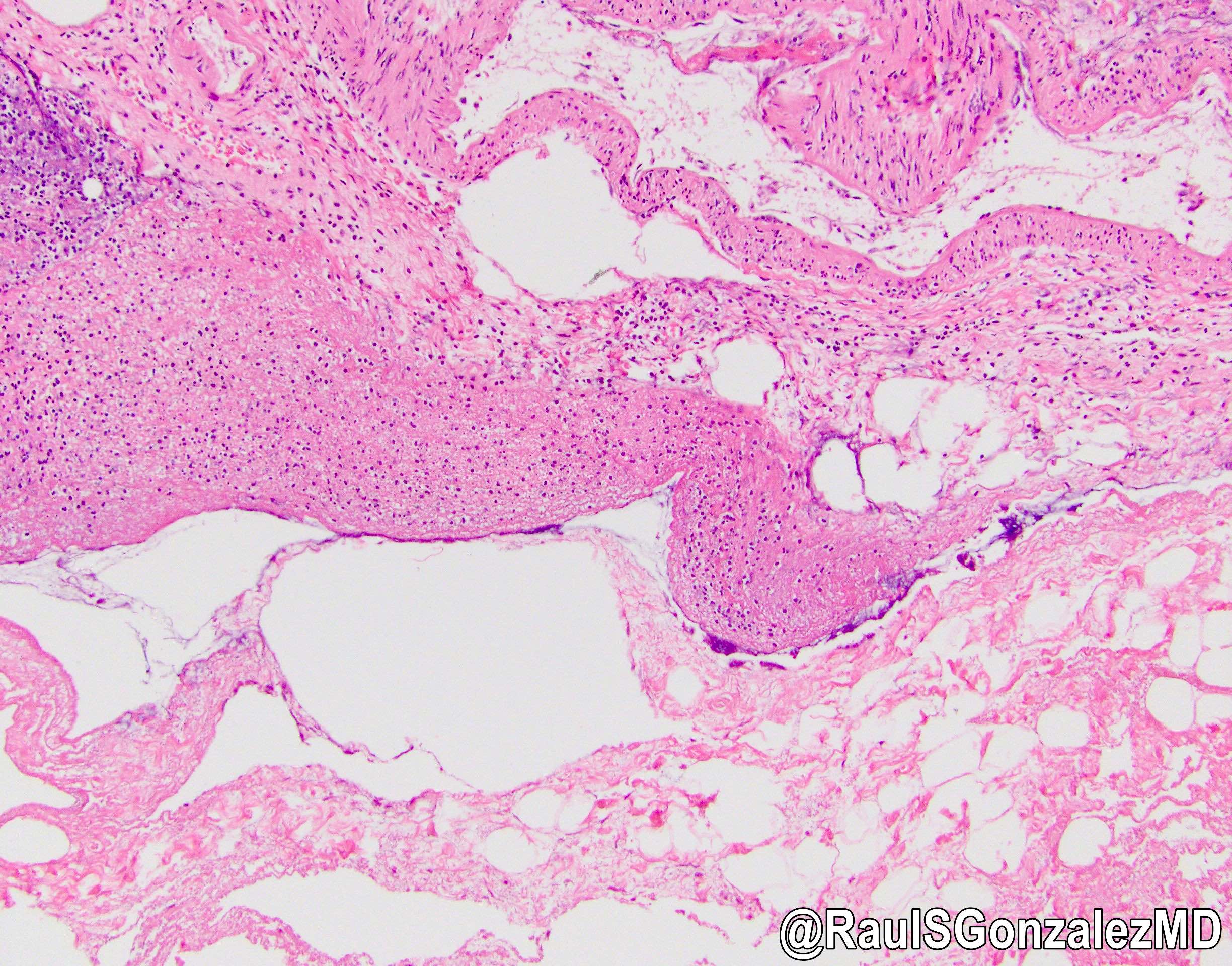
RA sinus
RA sinuses with inspissated bile
Mucosal atrophy
Focal intestinal metaplasia
Intestinal metaplasia
Pyloric gland metaplasia
Hyalinizing cholecystitis
Images hosted on other servers:

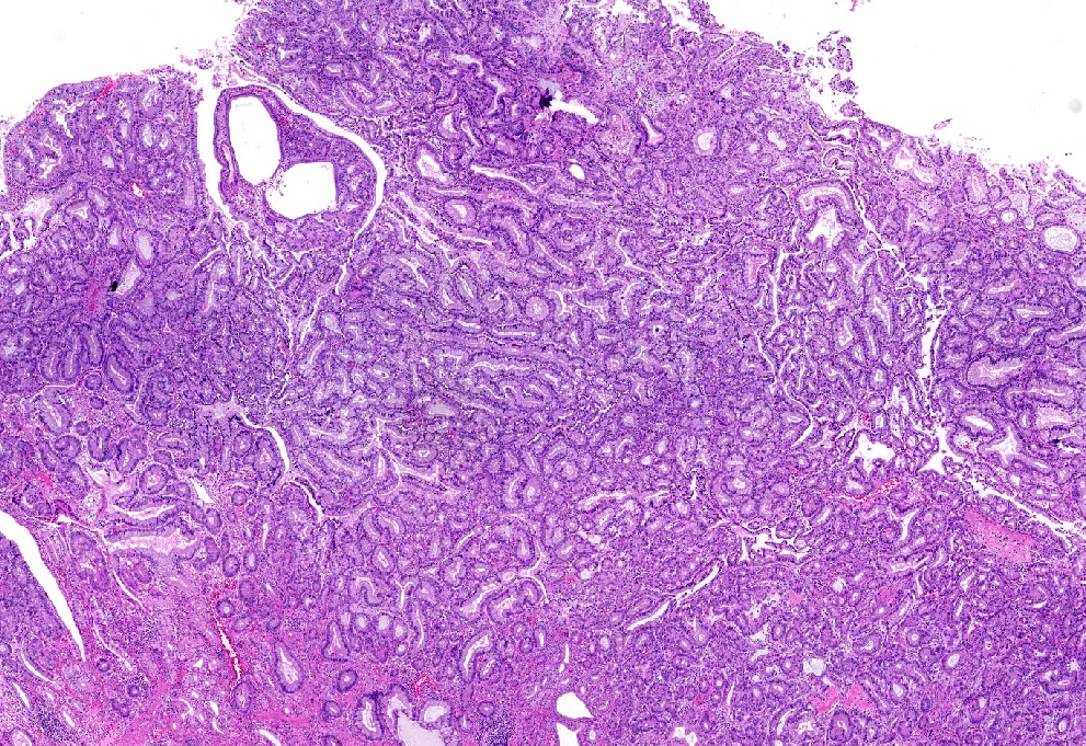
Low grade dysplasia distribution in gallbladder
Contributed by Raul S. Gonzalez, M.D. and Monica T. Garcia-Buitrago, M.D.
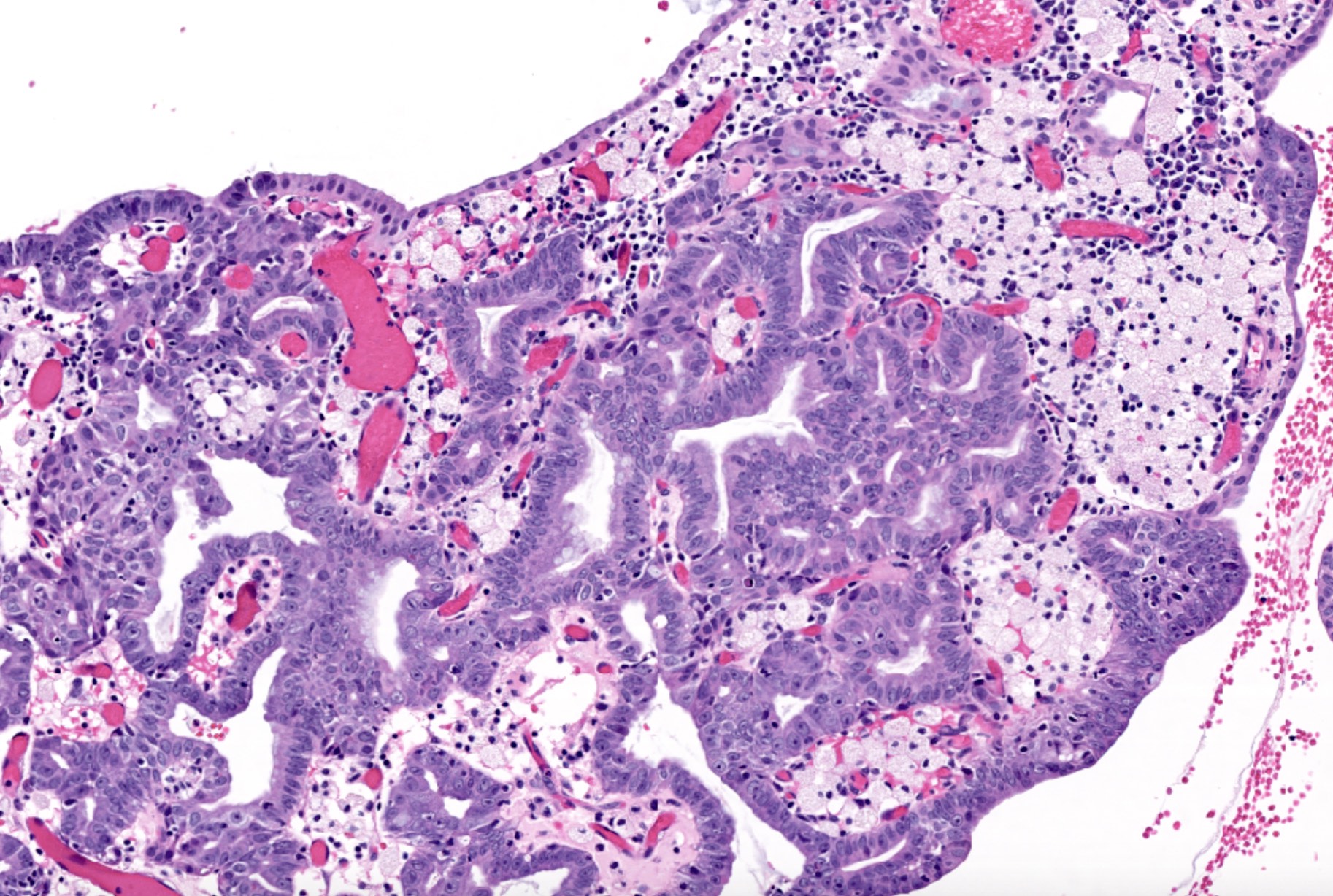
Cancer and dysplasia
High grade BilIN
Contributed by Raul S. Gonzalez, M.D.
Incidental finding

Prominent atypia

Associated with malignancy
Gallbladder and bile duct pathology by Dr. Adsay
Contributed by @RaulSGonzalezMD on Twitter
 Contributed by @RaulSGonzalezMD on Twitter (see original post here)">
Emphysematous cholecystitis
Contributed by Kelsey E. McHugh, M.D.
Follicular cholecystitis
Images hosted on other servers:

Heterogeneous mass
Contributed by Ashwin S. Akki, M.D., Ph.D.
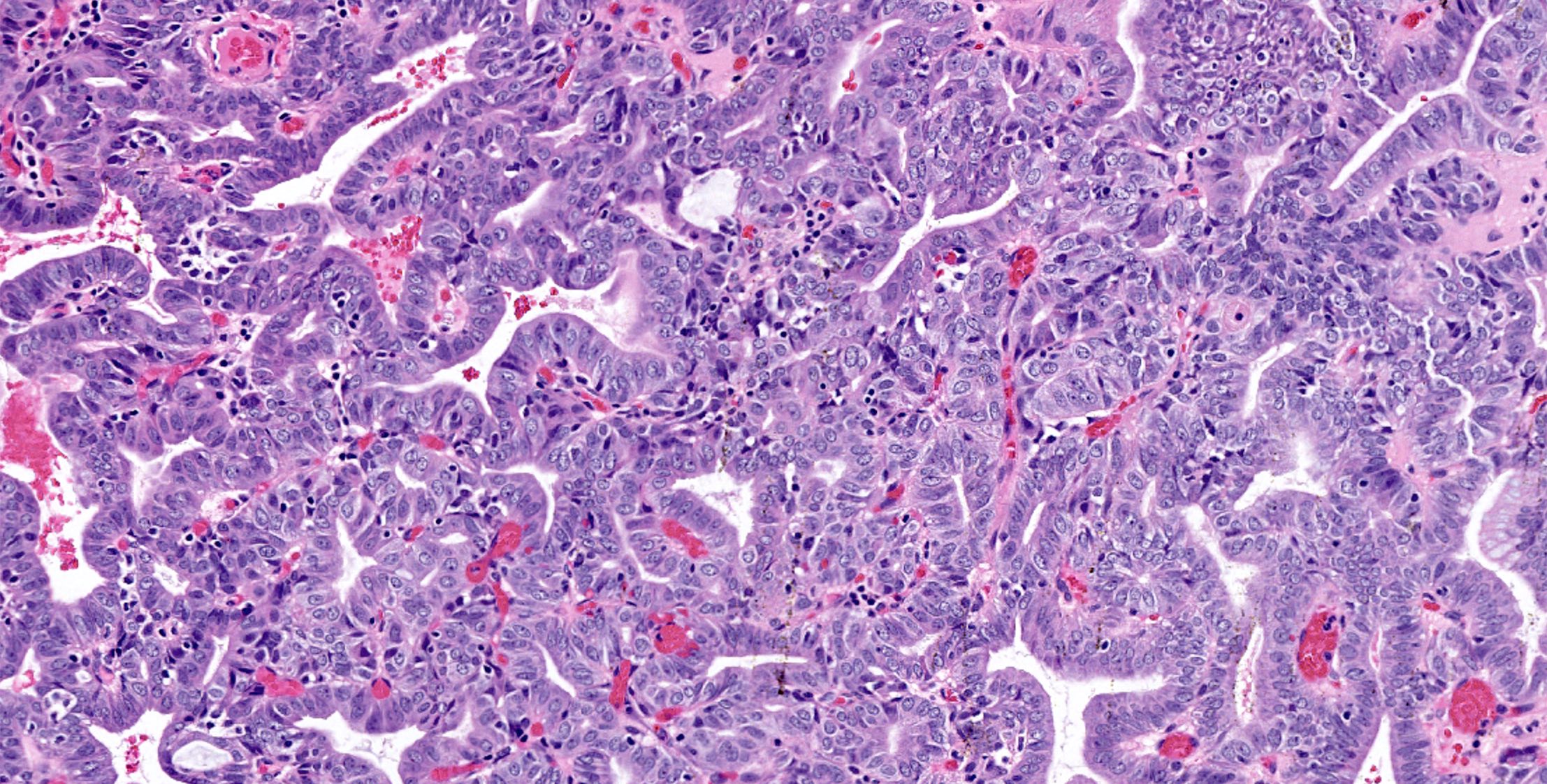
Papillary proliferation
Carcinoma in a polyp
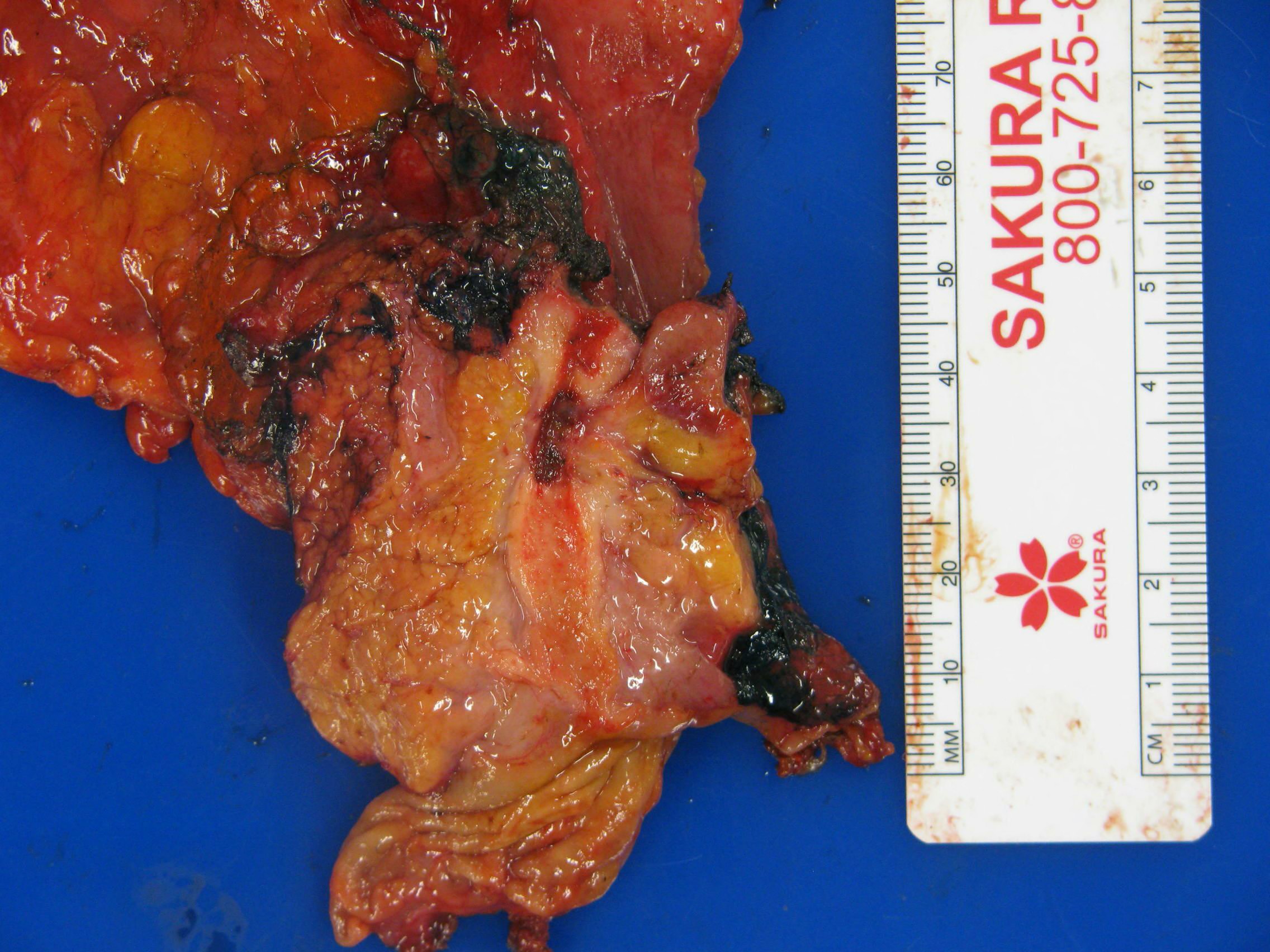
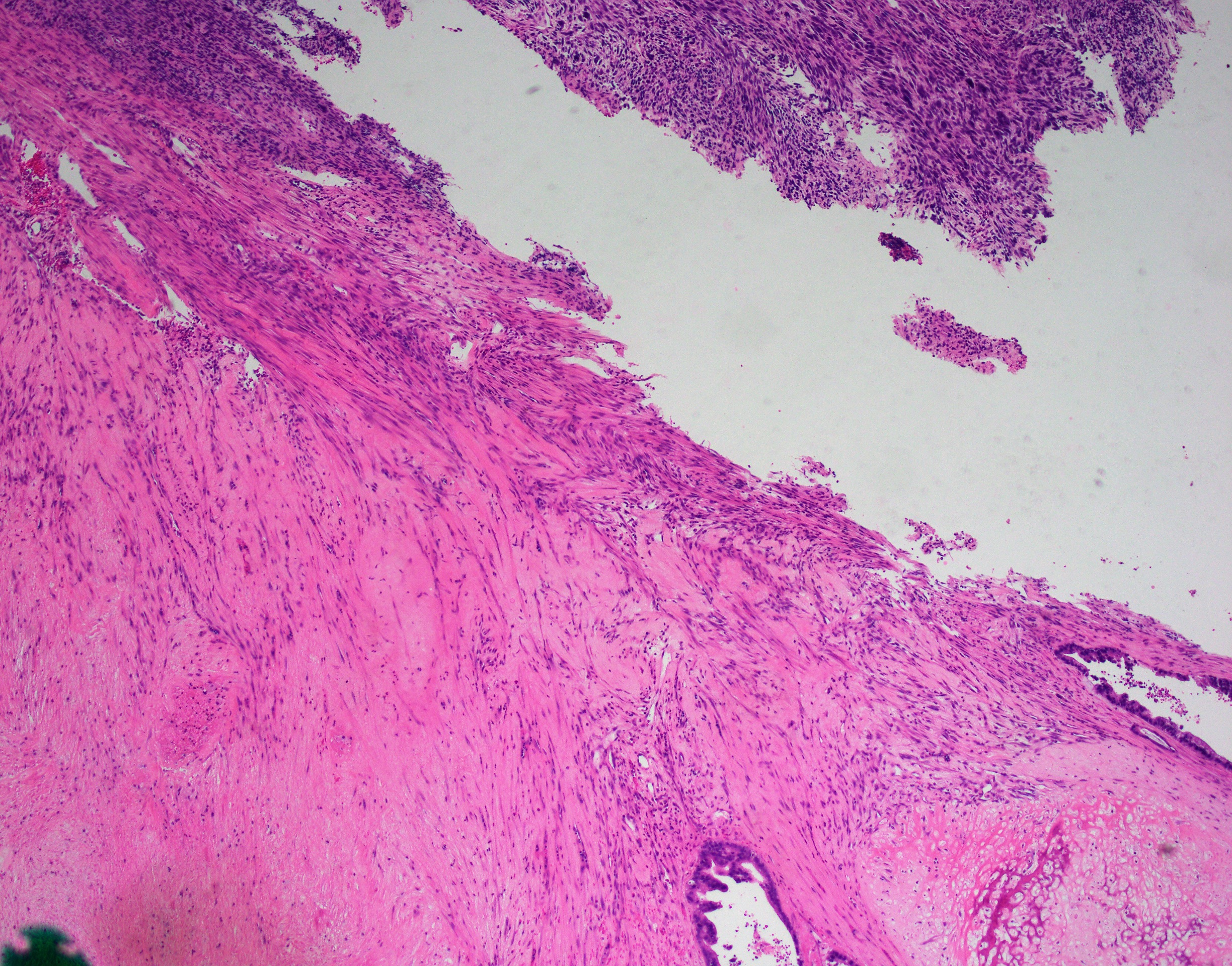
Gallbladder cancer invading liver
Diffusely thickened gallbladder wall
Contributed by Ashwin S. Akki, M.D., Ph.D.
Desmoplasia
Well formed glands

Intermediate gland formation
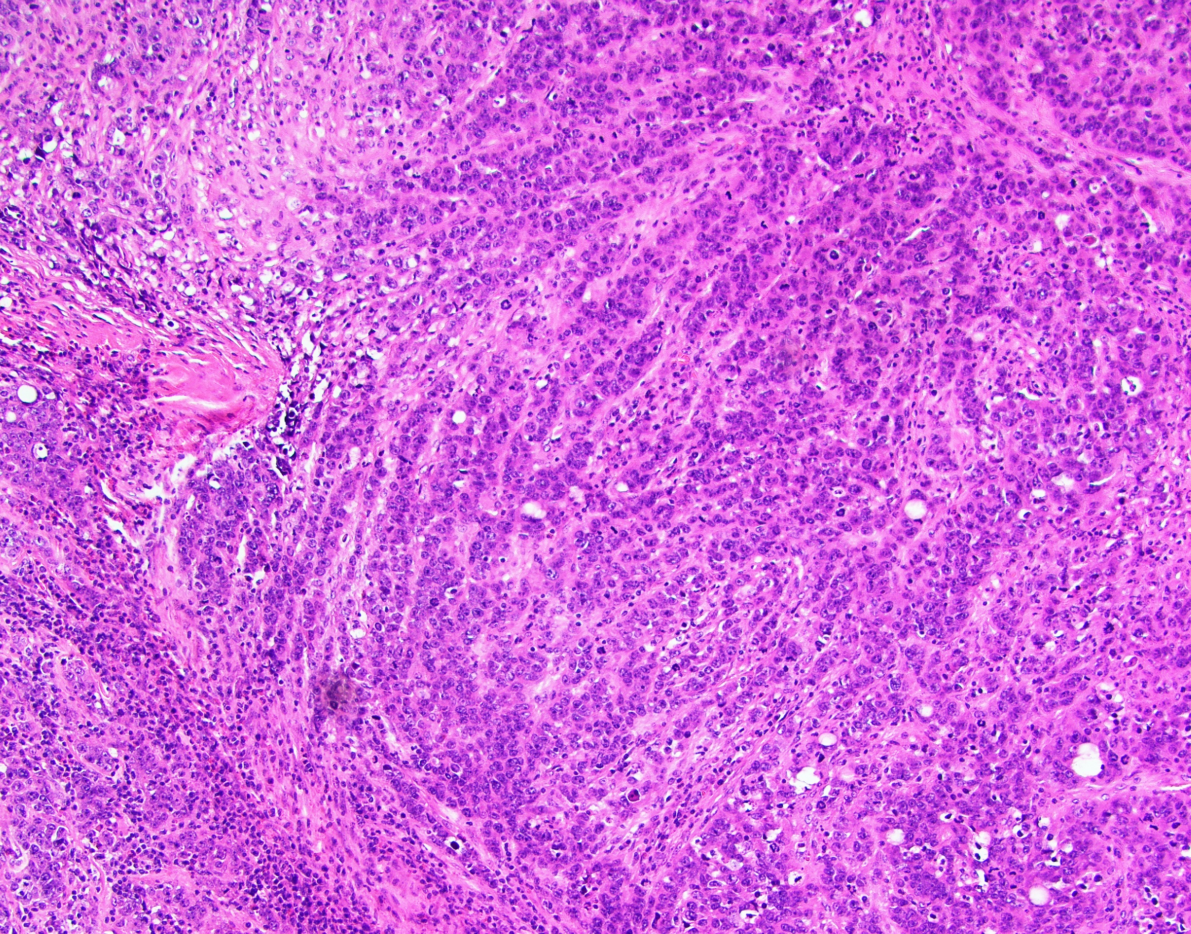
Poor gland formation
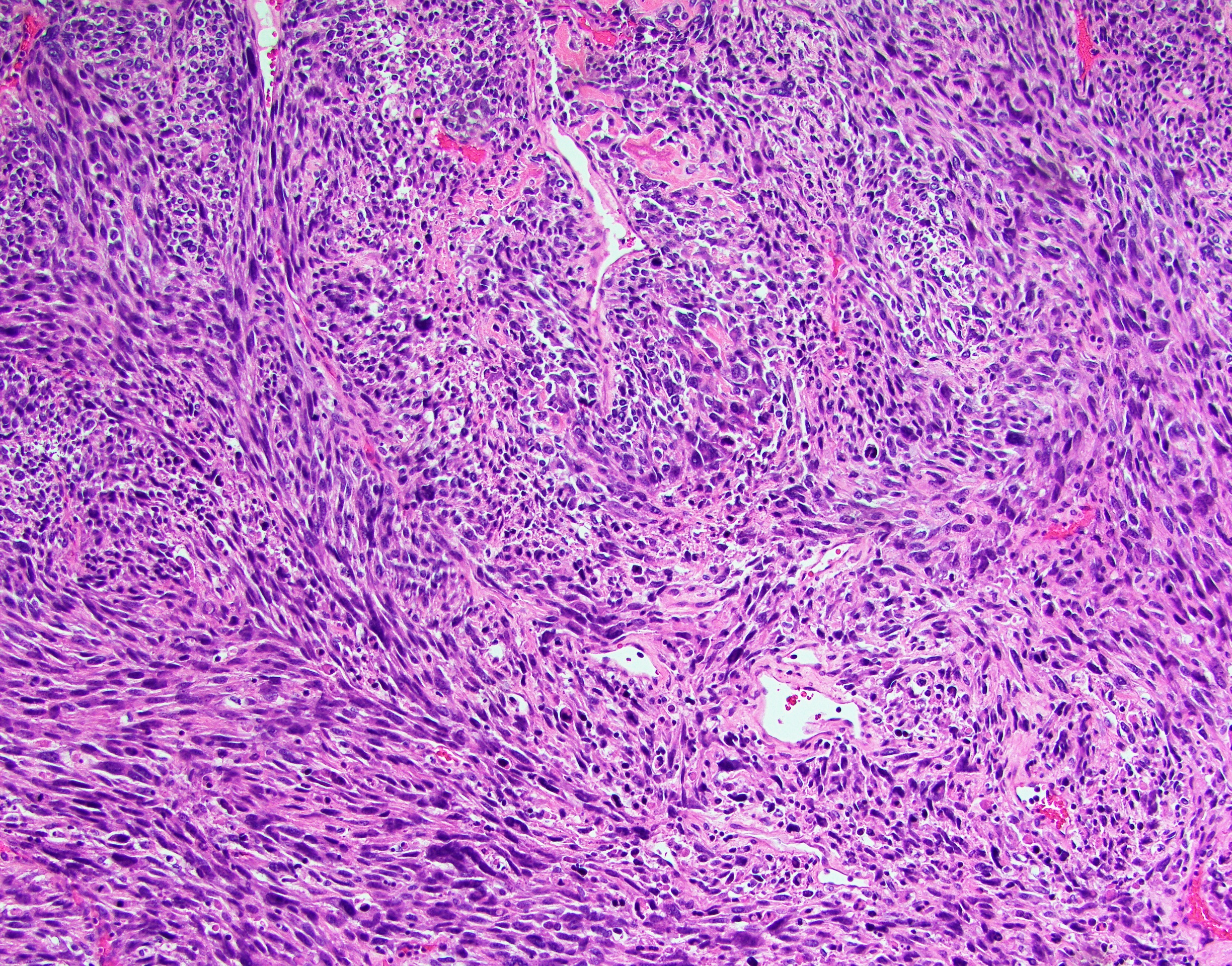
Sarcomatoid differentiation
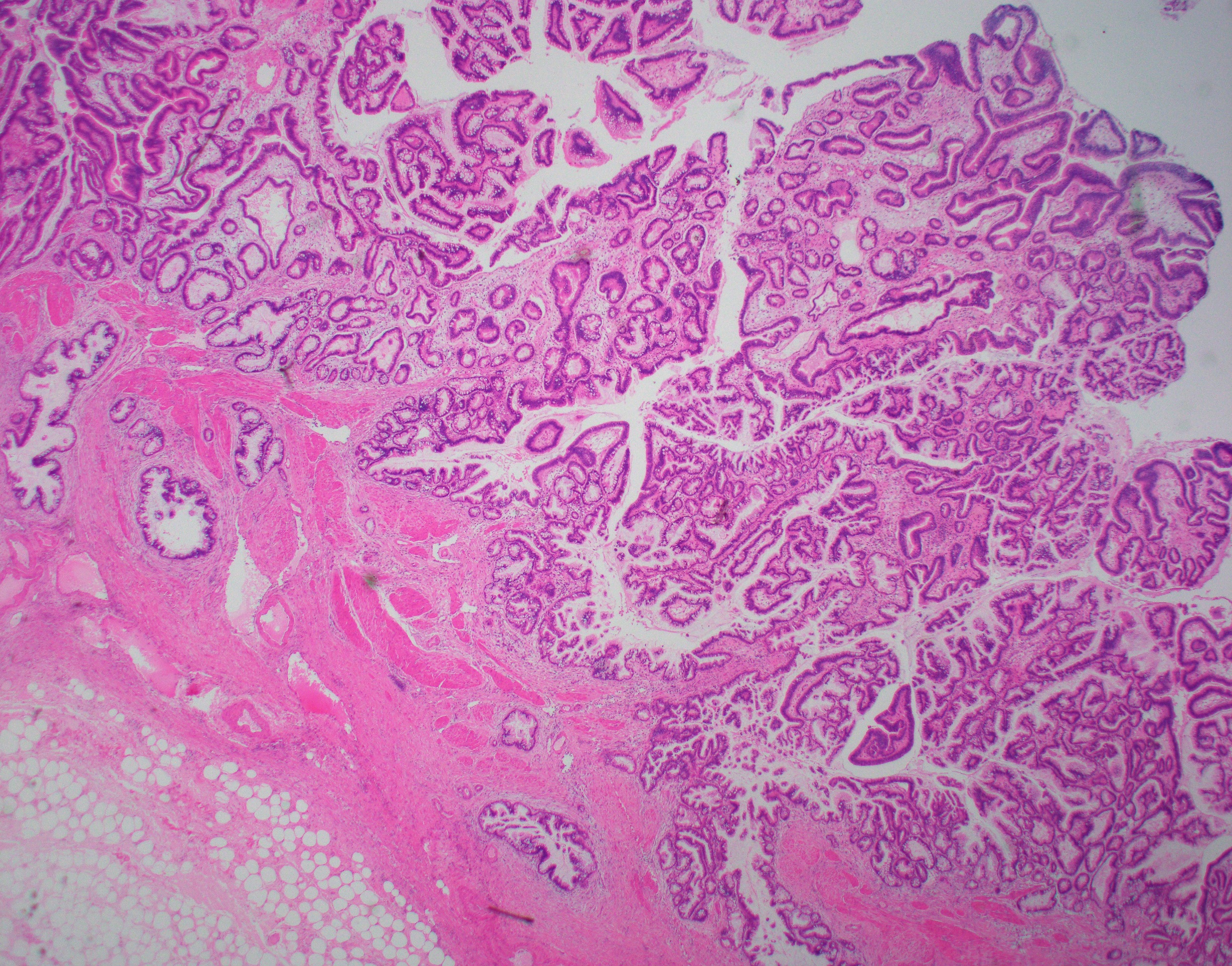
Papillary proliferation with invasion
Extended cholecystectomy
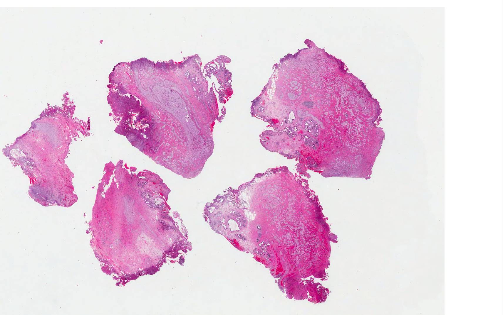
Images hosted on other servers:


Various images
Images hosted on other servers:

Tuberculosis
Images hosted on other servers:

Smooth surfaced polyp
Images hosted on other servers:

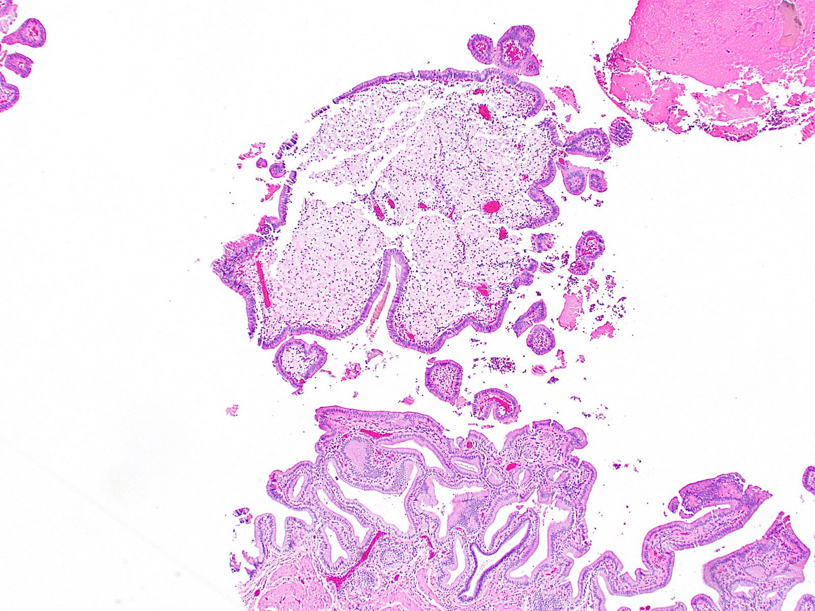
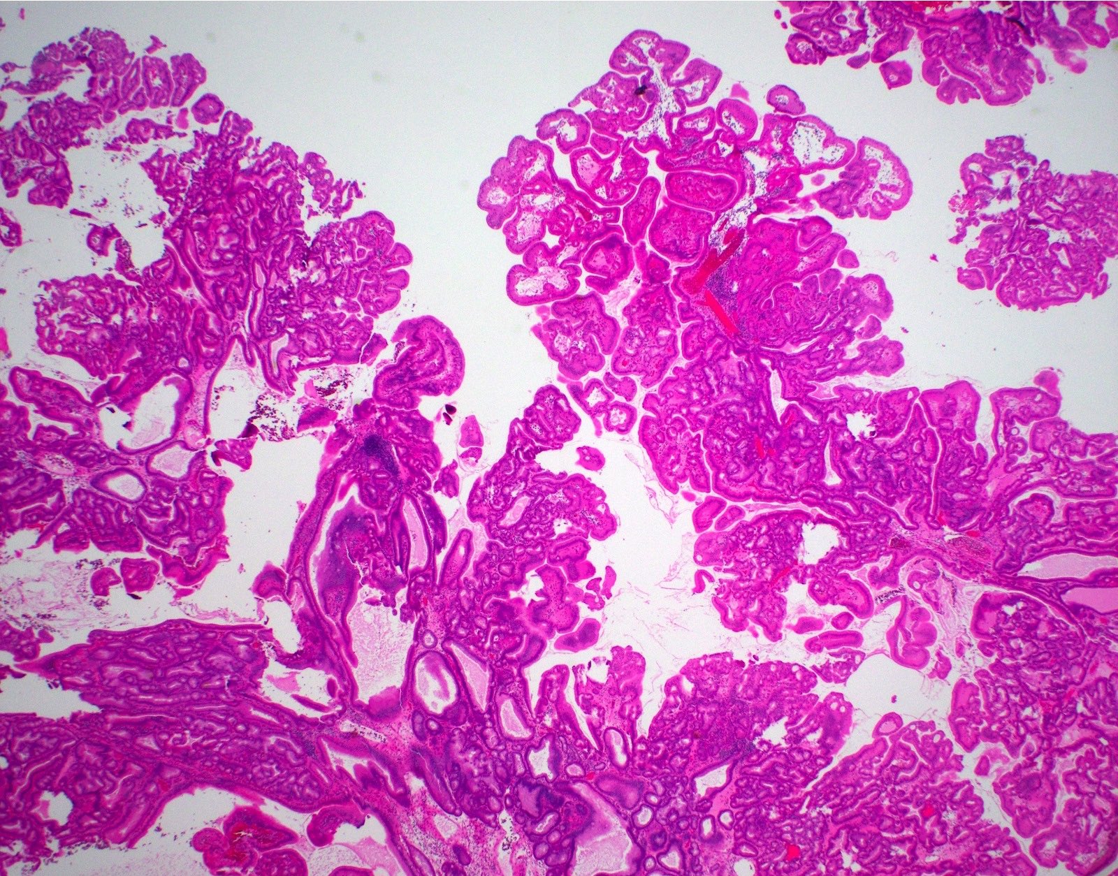
Nonneoplastic polyp with edematous stroma
Contributed by Gokce Askan, M.D. and Olca Basturk, M.D.
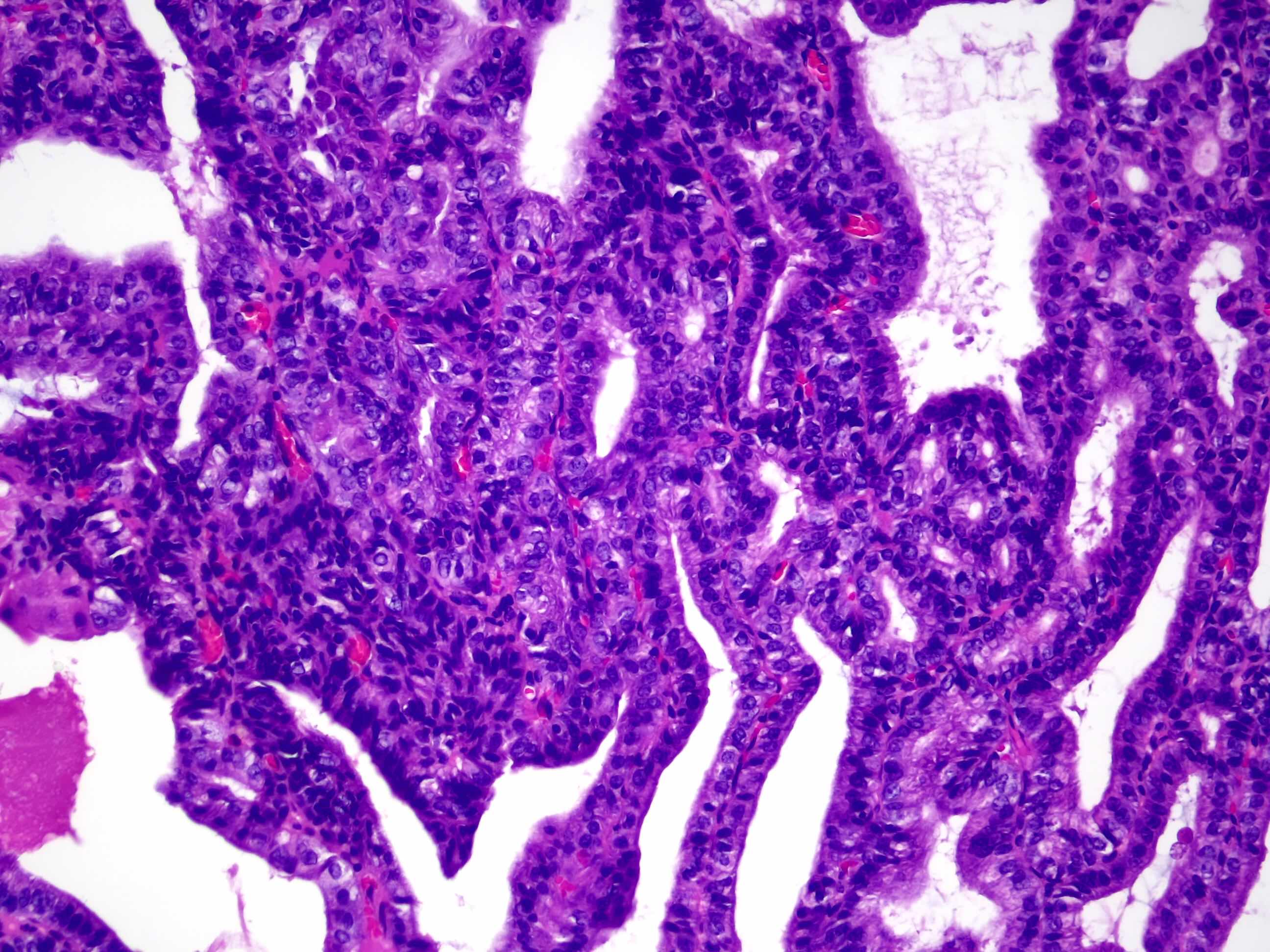
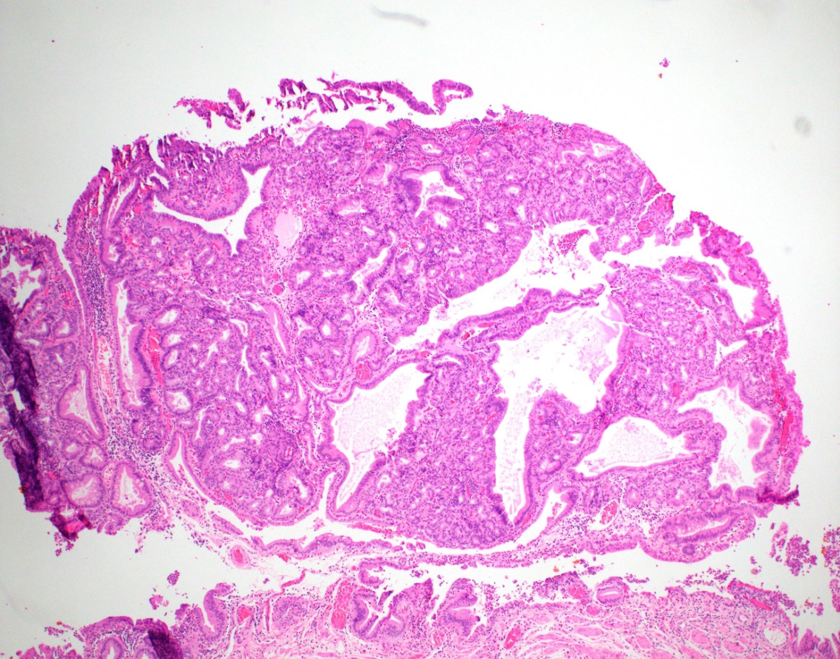
Prominent papillary exophytic lesion
Contributed by Gokce Askan, M.D. and Olca Basturk, M.D.
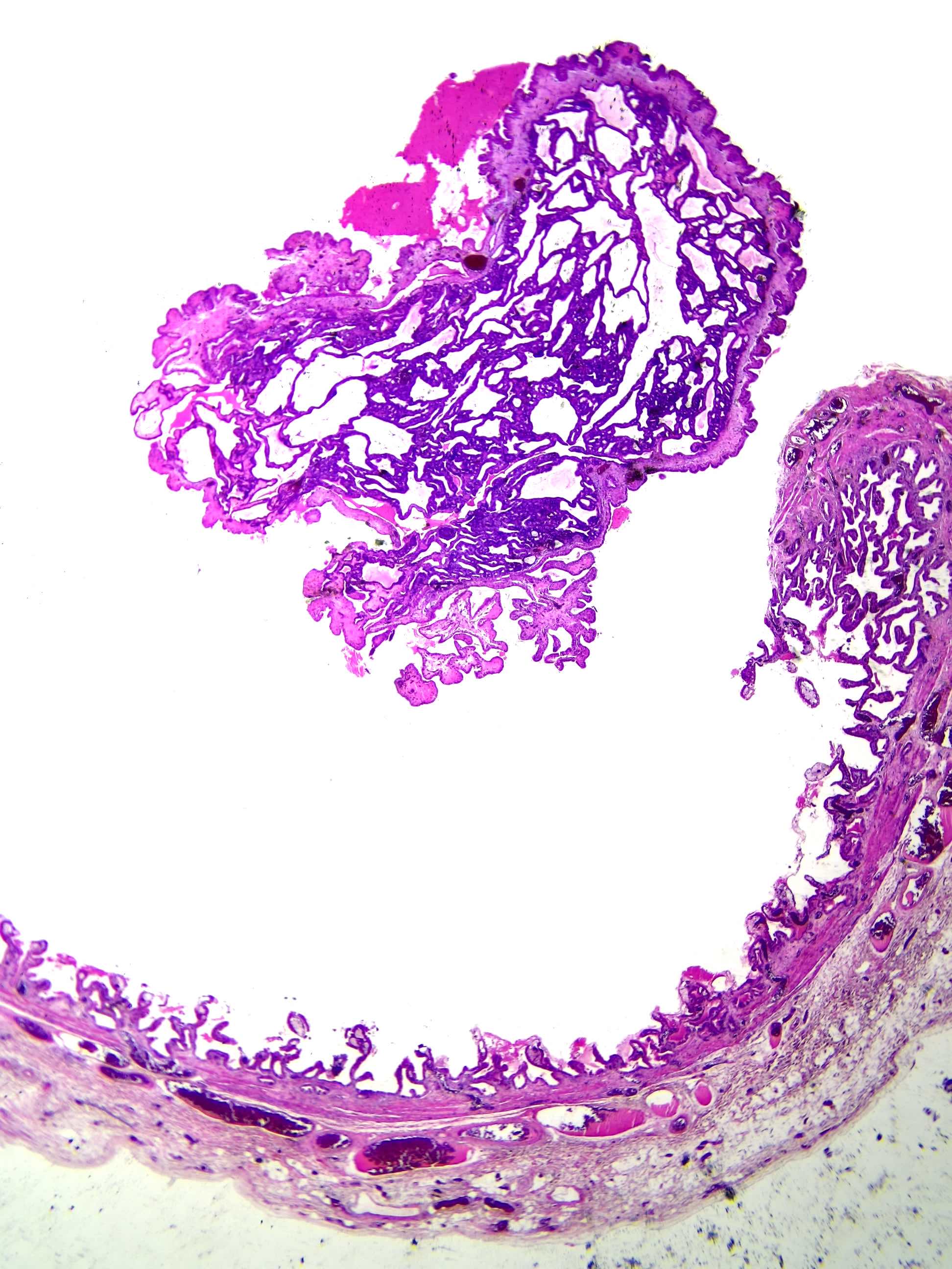
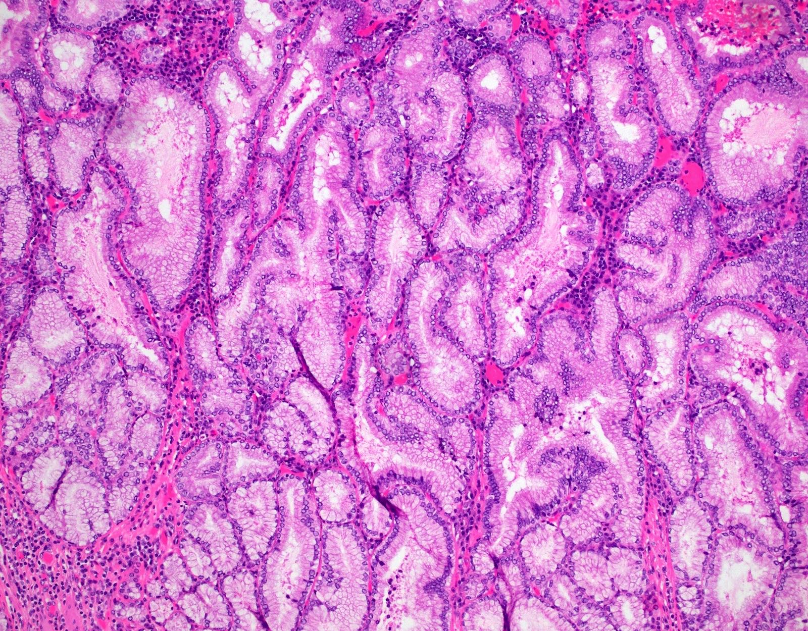
Papillary growth patterns
Tubular growth patterns
Biliary phenotype
Gastric pyloric phenotype
Gastric foveolar phenotype
Intestinal phenotype
ICPN with colloid type invasive carcinoma
ICPN with malignant cells in floating mucin
Images hosted on other servers:

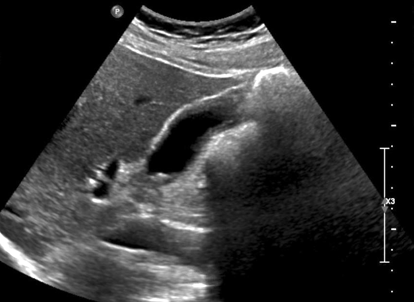
Abdominal ultrasound
Contributed by Burcin Pehlivanoglu, M.D. and Volkan Adsay, M.D.
Detached polyp in the lumen
Back to back, small nonmucinous tubules

Back to back, nonmucinous tubules
High grade dysplasia

Squamoid morules
Amyloid-like material in the stroma
Nuclei reminiscent of papillary thyroid carcinoma
Cholesterolosis in the stroma

Cholesterolosis in the background gallbladder
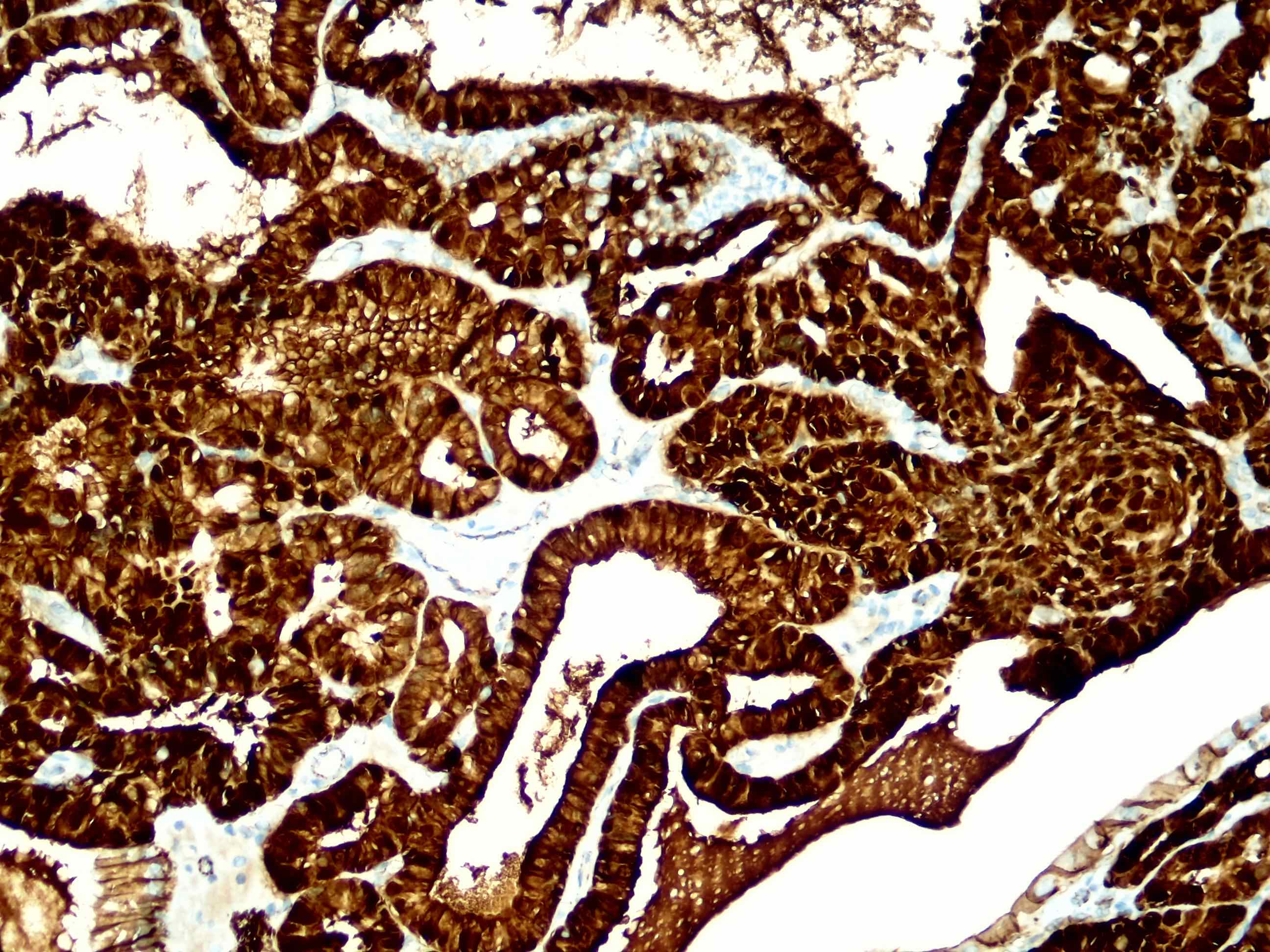
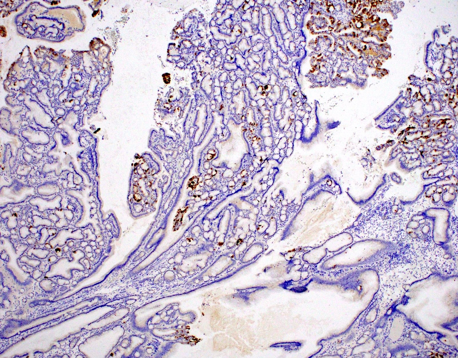
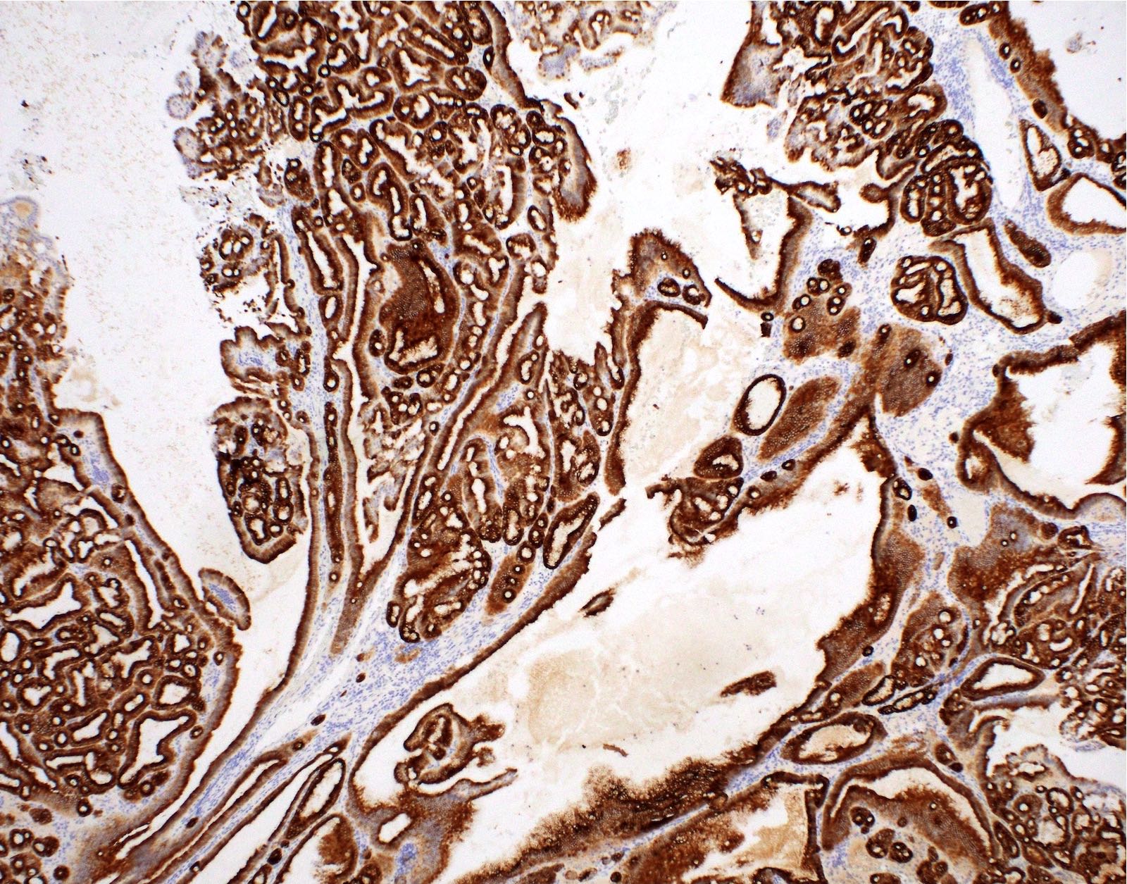
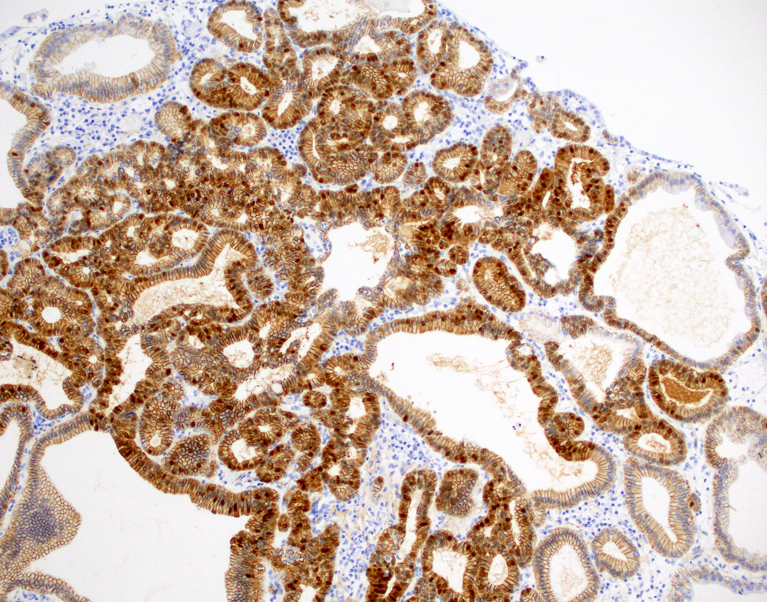
Nuclear beta catenin staining

MUC6 staining
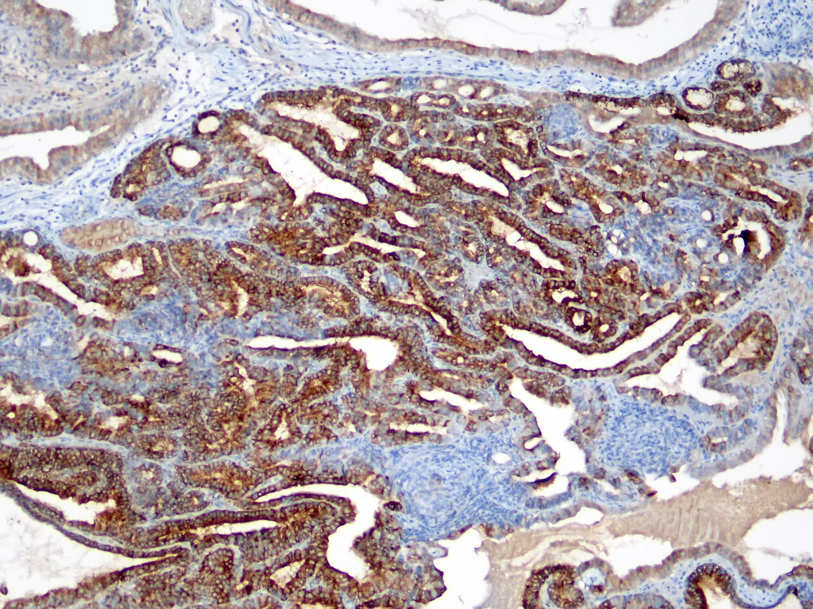
MUC1 staining

MUC5AC
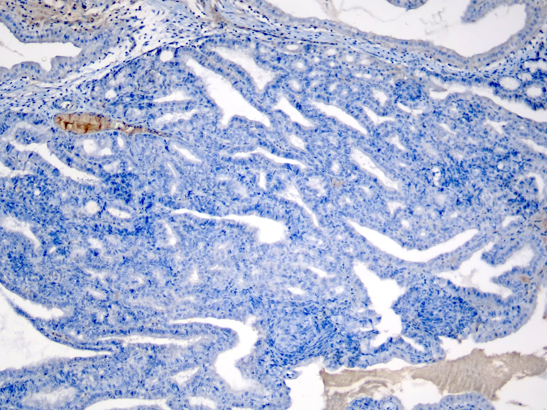
MUC2
Scattered neuroendocrine cells
Images hosted on other servers:

Hilar tumor whose
connective tissue
contains multiple
cysts
Images hosted on other servers:

Papillary
cytoarchitecture

Immunostains: CK7+, MUC1+, MUC6+, MUC5AC-

Various cysts
Images hosted on other servers:


Porcelain gallbladder
Images hosted on other servers:

Portal tract fibrosis

Bile duct with marked periductal sclerosis
Images hosted on other servers:

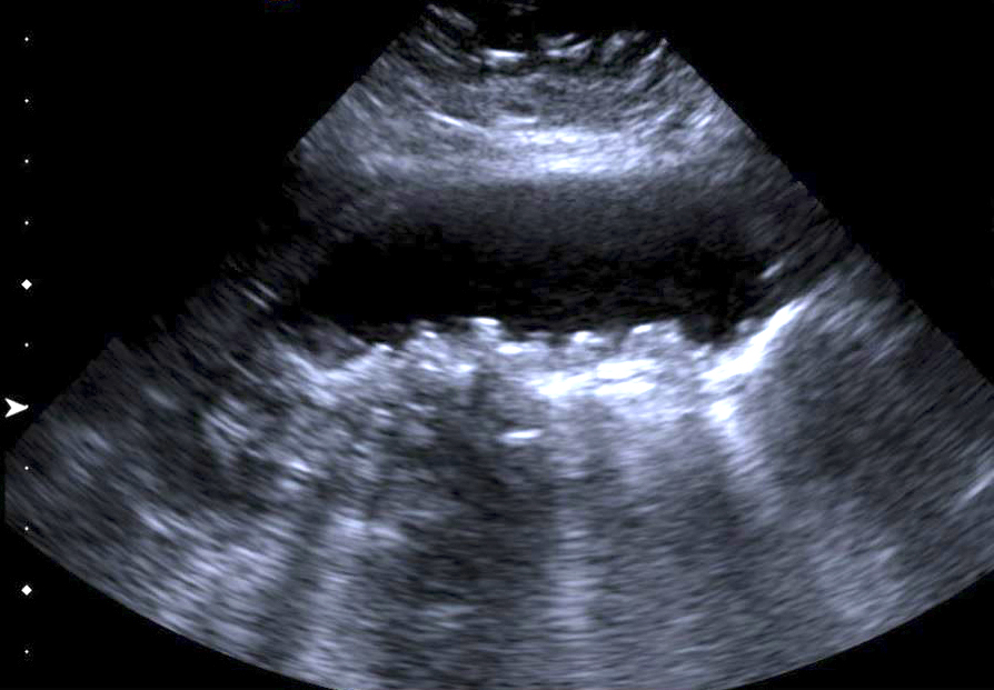
Transverse ultrasound

Sagittal ultrasound
Images hosted on other servers:

Intraluminal tumor
Contributed by Xiaoyan Liao, M.D.
Polypoid lesion
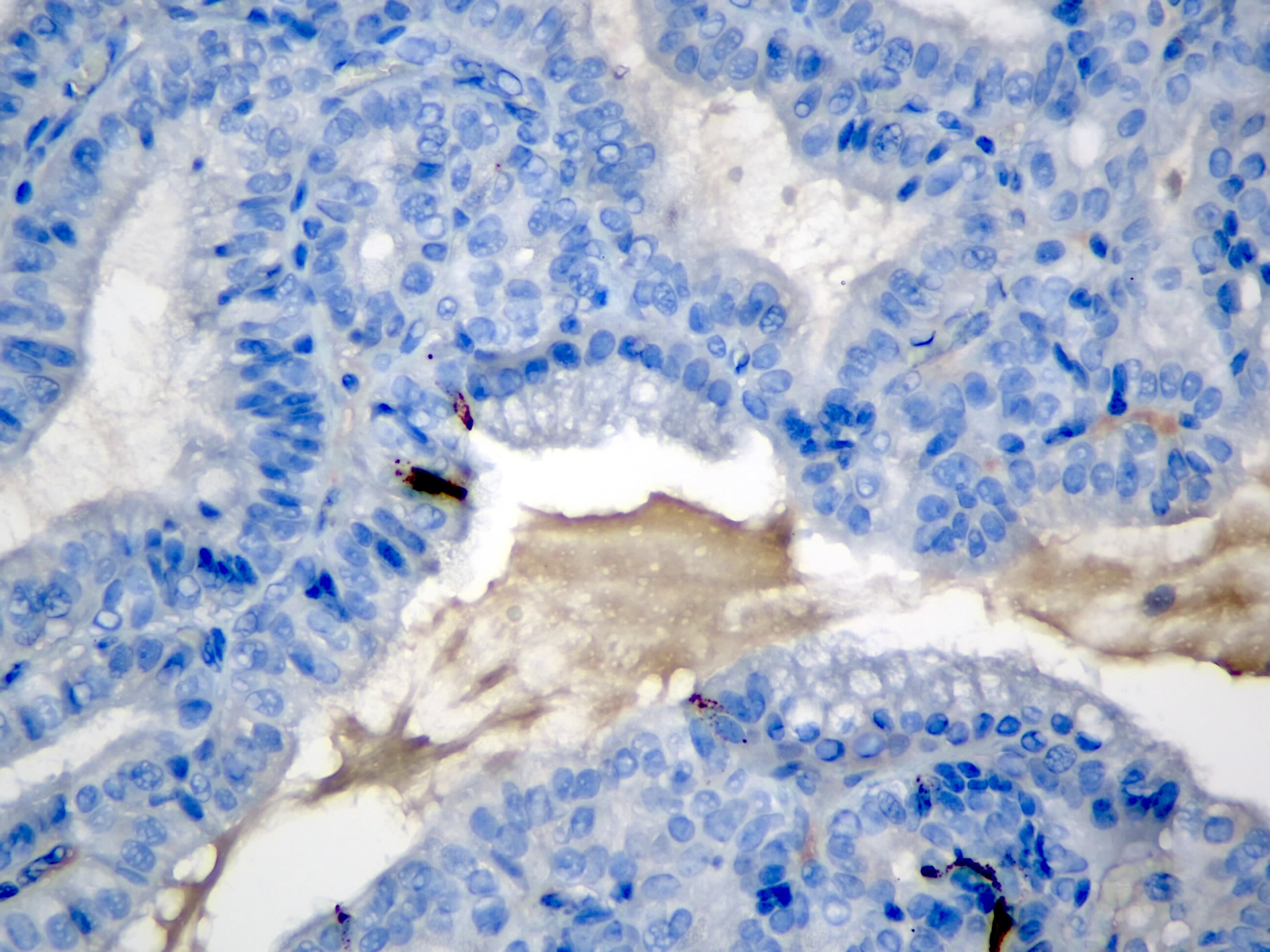
Eosinophilic cytoplasm
Intervening stroma
Tightly packed mucinous glands
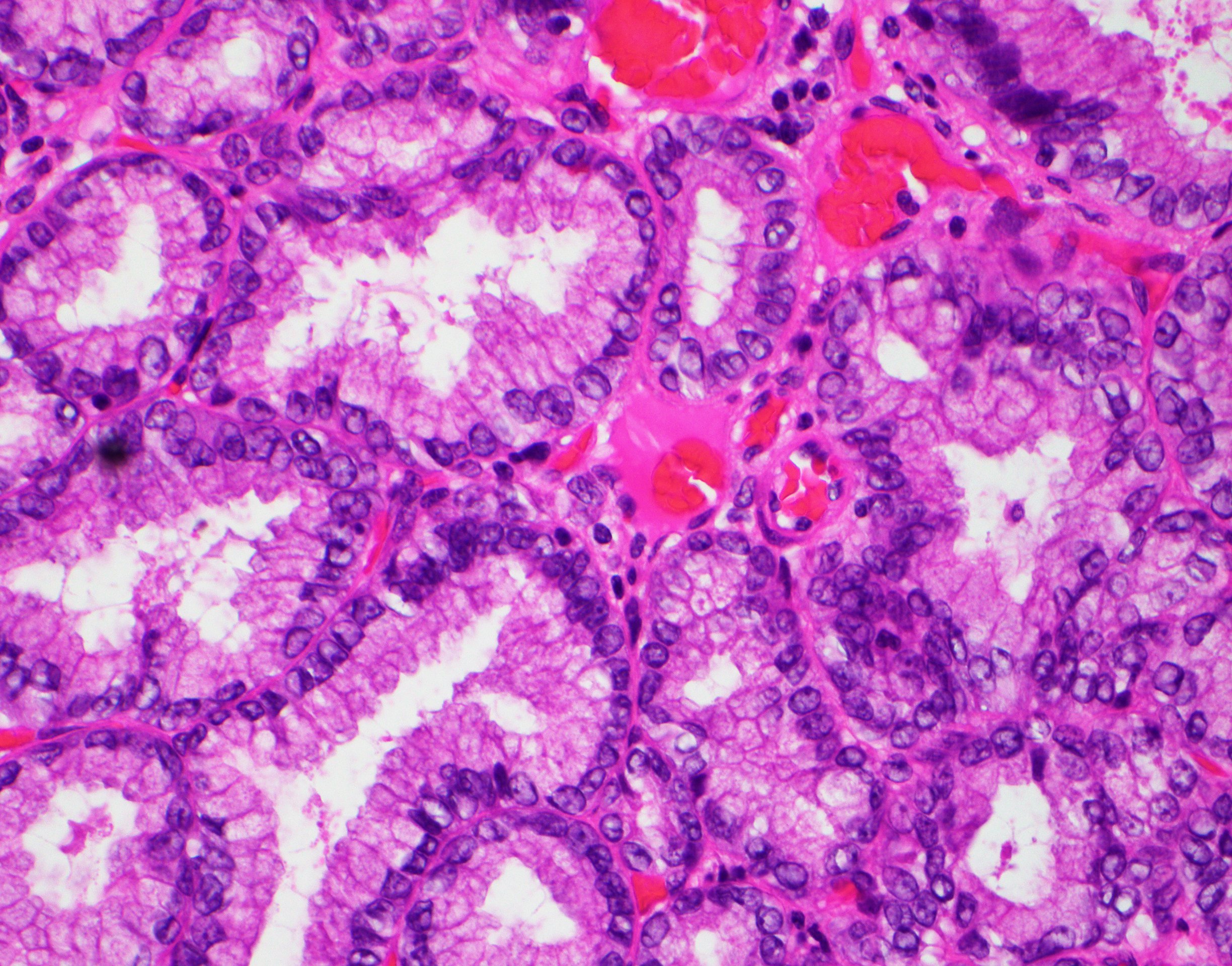
Bland cytologic atypia
MUC5AC
MUC6
Beta catenin
Images hosted on other servers:

Comparative genomic hybridization
Images hosted on other servers:
See Registry data collection variables

Bismuth-Corlette classification
Images hosted on other servers:

Endoscopic ultrasound and CT

MRCP with biliary stricture

Contrast enhanced CT

CT of tumor and adjacent CBD

MRCP of tumor
Images hosted on other servers:

Upper endoscopy
Images hosted on other servers:

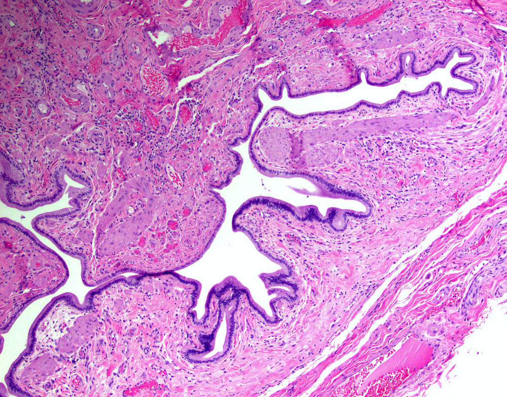
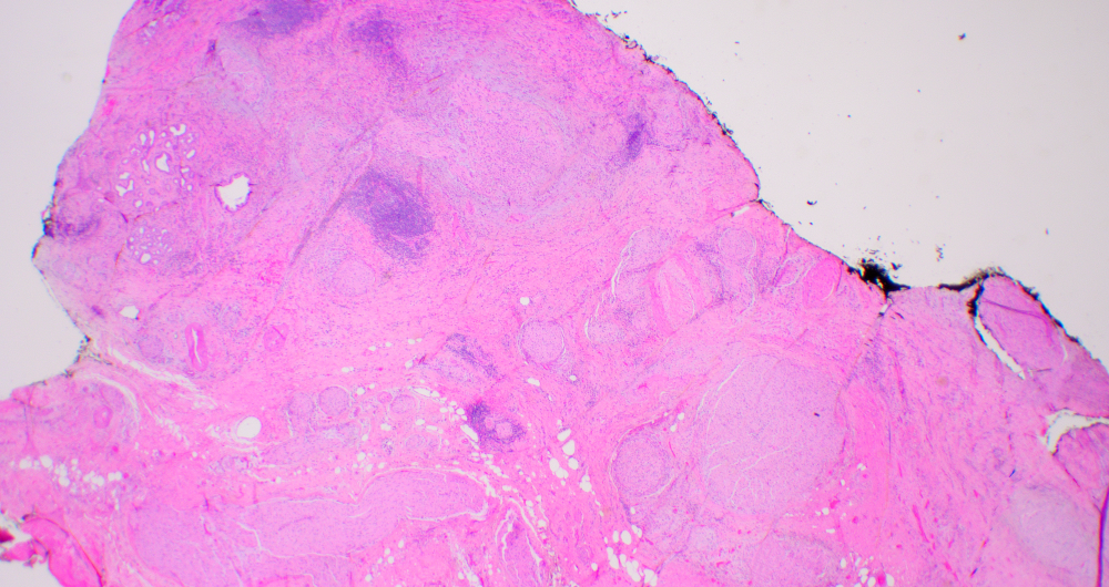
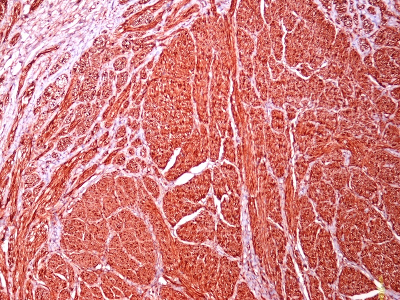
Hepatectomy with hilar nodules

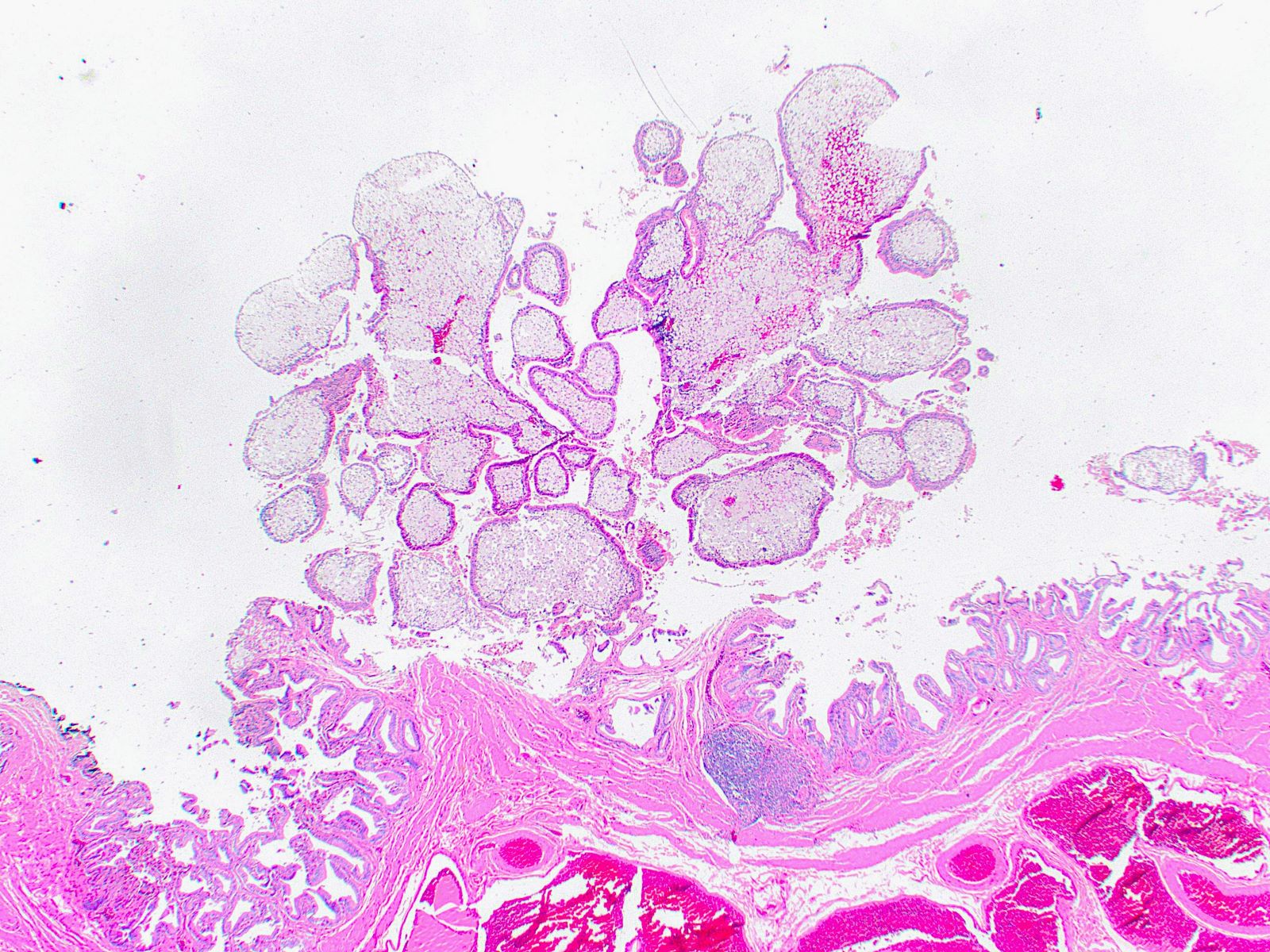
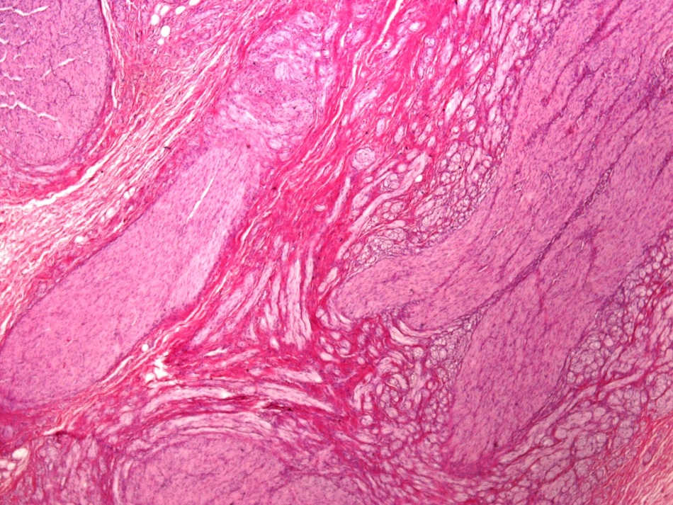
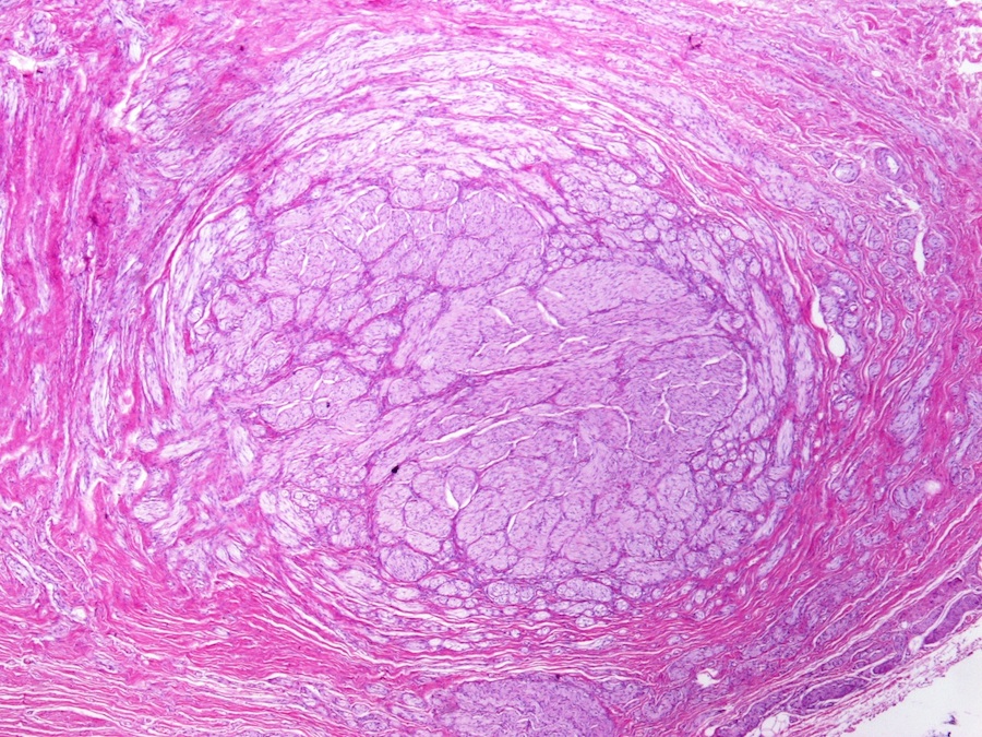
Gross features of neuroma

Remnant cystic duct with tumor
Contributed by Naziheh Assarzadegan, M.D., Saba Hassan, M.B.B.S. and Kristin Olson, M.D. (Case #329)
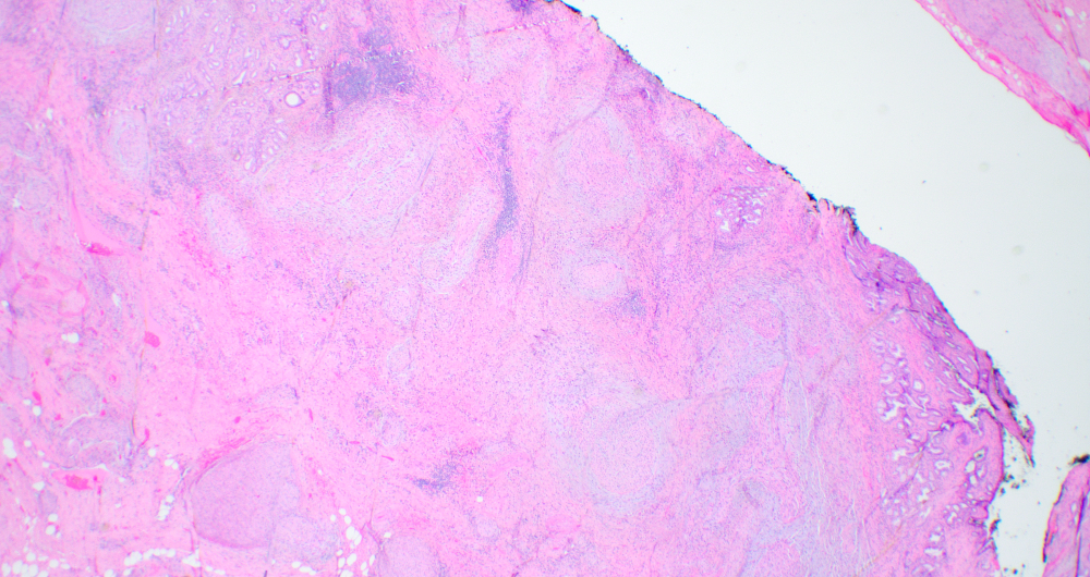
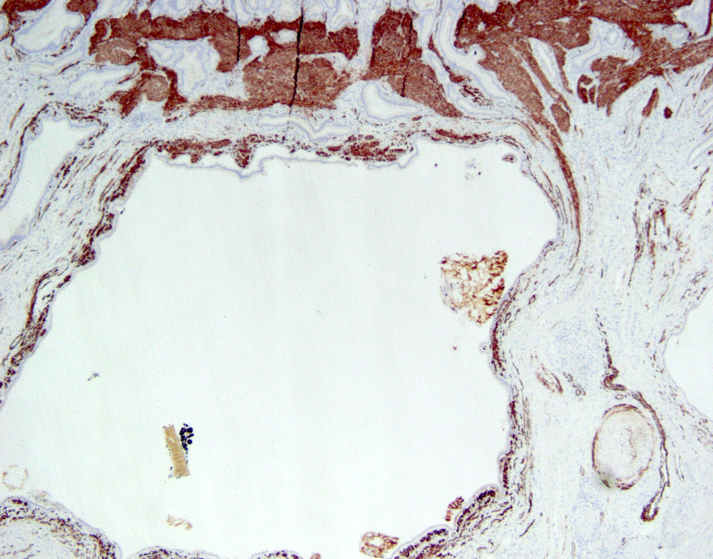
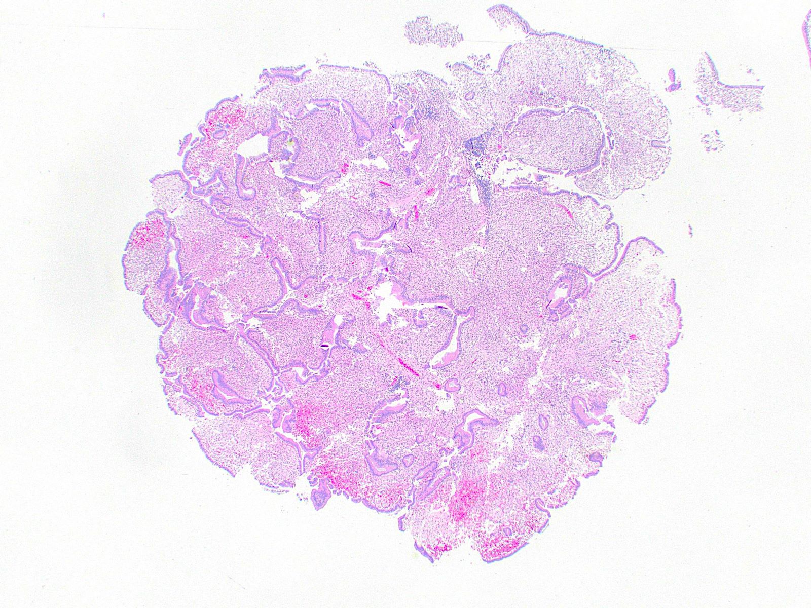
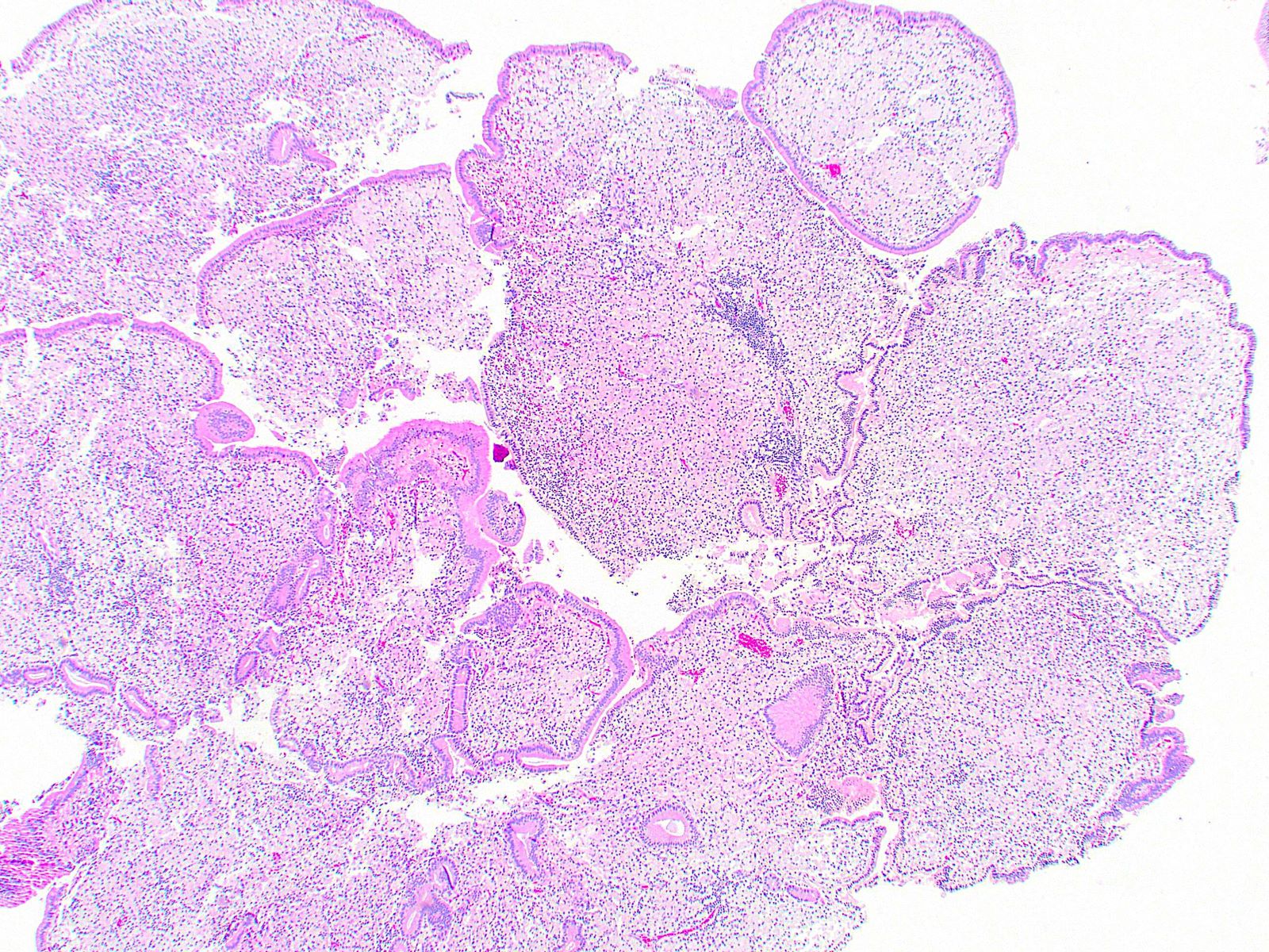
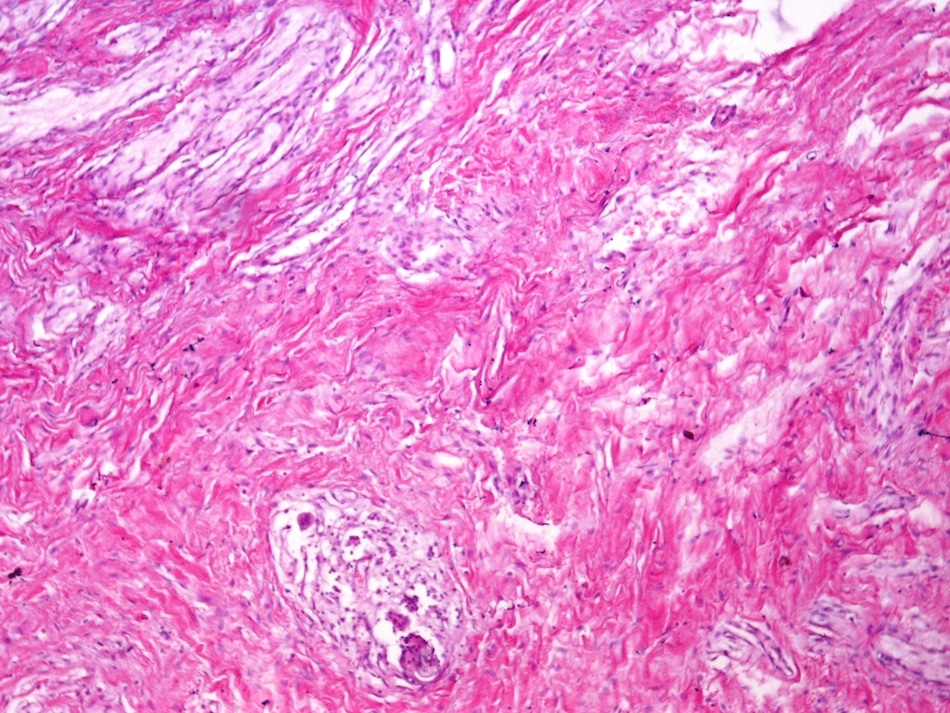
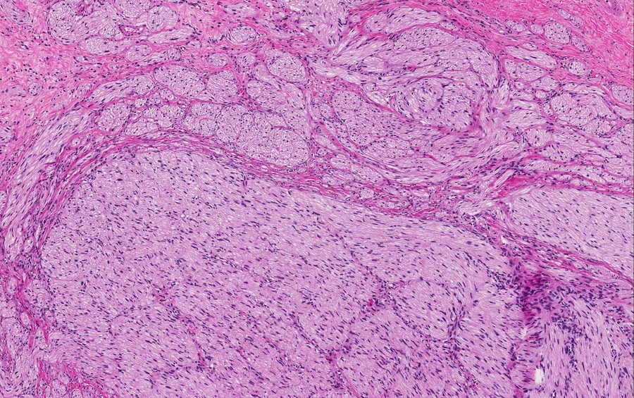
Haphazard proliferation of nerve fascicles
Haphazard nerve fascicles in collagenous stroma
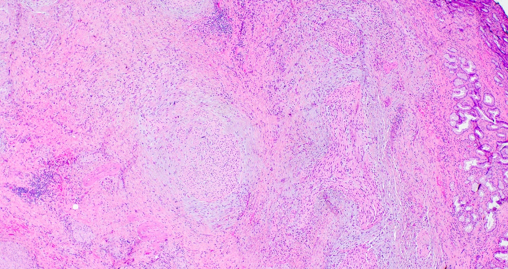
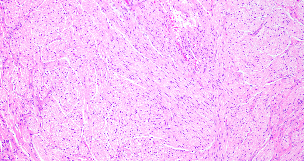
Nerve fascicles
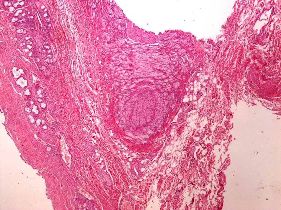
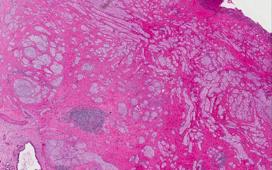
Solid, well circumscribed lesion
Disorganized nerve bundles
Disorganized, thickened nerve bundle
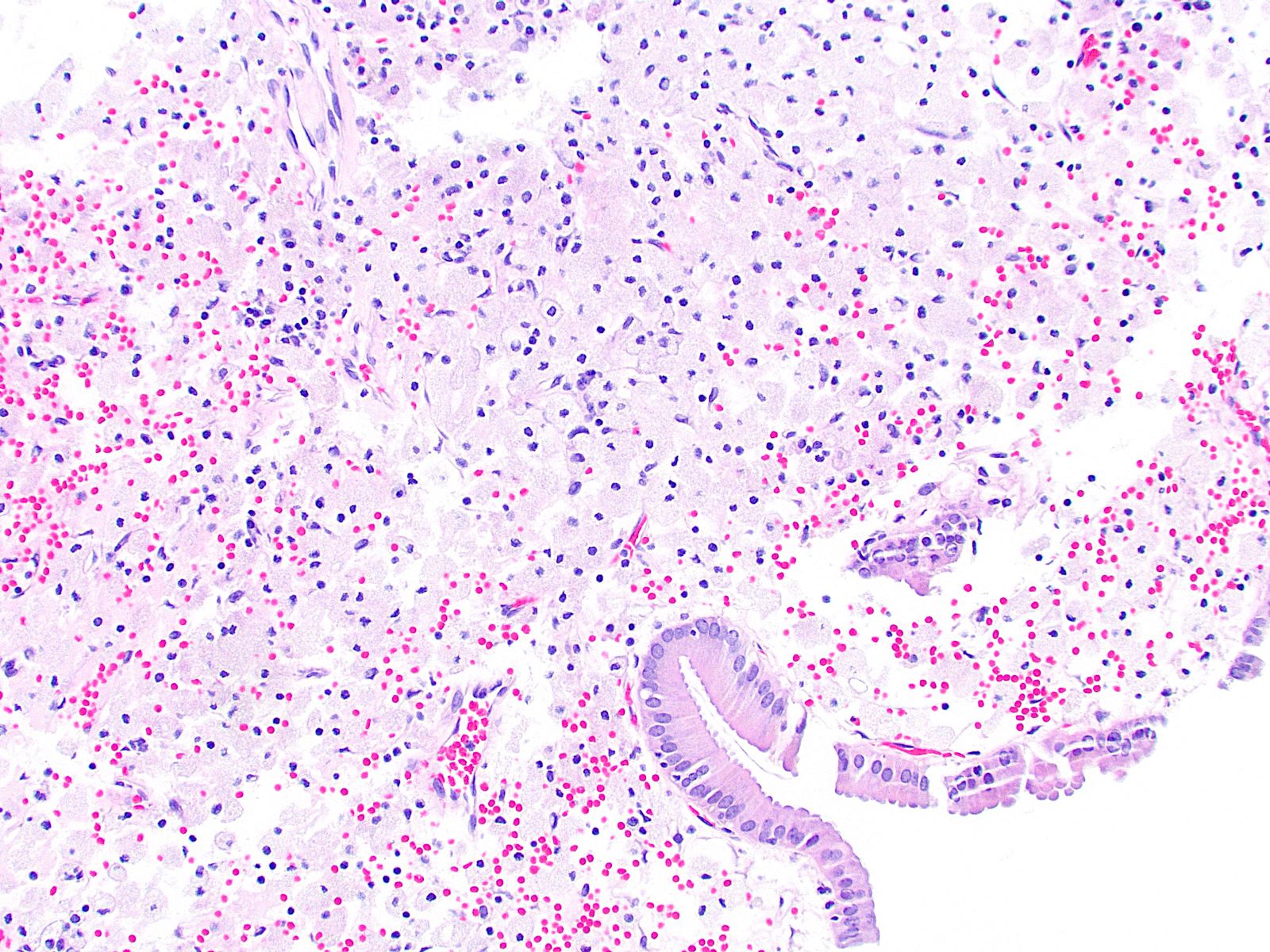
Foreign body type giant cell reaction
S100

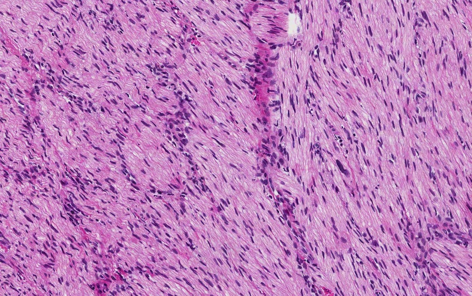
Haphazard proliferation of nerve fibers
Lesion with focus of inflammation
Interface with the adjacent glands
Lesion and adjacent bile ducts
Haphazard arrangement of thickened nerve bundles
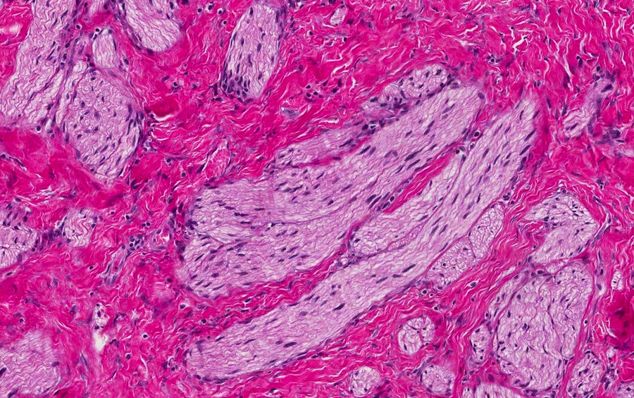
Nerve bundles
Cytologic features
Contributed by Amira Elbendary, M.B.B.Ch., M.Sc. and Soon Auck Hong, M.D., Ph.D.
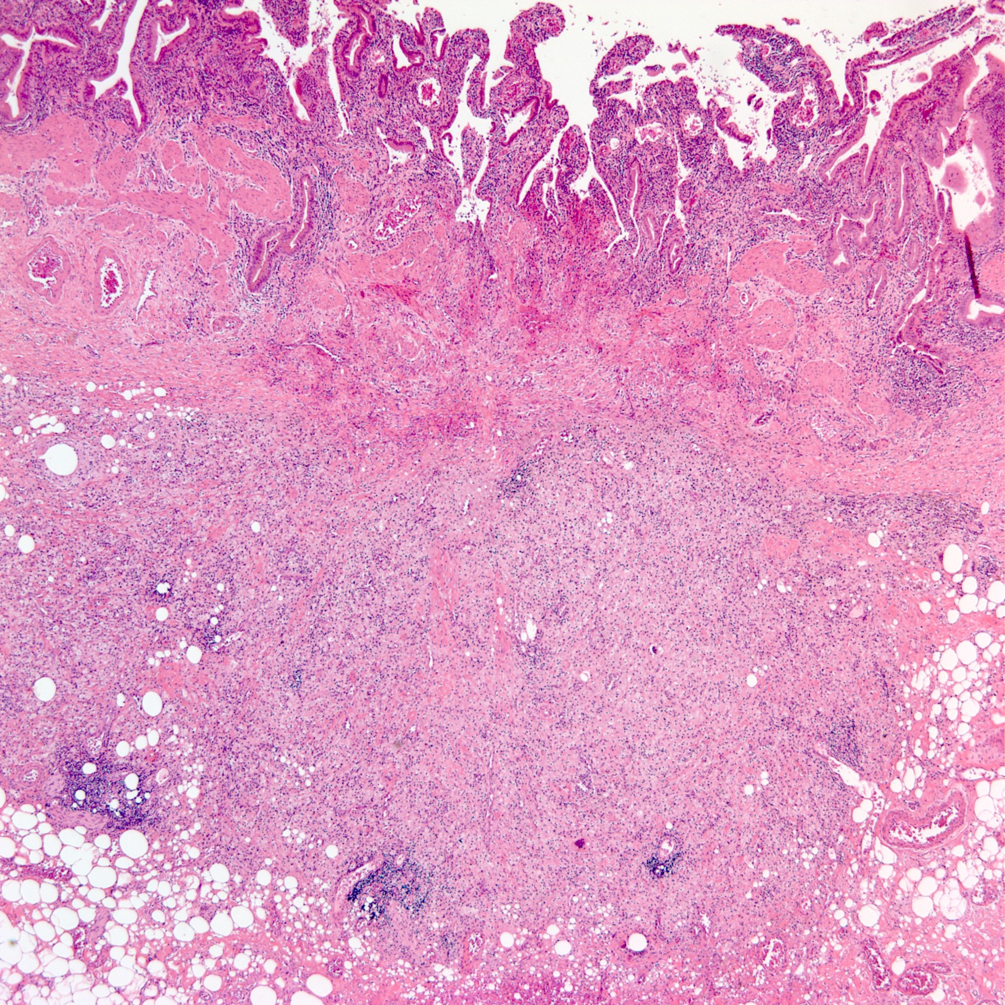
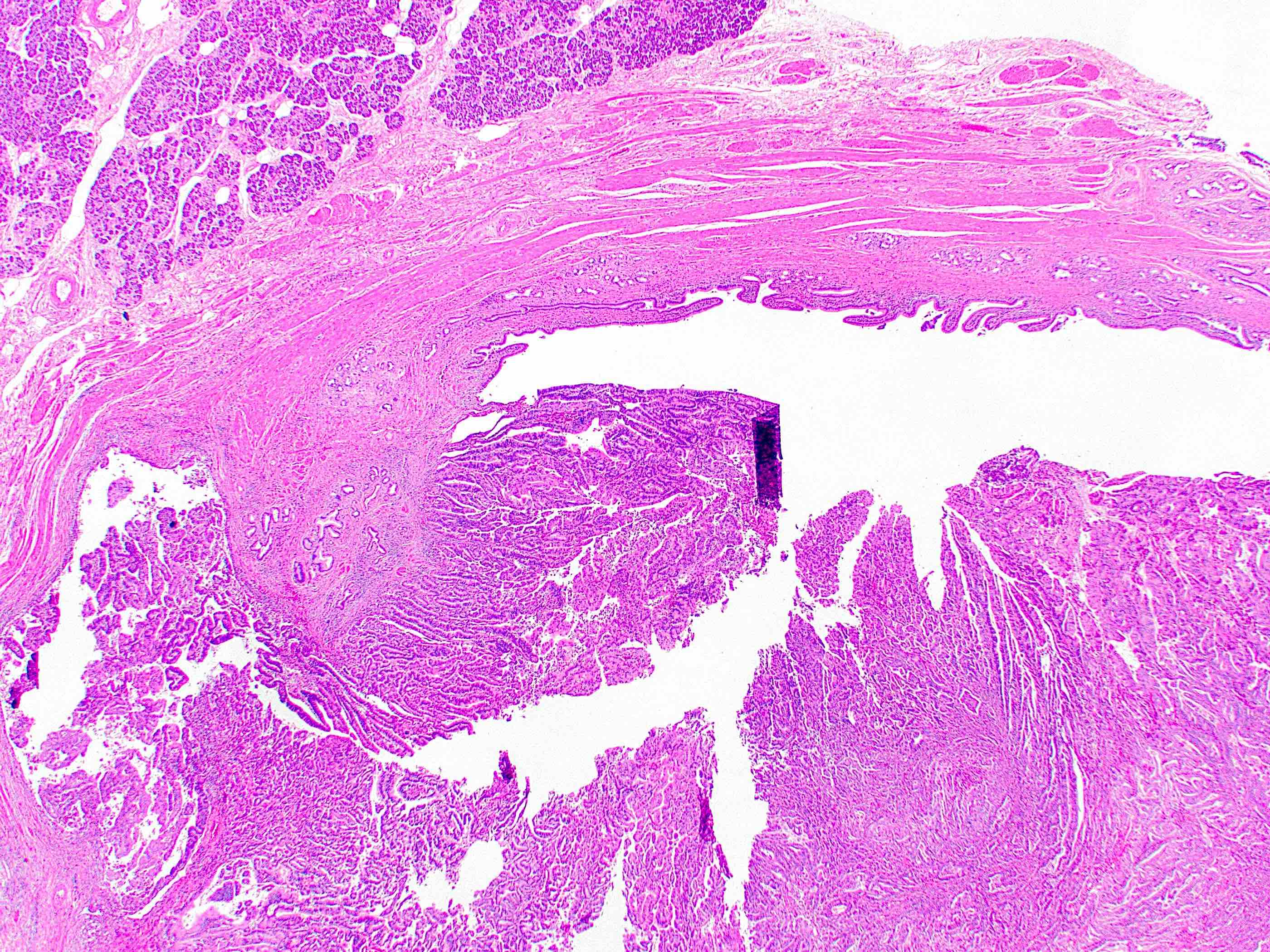
Transmural inflammation
Foamy histiocytes
Greenson: 2019

IARC: 2019
Odze: 2022
Saxena: 2017
Srivastava: 2023
Find related Pathology books: GI, liver
