

Adrenal gland & paraganglia
General
Features to report-adrenal cortical carcinoma
Author: Debra L. Zynger, M.D.

Last author update: 2 January 2019
Last staff update: 31 January 2024 (update in progress)
Copyright: 2002-2024, PathologyOutlines.com, Inc.
PubMed Search: Adrenocortical carcinoma pathology features to report
Table of Contents
Definition / general | Weiss criteria | Required features to report | Microscopic (histologic) imagesCite this page: Zynger D. Features to report-adrenal cortical carcinoma. PathologyOutlines.com website. https://www.pathologyoutlines.com/topic/adrenalreportadrenalcarcinoma.html. Accessed May 6th, 2024.
Definition / general
Weiss criteria
- Weiss criteria can be evaluated to determine malignant potential:
- High nuclear grade
- Mitotic rate > 5 mitoses per 50 high powered (40x) fields
- Atypical mitotic figures
- < 25% clear cells
- Diffuse architecture
- Necrosis
- Venous invasion
- Sinusoidal invasion
- Capsular invasion
- Reference: Am J Surg Pathol 1984;8:163
Required features to report
- If malignancy is established, the following items are required to be reported per the CAP Protocol for the Examination of Specimens From Patients With Carcinoma of the Adrenal Gland:
- Procedure
- Laterality
- Size (greatest dimension)
- Weight
- Histologic type (oncocytic, myxoid, sarcomatoid)
- Grade (low grade if 20 or fewer mitoses per 50 high powered [40x] fields; high grade if > 20 mitoses per 50 high powered fields)
- Lymphovascular invasion
- Tumor extent (into capsule, extra-adrenal tissue or adjacent organs)
- Margins
- Regional lymph node status
- Distant metastasis (if applicable)
- pTNM stage
- The following items are necessary to establish the 8th edition AJCC pTNM stage:
- Size (5 cm or less or > 5 cm)
- Tumor extent (into extra-adrenal tissue or adjacent organs, renal vein or vena cava)
- Regional lymph node status
- Distant metastasis (if applicable)
- References: CAP: Protocol for the Examination of Specimens From Patients With Carcinoma of the Adrenal Gland [Accessed 30 November 2022], Amin: AJCC Cancer Staging Manual, 8th Edition, 2017
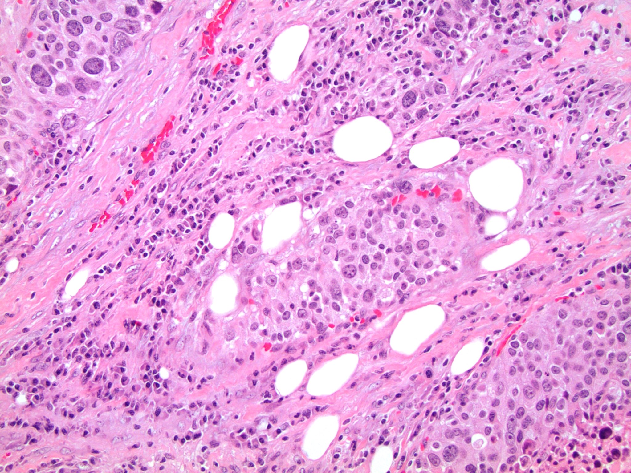
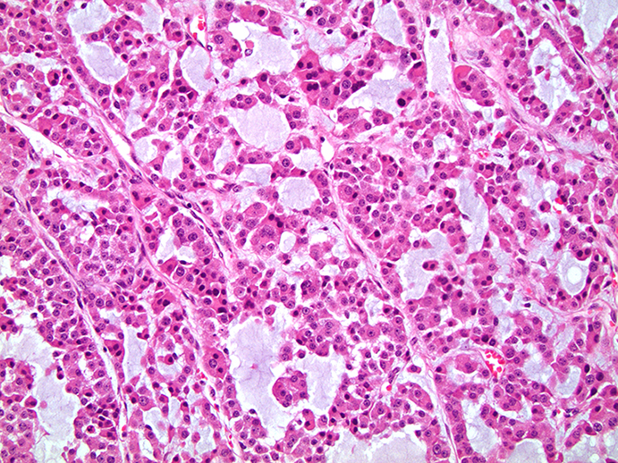
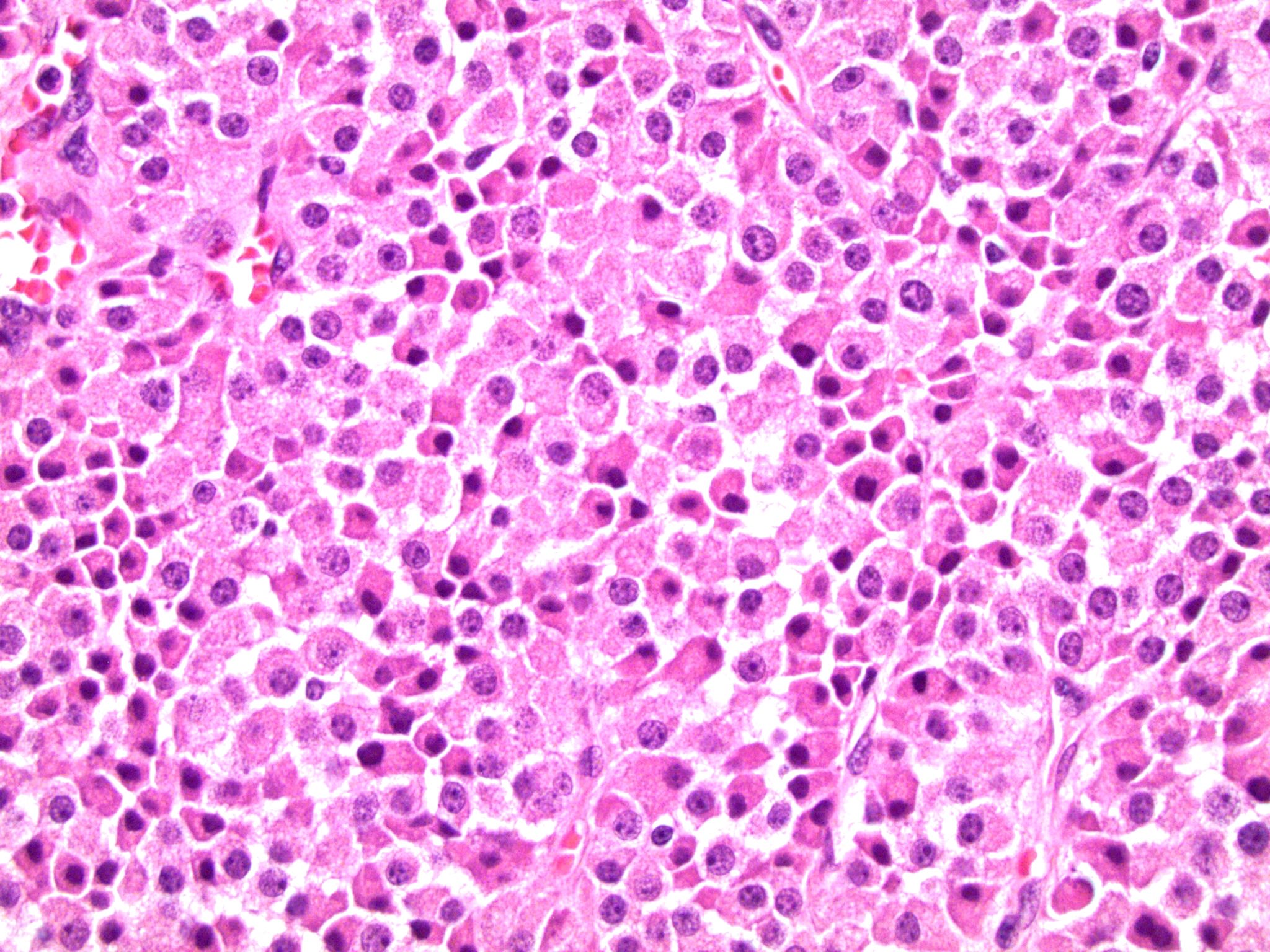
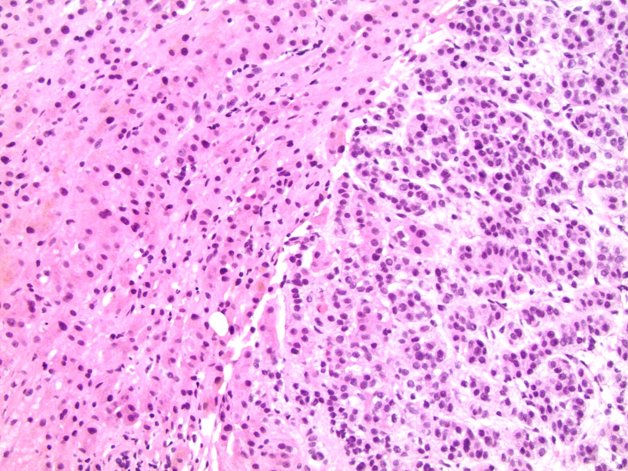
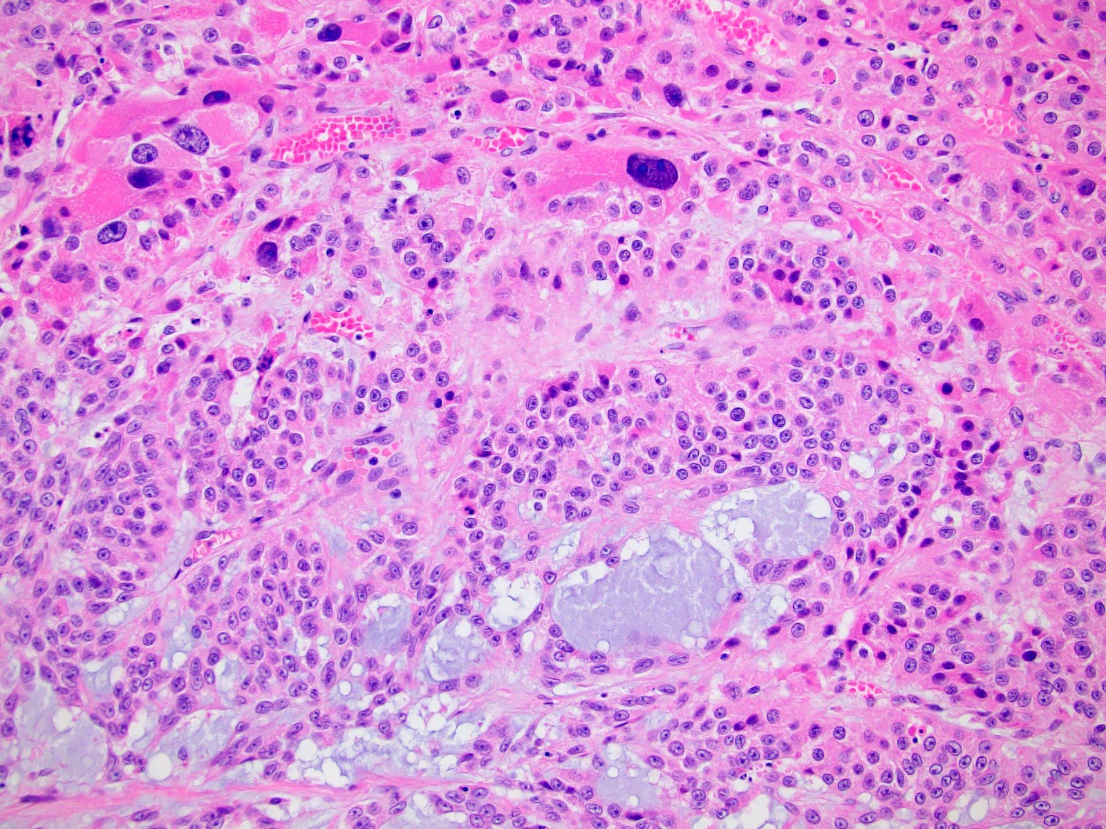
Microscopic (histologic) images
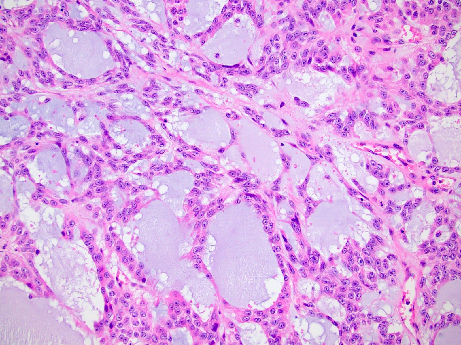
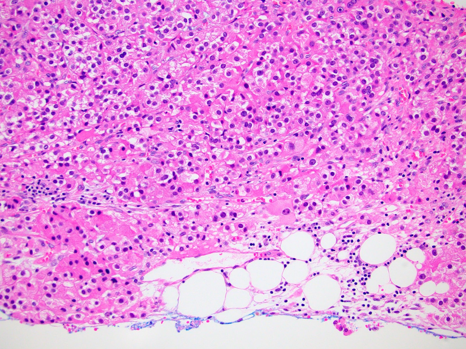
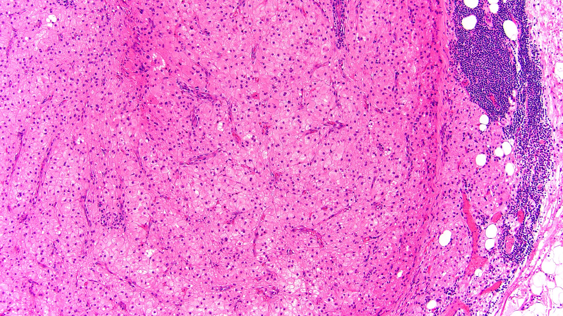
Contributed by Debra Zynger, M.D. and Maria Tretiakova, M.D., Ph.D.
Extra-adrenal adipose invasion (pT3)
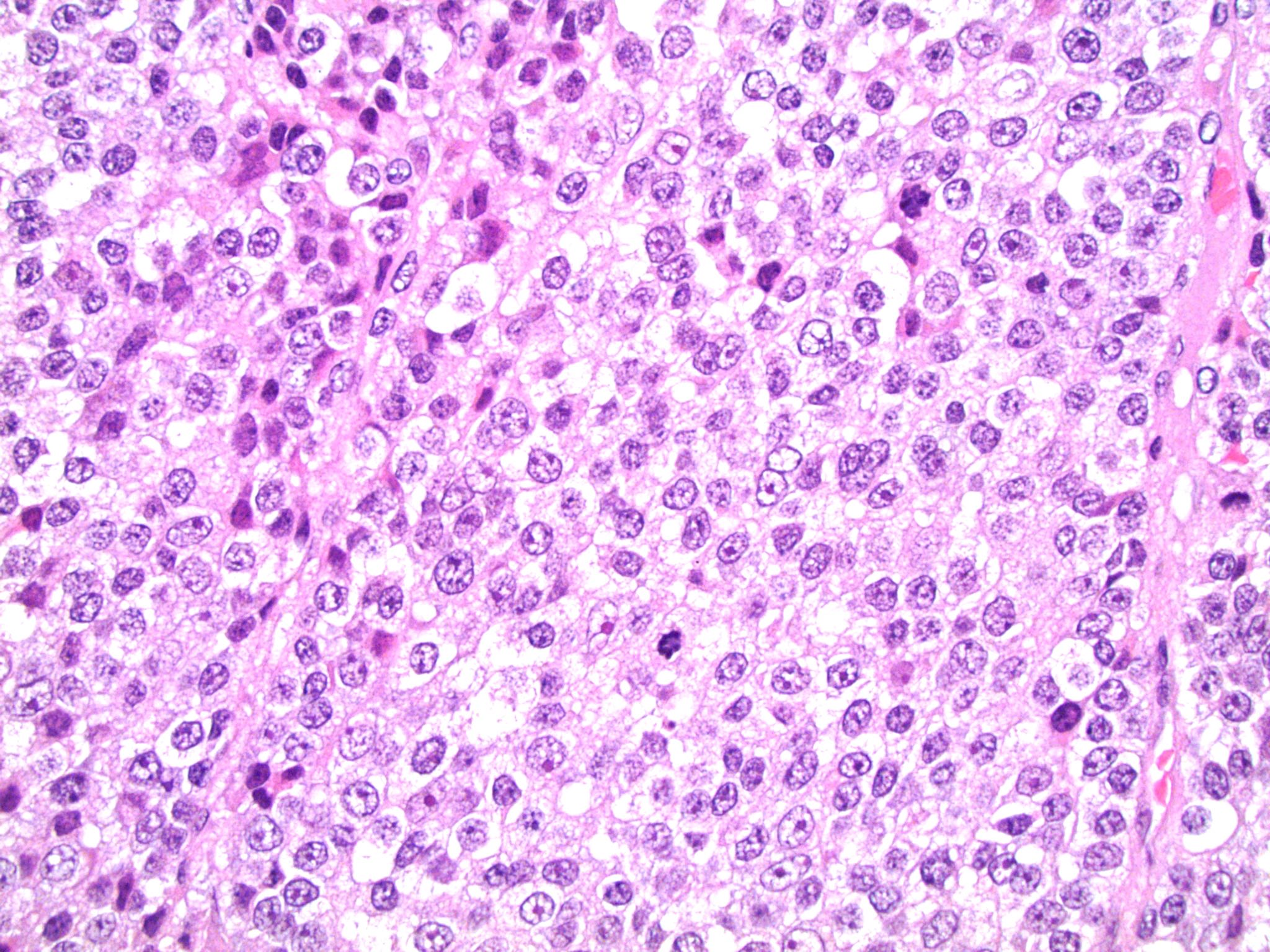
High mitotic rate
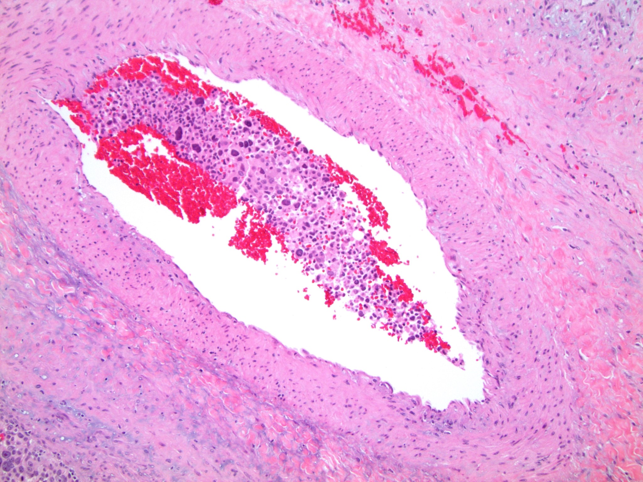
Lymphovascular invasion
Myxoid variant
Oncocytic variant
Liver involvement (pT4)
Transition to myxoid area
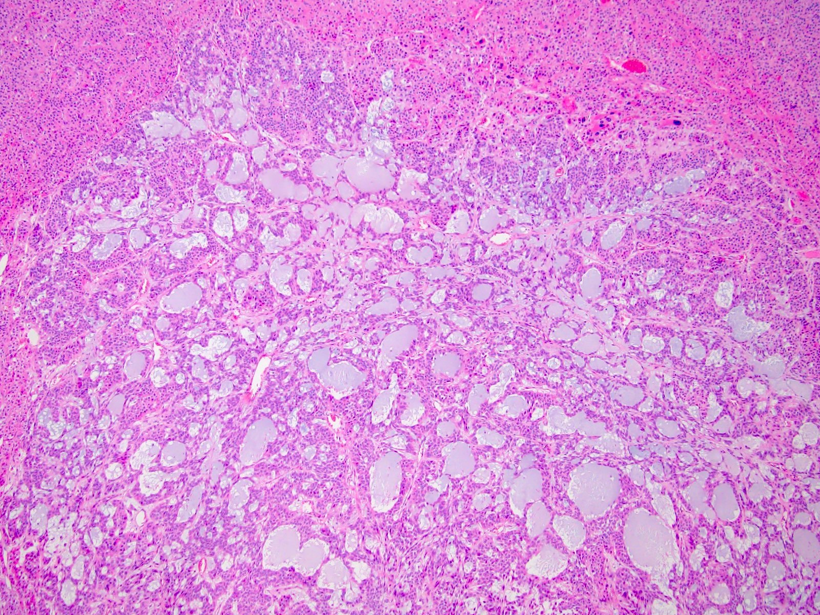
Myxoid variant
Extra-adrenal adipose invasion (pT3)
Positive margin
Regional node involvement (pN1)
