
Breast
Other nonneoplastic
Mucocele-like lesion
Author: Cansu Karakas, M.D.
Editorial Board Member: Gary Tozbikian, M.D.
Editor-in-Chief: Debra L. Zynger, M.D.

Last author update: 10 November 2020
Last staff update: 17 February 2021
Copyright: 2002-2024, PathologyOutlines.com, Inc.
PubMed Search: Mucocele-like lesion of breast

Table of Contents
Definition / general | Essential features | Terminology | Epidemiology | Sites | Etiology | Clinical features | Diagnosis | Radiology description | Radiology images | Prognostic factors | Case reports | Treatment | Microscopic (histologic) description | Microscopic (histologic) images | Cytology description | Positive stains | Sample pathology report | Differential diagnosis | Additional references | Board review style question #1 | Board review style answer #1 | Board review style question #2 | Board review style answer #2Cite this page: Karakas C. Mucocele-like lesion. PathologyOutlines.com website. https://www.pathologyoutlines.com/topic/breastmucocele.html. Accessed May 13th, 2024.
Definition / general
- Rare lesion characterized by dilated epithelium lined ducts filled with mucin; associated with extravasation of acellular mucin into the stroma
- Cysts lined by flat or low cuboidal epithelium
Essential features
- Characterized by extravasated acellular mucin in periductal stroma
- Although there is variable associated hyperplasia of the cyst lining in mucocele-like lesions, there are no epithelial cells floating within the luminal or extravasated mucin, which is a critical finding in distinguishing mucocele-like lesions from mucinous carcinoma of the breast
- Although originally described as a benign lesion, associations with atypical ductal hyperplasia (ADH), ductal carcinoma in situ (DCIS) and invasive carcinoma have been reported in several studies
- Searching for atypia when a mucocele-like lesion is present is important to exclude possibility of ADH, DCIS or invasive carcinoma
- Pure mucocele-like lesions without atypia typically have benign behavior
Terminology
- Mucocele-like lesion (MLL), mucocele-like tumor
Epidemiology
- Mean age 40 years, range 25 - 61 years (Am J Surg Pathol 1986;10:464)
Sites
- Anywhere in the breast
Etiology
- Pathogenesis is unclear but excessive mucinous secretions or ductal obstruction may be responsible
Clinical features
- Many reports have described an association between mucocele-like lesions and the simultaneous presence of ADH, DCIS or mucinous carcinoma (Hum Pathol 2016;49:33, Mod Pathol 2011;24:683, Diagn Pathol 2011;6:29, Histopathology 2013;62:894, Arch Pathol Lab Med 1991;115;137, Am J Clin Pathol 2012;138:783, N Eng J Med 2005;353:229)
- Mucin extravasation or mucocele-like lesions at core biopsy warrant radiological pathological correlation and close followup is recommended to rule out any atypical lesion (Histopathology 2009;55:609)
Diagnosis
- Diagnosis can be made on core biopsy or surgical specimen but careful evaluation of excised tissue, multiple H&E levels and clinicopathologic correlation may be helpful to rule out in situ lesions and invasive mucinous carcinoma
Radiology description
- There is no definitive test or imaging for differentiating mucocele-like lesions from other suspicious lesions
- Mammographic findings: heterogeneously dense breast tissue with mostly clustered round or pleomorphic calcifications (AJR Am J Roentgenol 2006;186:1356, AJR Am J Roentgenol 2005;185:1310)
- Ultrasonographic findings: usually multiple, oval shaped cysts with calcified or noncalcified solid areas; clustered cysts with thick septations and complex masses are associated with atypical proliferations or malignancy (Ultrasonography 2015;34:133, AJR Am J Roentgenol 2011;196:1424)
Radiology images
Images hosted on other servers:

Microcalcifications

Mucocele-like lesion with focal atypical proliferation

Pleomorphic calcifications

Palpable mass with tubular cystic structures
Prognostic factors
- Pure mucocele-like lesions without atypia are usually associated with a benign outcome (Histopathology 2013;62:894, Ultrasonography 2015;34:133, Breast J 2016;22:173)
- Risk of associated malignancy is much higher if atypia is present
Case reports
- 34 year old woman with associated ductal carcinoma in situ and mucinous carcinoma (J Korean Med Sci 2001;16:516)
- 47 year old woman with mucocele-like lesion on core biopsy (Bezmialem Science 2018;6:147-9)
- 71 year old woman with associated ductal carcinoma in situ and ductal carcinoma (J Clin Diagn Res 2016;10:ED16)
- 2 cases of mucocele-like lesions with cytologic findings that illustrate the difficulty of separating those lesions from colloid carcinoma (Am J Clin Pathol 1991; 95:875)
- Mucocele-like tumor associated with ductal carcinoma in situ diagnosed as mucinous carcinoma by fine needle aspiration cytology (Surg Today 2012;42:280)
Treatment
- Several studies have recommended close clinical followup as an alternative to surgical excision in women with a core biopsy of a mucocele-like lesion without atypia and no associated mass (Am J Clin Path 2012;138:783, Ultrasonography 2015;34:133, Ann Surg Oncol 2019;26:3478)
- Complete surgical excision recommended in certain situations where core needle biopsy reveals the presence of associated ADH / DCIS or where a mass with indistinct and irregular margins is shown by mammography or sonography (Clin Imaging 2011;35:94, Diagnostic Pathol 2011;6:29)
Microscopic (histologic) description
- Cysts and dilated ducts filled with mucin
- Mucin containing cysts that often rupture with extravasation of mucin into surrounding stroma
- Myoepithelial cells adhere to strips of cells floating in lakes of mucin
- Calcifications are often present
- Epithelium lining the cysts may show typical and atypical proliferative changes including benign / flat, hyperplasia, ADH, DCIS or mucinous carcinoma
- Microscopic examination of the entire specimen is important to rule out any atypia / malignancy
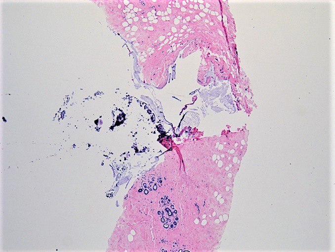
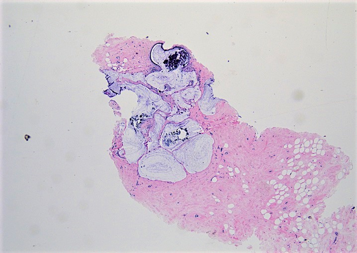
Microscopic (histologic) images
Contributed by Cansu Karakas, M.D.
Mucin filled cyst
Multiple cysts
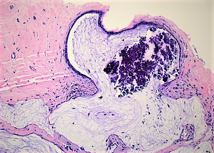
Coarse calcifications
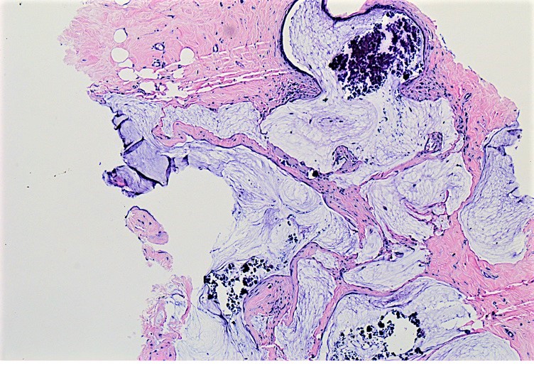
Extravasation of mucin
Cytology description
- Poorly cellular with cohesive clusters of bland cells in 2 dimensional sheets with abundant mucoid background, no / rare intact single tumor cells, no atypia if mucocele only (Am J Surg Pathol 1999;23:552, Am J Clin Pathol 1991; 95:875)
- Excisional biopsy is necessary to confirm diagnosis (Breast Cancer 2009;16:77)
- Most important features favoring a benign mucocele-like lesion over mucinous carcinoma on FNA are:
- Younger patient
- Cells arranged in cohesive monolayers
- No significant nuclear atypia
- Scant cellularity
- No or rare single, intact tumor cells
- Mucinous carcinomas are usually more cellular with more single tumor cells, 3 dimensional clusters, mild / moderate nuclear atypia and a solid mass by imaging (Cytopathology 2004;15:104, Acta Cytologica 2000;44:765)
Positive stains
- Mucicarmine
- PAS diastase
- Alcian blue
- At pH 2.7: positive
- At pH 0.9: equivocal
- Myoepithelial markers: smooth muscle myosin heavy chain (SMMHC), p63, calponin
Sample pathology report
- Left breast, upper outer quadrant, core needle biopsy:
- Mucocele-like lesion
Differential diagnosis
- Cystic mastopathy:
- Associated with prominent apocrine differentiation
- Florid duct ectasia with luminal mucin:
- Generally contains lipid rich material within ducts with prominent foamy histiocytes
- Mucinous carcinoma:
- Prominent luminal cell proliferation and variable number of tumor cells floating within the mucin
- Luminal cells in mucin are not associated with myoepithelial cells
- Nodular mucinosis:
- Rare lesion with accumulation of stromal mucin, typically located under or adjacent to nipple
- Can be distinguished from mucocele-like lesions by location and staining feature of mucous substance
Additional references
Board review style question #1
Which of the following statements is true about mucocele-like lesions of the breast?
- Frequently found in a retroareolar location
- Mammographically they usually form a palpable mass
- Most of the patients are elderly
- Usually benign, although they can be associated with atypia or malignancy
Board review style answer #1
D. Mucocele-like lesions are typically benign, although they can be associated with atypia or malignancy. Mucocele-lesions without atypia or carcinoma are benign lesions. The other statements are false. They can arise anywhere in the breast. They commonly present with calcifications with variable circumscribed mass on mammography. There is a wide range at presentation with mean age of 40.
Comment Here
Reference: Mucocele-like lesion (MLL) of breast
Comment Here
Reference: Mucocele-like lesion (MLL) of breast
Board review style question #2
A 35 year old woman had a mammogram showing polymorphous, grouped microcalcifications. The histologic details of the core needle biopsy are shown in the image above. Which of the following statements are true about this entity?
- CK5/6 and p63 are negative
- Likelihood of carcinoma is very high
- Mucicarmine staining is negative
- When there is a rupture to stroma, the possibility of mucinous carcinoma must always be considered
Board review style answer #2
D. When there is a rupture to stroma, the possibility of mucinous carcinoma must always be considered. A mucocele-like lesion may show CK5/6 and p63 expression and highlight the myoepithelial cells surrounding the benign cystic lining of mucocele-like lesion. Mucin in the cyst and stroma usually show strong and diffuse mucicarmine staining in mucocele-like lesion. Mucocele-like lesion without associated carcinoma is a benign lesion.
Comment Here
Reference: Mucocele-like lesion (MLL) of breast
Comment Here
Reference: Mucocele-like lesion (MLL) of breast
