
Microbiology & infectious diseases
Filamentous bacteria
Nocardia
Editorial Board Member: Patricia Tsang, M.D., M.B.A.


Last author update: 10 August 2020
Last staff update: 13 April 2022
Copyright: 2020-2024, PathologyOutlines.com, Inc.
PubMed Search: Nocardia[TI] full text[sb] review[ptyp]
Table of Contents
Definition / general | Essential features | Epidemiology | Sites | Pathophysiology | Clinical features | Laboratory | Case reports | Treatment | Clinical images | Microscopic (histologic) description | Microscopic (histologic) images | Molecular and mass spectrometry techniques | Differential diagnosis | Additional references | Board review style question #1 | Board review style answer #1 | Board review style question #2 | Board review style answer #2Cite this page: Larsen N, Farr GA, Leal SM. Nocardia. PathologyOutlines.com website. https://www.pathologyoutlines.com/topic/microbiologynocardia.html. Accessed May 13th, 2024.
Definition / general
- Gram positive genus containing over 50 species
- Taxonomy: order Actinomycetales, family Nocardiaceae
- Common species:
- N. asteroides
- N. farcinica
- N. brasiliensis
- N. asteroides
Essential features
- Gram positive, catalase positive bacilli with thin filamentous branching, often with a beaded appearance
- Microbiology: negative on Ziehl-Neelsen and Kinyoun staining; positive on modified AFB stain
- Histopathology: AFB stain negative; Fite stain positive
- Diseases range from cutaneous infection to severe pulmonary or central nervous system disease (Int J Infect Dis 2020;90:161)
- Disseminated disease has poor prognosis
Epidemiology
- Ubiquitous saprophytes; present in soil
- Immunocompromised patients at higher risk
- N. asteroides and N. farcinica more widespread in the United States; N. brasiliensis favors Southwestern or Southeastern United States (Clin Microbiol Rev 2006;19:259)
Sites
- Immunocompromised host
- Lung / pneumonia (Int J Infect Dis 2020;90:161)
- Brain abscesses
- Hematogenous spread
- Skin involvement
- Immunocompetent hosts (N. brasiliensis)
- Lung / pneumonia (Int J Infect Dis 2020;90:161)
- Lymphocutaneous infection (sporotrichoid lesion)
- Mycetoma
Pathophysiology
- Virulence factors include filamentous growth, mycolic acid, catalases and superoxide dismutases
Clinical features
- Most commonly transmitted via inhalation from environmental sources
- Traumatic inoculation possible from environmental sources
- Hospital acquired infections due to contaminated equipment or post surgical wounds (Centers for Disease Control: How is Nocardiosis Spread? [Accessed 10 August 2020])
- Pulmonary disease can be subacute and indolent, symptoms may be present for weeks (Clin Microbiol Rev 2006;19:259)
- Cough with or without thick, purulent sputum
- Chest Xray infiltrates, nodules, cavitation
- CNS disease presents with brain abscess and signs of increased intracranial pressure
- More common in immunocompromised patients (BMC Infect Dis 2020;20:56)
- Skin disease: granules may be present and should be examined microscopically
- Also seen with Actinomyces; modified AFB stain / Fite stain is helpful
- May present similarly to other mycobacterial diseases with fever, weight loss and malaise
- Environmental source (soil); not considered communicable
Laboratory
- Aerobic growth on sheep blood and chocolate agar
- Optimal growth on charcoal buffered yeast extract at 35 ± 2°C
- Biochemical methods unable to differentiate between clinically relevant species
- Dry, crinkly colonies at 48 - 72 hours, may take up to 14 days; not ideal for timely identification
- Variable colony morphology (Clin Microbiol Rev 2006;19:259):
- N. farcinica turns orange with age
- N. brasiliensis has yellow discoloration
- N. asteroides type VI produces brown pigment
- Species level identification via MALDI-TOF mass spectrometry or sequencing is most timely and accurate technique for identification
- If MALDI-TOF is not available, make presumptive diagnosis and send to reference lab
- CLSI M62 document provides susceptibility testing breakpoints (CLSI: M62 - Mycobacteria Susceptibility Testing Standards [Accessed 10 August 2020])
Case reports
- 40 year old man with AIDS and rare N. abscessus infection (BMJ Case Rep 2016;2016:bcr2016215649)
- 41 year old man with Nocardia farcinica infection mimicking pulmonary neoplasm, pneumonia, SVC syndrome and pericarditis (Thorax 1997;52:492)
- 45 year old man with history of kidney transplant and disseminated Nocardia infection (Transpl Infect Dis 2011;13:385)
- 52 year old chalk miner with chronic meningitis due to Nocardia farcinica (BMC Infect Dis 2020;20:56)
- 67 year old man with Nocardia cerebral abscess mimicking a high grade tumor (J Clin Neurosci 2010;17:1080)
Treatment
- Empiric therapy often includes high dose sulfamethoxazole with trimethoprim (TMP-SMX) and amikacin, ceftriaxone or imipenem
- Variable resistance patterns necessitate organism identification and susceptibility testing
Clinical images
Contributed by Sixto M. Leal Jr., M.D., Ph.D.
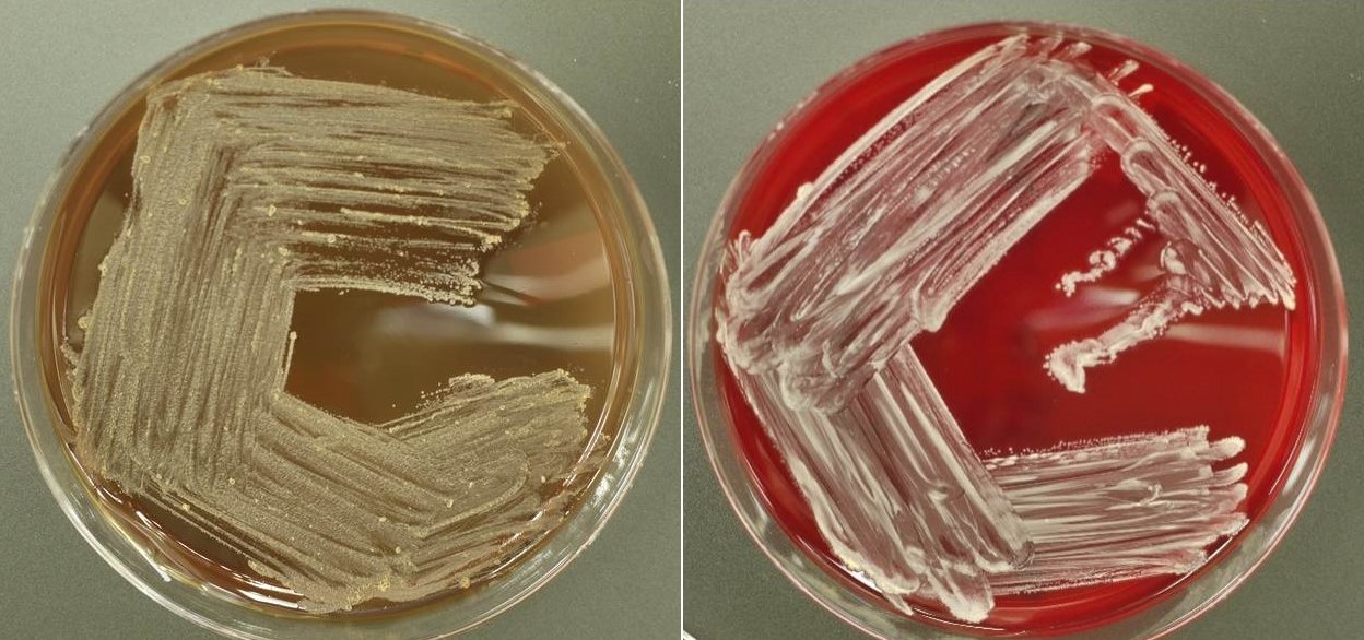
N. asteroides colonies,
chocolate and sheep
blood agar
Microscopic (histologic) description
- Thin filamentous branching rods with a gram positive beaded appearance
- Fite and Gomori methenamine silver (GMS) stain positive
- Width: 1 - 2 μm; length variable
- Host response; acute inflammation or pyogranulomatous
Microscopic (histologic) images
Contributed by Sixto M. Leal Jr., M.D., Ph.D.
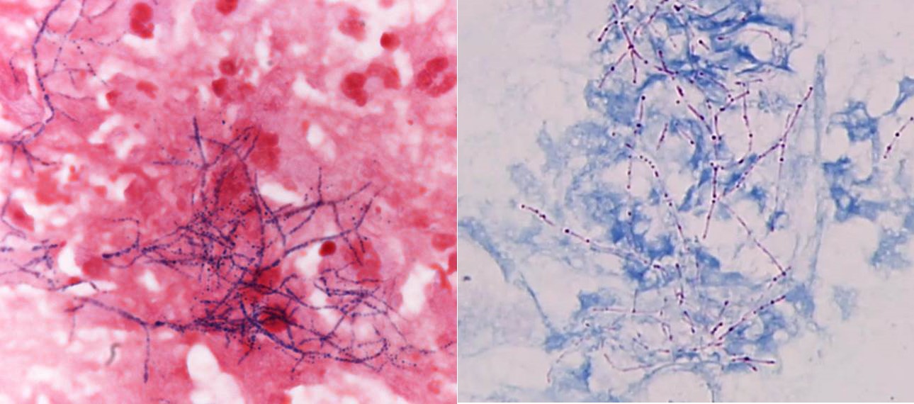
Filamentous branching
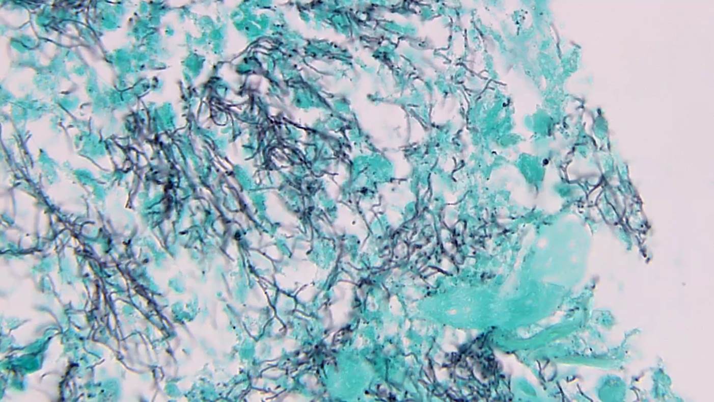
Filamentous branching on GMS stain
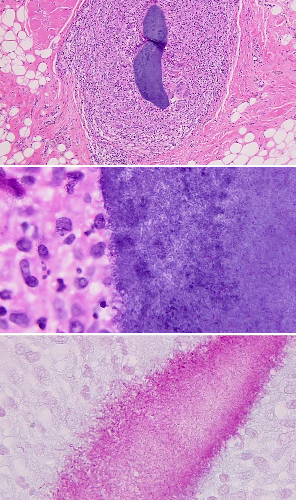
Skin nodule with Nocardia growth
Molecular and mass spectrometry techniques
- 16S highly sensitive and specific for species level identification and antimicrobial susceptibility (New Microbes New Infect 2017;19:96)
- rrs gene used as reference but does not have enough polymorphisms to differentiate at species level
- secA1 gene, part of protein translocase complex, was a novel target but still not perfect identification at species level
- sodA gene, encoding for superoxide dismutase, has variable regions that allow discrimination of closely related species (New Microbes New Infect 2017;19:96)
- Next generation sequencing has good diagnostic value, decreased turnaround time and can aid with clinical decision making (Front Cell Infect Microbiol 2020;10:13)
- No need for target specific primers, advantage over Sanger sequencing
- MALDI-TOF generates mass spectra based on whole cell material or extracted intracellular content matched to known database references (Front Microbiol 2018;9:1294)
- Bruker Daltonics and Vitek MS are most commonly used databases
Differential diagnosis
- Mimics filamentous Actinomyces bacteria
- Nocardia is aerobic not anaerobic and modified acid fast positive
- Indolent pulmonary disease mimics
- Mycobacterium tuberculosis
- Mold infection
- Malignancy
- Brain abscess mimics
- Mold
- Toxoplasma
- Malignancy
Additional references
Board review style question #1
- A 56 year old man presents to the emergency department with weight loss and a thick, productive cough. He has a past medical history of nonalcoholic steatohepatitis (NASH) cirrhosis and a liver transplant 2 years ago. A sputum culture Gram stain shows Gram positive filamentous bacteria. Which stain is helpful for distinguishing the 2 major genera of filamentous bacteria?
- GMS
- Gram stain
- Modified Kinyoun
- Ziehl-Neelsen
Board review style answer #1
C. Modified Kinyoun. Nocardia species have a thin layer of mycolic acid which retains the carbol fuchsin dye when using a weaker acid for decolorization (i.e. modified acid fast stain). This feature helps distinguish from other filamentous bacteria that stain negative with this procedure.
Comment Here
Reference: Nocardia
Comment Here
Reference: Nocardia
Board review style question #2
- A patient from Kenya presented with a chronic subcutaneous lesion in the right foot with significant swelling, fistula formation and purulence. On examination, a granule was expressed from this lesion and sent to microbiology. Half of the sample was crushed between 2 slides and stained, the other half submitted for culture. A Gram stain of the slide revealed filamentous structures with a diameter of 5 μm. Which culture conditions are optimal to isolate the likely etiologic agent?
- BCYE agar under aerobic conditions
- Blood plate under microaerophilic conditions
- CDC anaerobic agar under anaerobic conditions
- Fungal culture media under aerobic conditions
Board review style answer #2
D. Fungal culture media under aerobic conditions. Filamentous bacteria are 1 - 2 μm in diameter whereas fungal hyphae are 4 - 15 μm thick. Size differences are the key to making this distinction with significant implications on treatment and patient outcome.
Comment Here
Reference: Nocardia
Comment Here
Reference: Nocardia
