
Microbiology & infectious diseases
Parasites-gastrointestinal (not liver)
Strongyloides
Author: Nat Pernick, M.D.

Last author update: 1 July 2018
Last staff update: 15 February 2024
Copyright: 2018-2024, PathologyOutlines.com, Inc.
PubMed Search: Strongyloides
Table of Contents
Definition / general | Pathophysiology | Diagrams / tables | Clinical features | Diagnosis | Case reports | Treatment | Microscopic (histologic) description | Microscopic (histologic) images | Videos | Differential diagnosis | Additional referencesCite this page: Pernick N. Strongyloides. PathologyOutlines.com website. https://www.pathologyoutlines.com/topic/parasitologystrongyloides.html. Accessed April 26th, 2024.
Definition / general
- See also these topics
Pathophysiology
- Nematode whose larvae buries into the mucosa of the duodenum and jejunum where they mature into adults
- Females then lay eggs which develop into larvae that pass into the stool, where they mature and become infective
- Infective larvae penetrate intact skin, usually through the feet
- Larvae enter the circulatory system, are transported to the lungs and enter the alveolar spaces
- Larvae are carried to the trachea and pharynx, are swallowed and enter the intestinal tract, where the process is repeated
- If the larvae become infective before leaving the body, they may invade the intestinal mucosa or perianal skin, causing autoinfection
Diagrams / tables
Images hosted on other servers:

Life cycle
Clinical features
- Most patients suffer diarrhea, malabsorption or no symptoms
- Immunocompromised individuals can acquire disseminated strongyloidiasis; a possibly fatal condition in which worms move into other organs (WormBook 2015:1)
- Prevention is by wearing shoes in endemic areas
Diagnosis
- Stool exam looking for larvae or biopsy of small intestinal mucosa looking for the adult female or eggs
Case reports
- 43 year old Honduran man with diarrhea, abdominal pain and duodenal biopsy (Case #133)
Treatment
- Antihelminths such as thiabendazole (Ann Pharmacother 2007;41:1992)
Microscopic (histologic) description
- Larvae, adult female or eggs
- In female worms, the intestine or ovaries may be prominent
- In gravid females, an egg may be identified within the uterus
- There is often granulomatous or eosinophilic inflammation
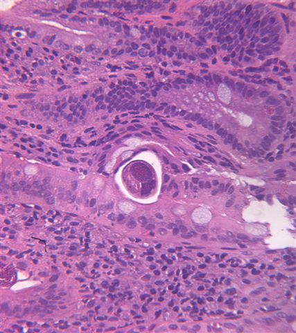
Microscopic (histologic) images
Contributed by Bobbi Pritt, M.D.
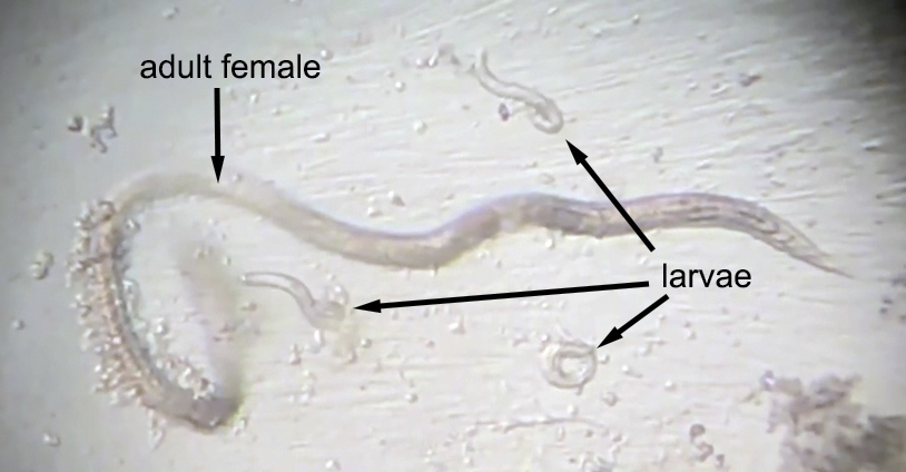
Adult female and larvae
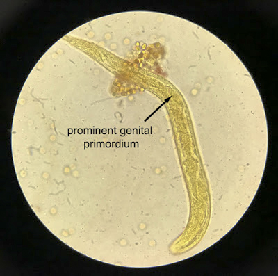
Prominent genital primordium
Case #133
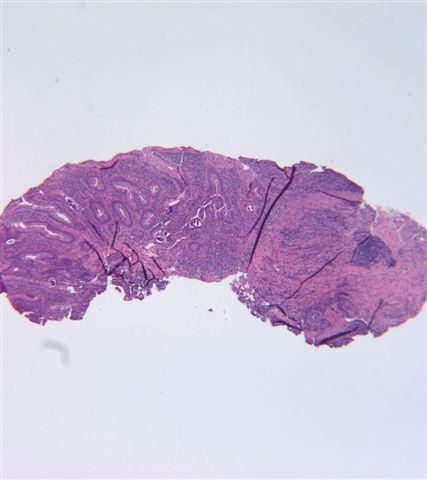
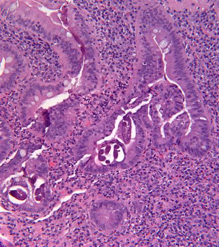
Duodenal biopsies of a 43 year old Honduran man
Videos
Contributed by Bobbi Pritt, M.D.
Endodoscopy of elderly woman with hematemesis (parasite case #499)
Differential diagnosis
- Capillaria philippinensis:
- May present similar to S. stercoralis with intestinal adults and larvae
- However, Capillaria is an obligate parasite and usually does not survive in viral culture media for very long
- In wet preparations Capillaria adults have a prominent stichosome but Strongyloides adults do not
- Capillaria rhabditiform larvae lack the prominent genital primordium and clavate eosphagus of Strongyloides
