

Soft tissue
Adipose tissue tumors
Lipoma and variants
Myolipoma
Author: Vijay Shankar, M.D.

Last author update: 1 August 2012
Last staff update: 1 April 2024 (update in progress)
Copyright: 2002-2024, PathologyOutlines.com, Inc.
PubMed Search: Myolipoma
Table of Contents
Definition / general | Terminology | Epidemiology | Radiology images | Case reports | Treatment | Gross description | Gross images | Microscopic (histologic) description | Microscopic (histologic) images | Positive stains | Differential diagnosis | Board review style question #1 | Board review style answer #1Cite this page: Shankar V. Myolipoma. PathologyOutlines.com website. https://www.pathologyoutlines.com/topic/softtissueadiposemyolipoma.html. Accessed April 25th, 2024.
Definition / general
- Benign tumor of mature adipocytes and mature smooth muscle
- First described in 1991 (Am J Surg Pathol 1991;15:121)
Terminology
- Called lipoleiomyoma in uterus
- Note: myelolipoma is tumor with hematopoietic (including myeloid) elements, often in adrenal gland
Epidemiology
- Very rare tumor of adults in abdomen, retroperitoneum or abdominal wall
- Other rare sites include eyelid, pericardium, base of tongue
Radiology images
Contributed by Drs. Arno Vanstapel and Raf Sciot, Case #452
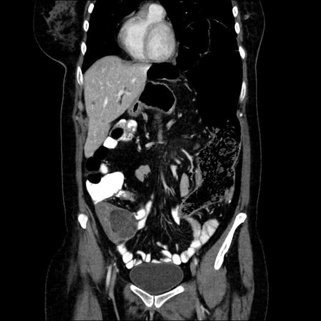
CT abdomen frontal
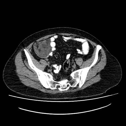
CT abdomen transverse
Case reports
- 4 year old boy presented with paraspinal tumor (Case Report Med 2009;2009:520126)
- 34 year old woman with tumor of iliac fossa (World J Surg Oncol 2005;3:72)
- 51 year old woman with pain in the right iliac fossa (Case #452)
- 55 year old woman with tumor presenting as inguinal hernia (Radiology Case Reports 2006, Vol 1, No 1)
Treatment
- Excision
- Does not recur, metastasize or transform
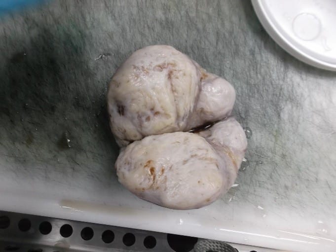
Gross description
- Often 9 cm or more, completely or partially encapsulated, yellow-white
Gross images
Contributed by Drs. Arno Vanstapel and Raf Sciot, Case #452

Images hosted on other servers:

Fatty tissue with bands and nodules of firm white tissue
Microscopic (histologic) description
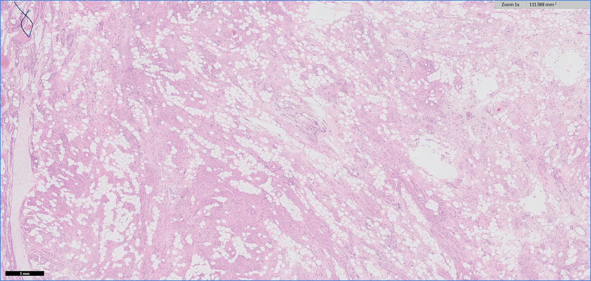
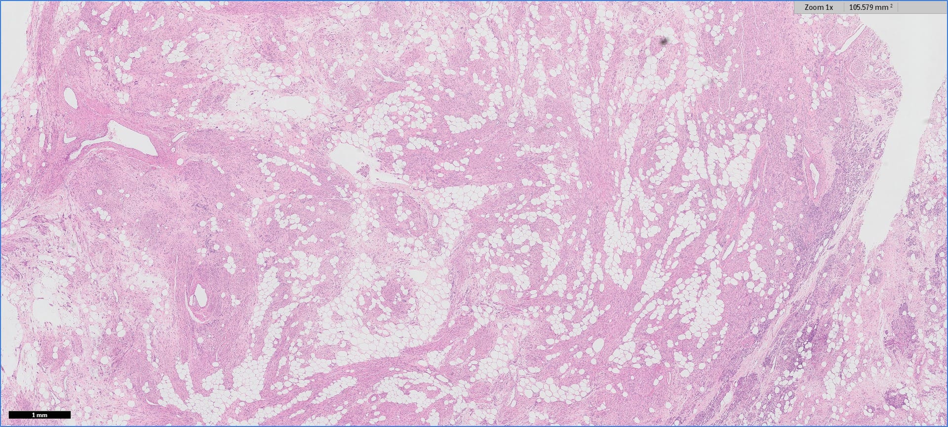
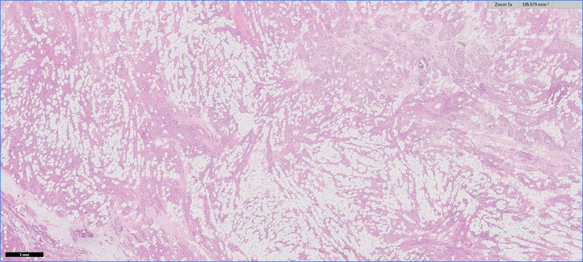
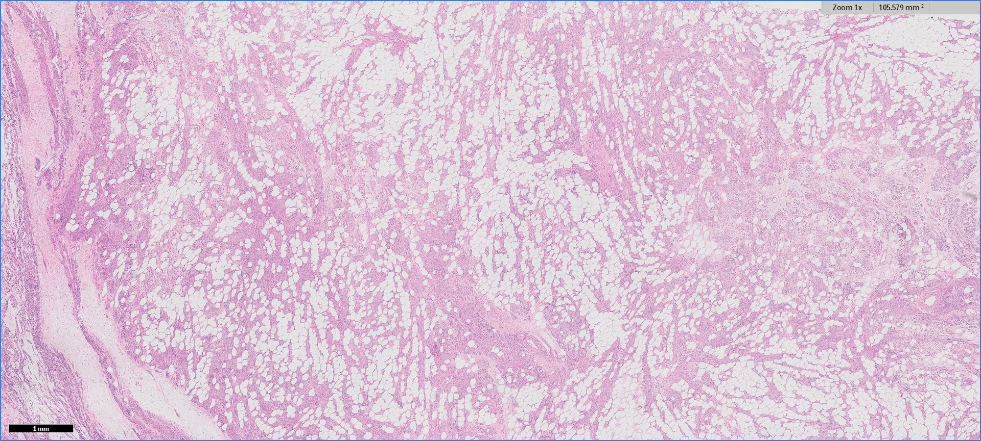
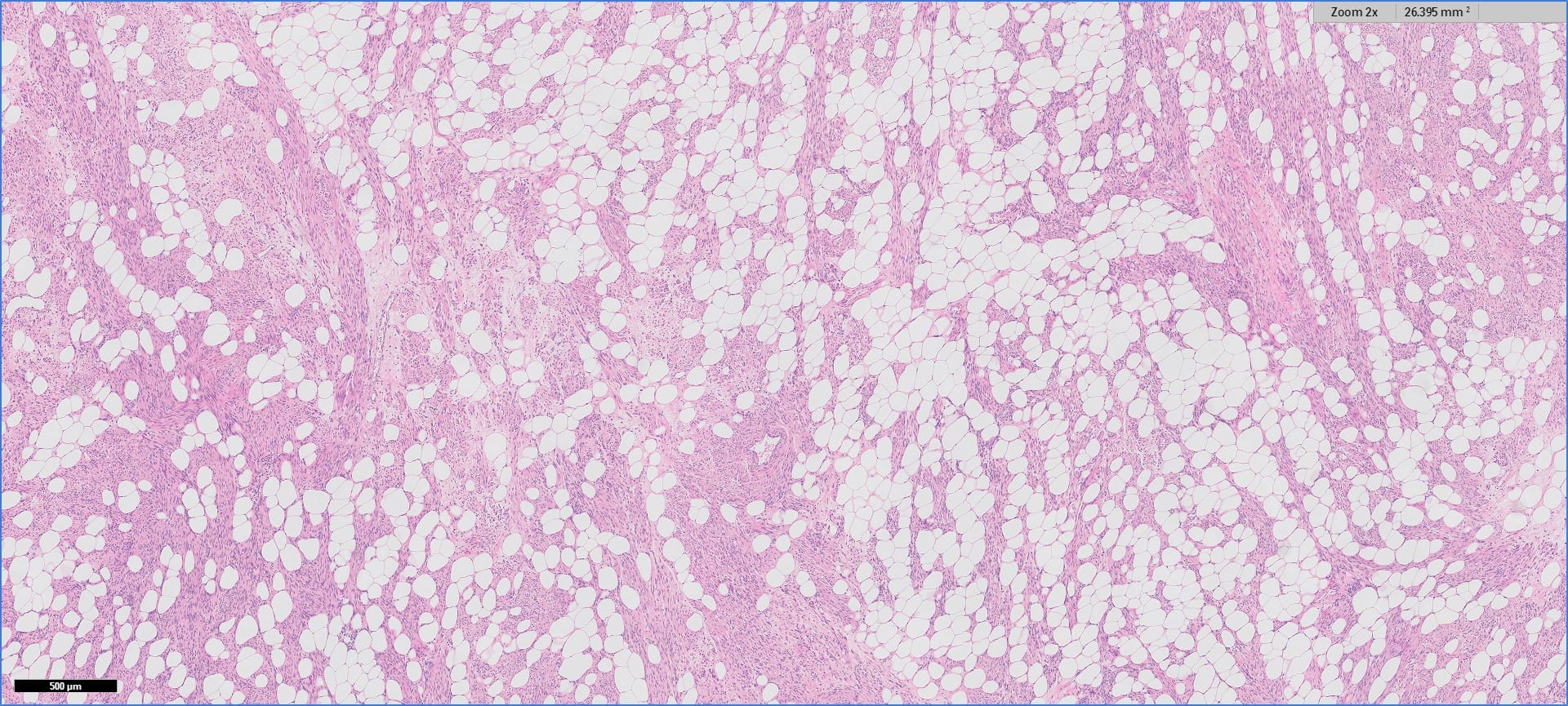
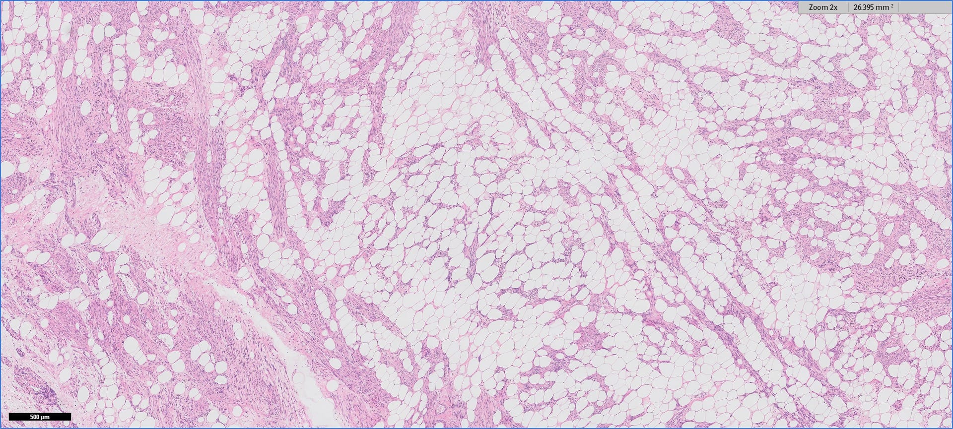
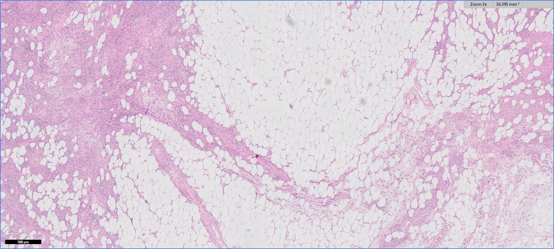
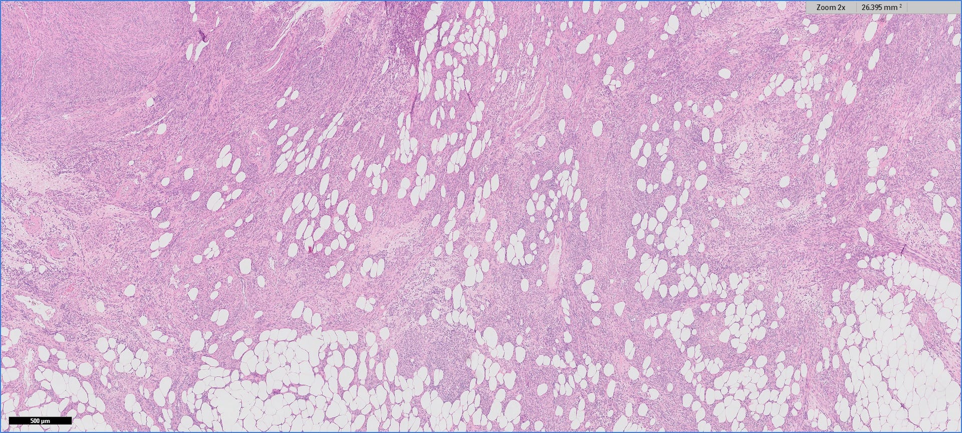
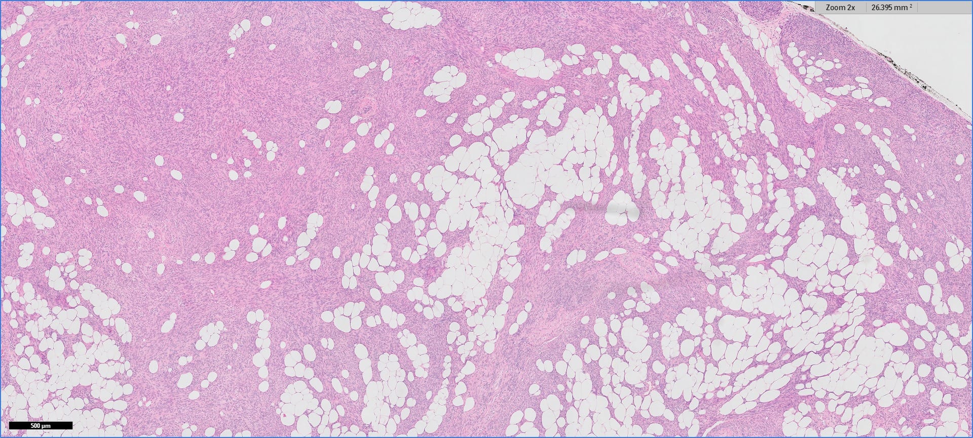
- Mature adipose tissue and mature (well differentiated) smooth muscle in short fascicles (Stanford University)
- Variable fibrosis and inflammation
- No floret cells, no thick walled vessels, no necrosis, no mitotic figures
Microscopic (histologic) images
Contributed by Drs. Arno Vanstapel and Raf Sciot, Case #452
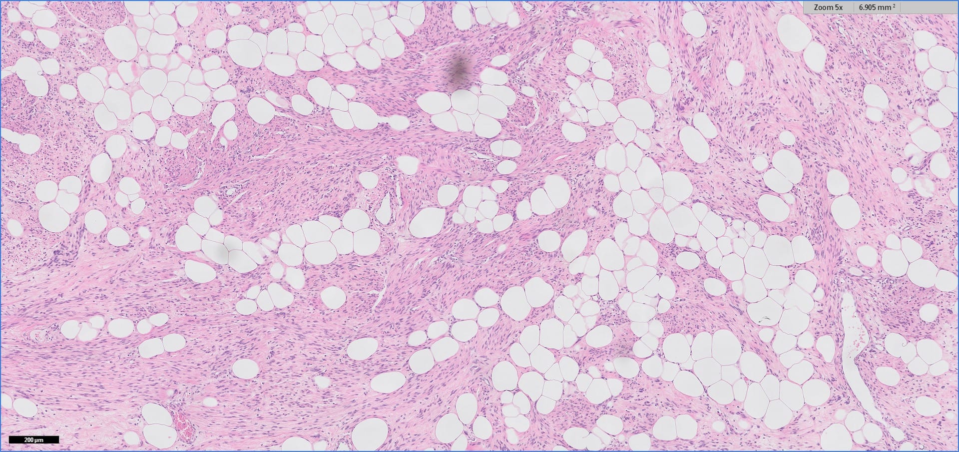
5x
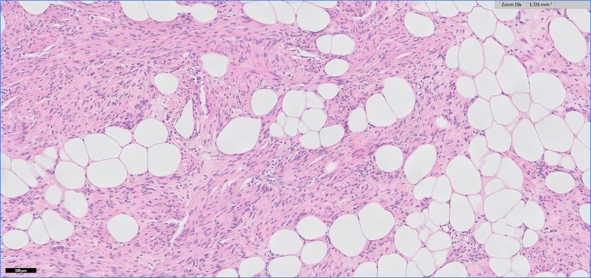
10x
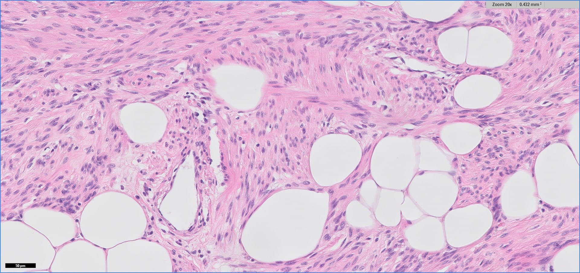
20x


Images hosted on other servers:

Mature fat and smooth muscle fibers

Low magnification view of myolipoma


Smooth muscle fibers are SMA+ (left), desmin+ (right)
Positive stains
- Smooth muscle actin, desmin
- Occasionally ER, PR
Differential diagnosis
- Angiomyolipoma: also thick walled vessels, HMB45+
- Liposarcoma with heterologous elements: has lipoblasts and atypia
- Low grade leiomyosarcoma infiltrating fat: smooth muscle component has atypia and mitotic figures, adipose tissue is not part of tumor
Board review style question #1
Which of the following immunohistochemical stains would help distinguish retroperitoneal myolipoma from a well differentiated liposarcoma invading smooth muscle?
A. CDK4
B. HMB45
C. S100
D. Smooth muscle actin
E. Vimentin
A. CDK4
B. HMB45
C. S100
D. Smooth muscle actin
E. Vimentin
Board review style answer #1
