

Soft tissue
Fibroblastic / myofibroblastic
Fibrosarcoma
Fibrosarcoma-infantile
Author: Komal Arora, M.D.

Last author update: 1 July 2012
Last staff update: 6 December 2021
Copyright: 2002-2024, PathologyOutlines.com, Inc.
PubMed Search: Fibrosarcoma [title] infantile
Table of Contents
Definition / general | Epidemiology | Clinical features | Case reports | Treatment | Gross description | Gross images | Microscopic (histologic) description | Microscopic (histologic) images | Positive stains | Electron microscopy description | Molecular / cytogenetics description | Differential diagnosis | Additional referencesCite this page: Arora K. Fibrosarcoma-infantile. PathologyOutlines.com website. https://www.pathologyoutlines.com/topic/softtissuefibrosarcomainfantile.html. Accessed May 12th, 2024.
Definition / general
- Resembles adult fibrosarcoma morphologically but better prognosis
- Age cutoff between infantile and adult forms usually varies between 5 and 10 years
Epidemiology
- Usually presents before age 2 years in axial regions or extremities with vary rapid growth
- Related to congenital mesoblastic nephroma, which has same translocation
Clinical features
- 40 - 50% recur but only rarely metastasizes
- Survival is 90%+
Case reports
- Premature newborn with large facial mass (Arch Pathol Lab Med 2003;127:e281)
- Patient with shoulder mass clinically diagnosed as benign vascular lesion (Cutis 2012;89:61)
Treatment
- Surgery and chemotherapy (J Pediatr Hematol Oncol 2002;24:722, Pediatr Blood Cancer 2009;53:23)
Gross description
- May exceed 30 cm (grotesquely large compared to size of child) with tense erythematous and ulcerated overlying skin
- Firm to soft cut surface is fleshy, gray-tan with myxoid change, cystic degeneration, hemorrhage and necrosis
Gross images
AFIP images
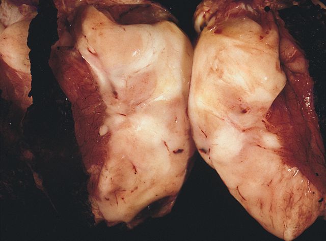
Fleshy white
mass similar
to adult
fibrosarcoma
Microscopic (histologic) description
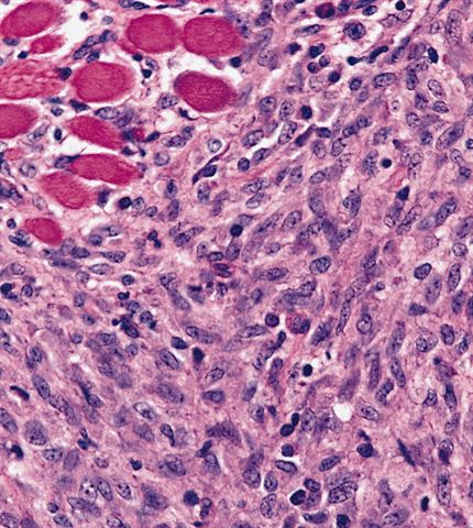
- Poorly circumscribed, lobulated mass of small to large spindled cells in fascicles or herringbone pattern with high cellularity, nuclear atypia and pleomorphism
- Increased mitotic figures, hemorrhage and necrosis
- Resembles adult fibrosarcoma
- May have prominent hemangiopericytoma-like areas, dystrophic calcification, extramedullary hematopoiesis
- Infiltrates adjacent soft tissue with irregular margins
Microscopic (histologic) images
AFIP images

Biphasic pattern with fibroblastic and cellular myxoid areas
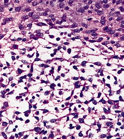
High power of myxoid area
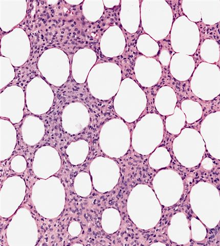
Infiltration of fat
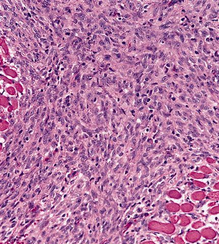
Infiltration of muscle
Spindle cells
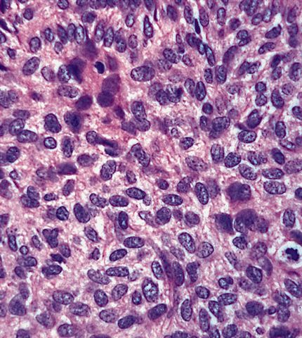
Plump cells have granular chromatin
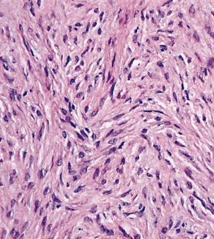
Less cellular tumor which overlaps with infantile fibromatosis, although it almost never metastasizes
Images hosted on other servers:


Leg tumor
Positive stains
- Vimentin
- Variable focal smooth muscle actin, desmin, S100 and CD34
Electron microscopy description
- Fibroblastic and myofibroblastic features
Molecular / cytogenetics description
- 70% have t(12;15)(p13;q26), causes ETV6-NTRK3 gene fusion transcript (ETS variant gene 6 and neurotrophic tyrosine receptor kinase type 3) detectable by FISH (Mod Pathol 2001;14:1246) or RT-PCR (Am J Surg Pathol 2000;24:937, Am J Clin Pathol 2001;115:348)
- Similar translocation also present in secretory breast carcinoma (Mod Pathol 2009;22:291)
- Also trisomy 8, 11, 17 and 20
Differential diagnosis
- Adult type fibrosarcoma: usually age 10+, no t(12;15)
- Infantile fibromatosis: no pleomorphism, no mitotic figures, no t(12;15)
- Myofibromatosis: myofibroblastic features, no t(12;15) (Pediatr Dev Pathol 2008;11:355)
Additional references
