
14 October 2020 - Case of the Month #496
All cases are archived on our website. To view them sorted by case number, diagnosis or category, visit our main Case of the Month page. To subscribe or unsubscribe to the Case of the Month or our other email lists, click here.
Thanks to Dr. Cheng Wang, Dalhousie University and Nova Scotia Health Authority, Halifax, NS (Canada) and the Genitourinary Pathology Society (GUPS) for contributing this case and discussion. The discussion was reviewed by Dr. Maria Tretiakova, University of Washington.

Advertisement
Case of the Month #496
Clinical history:
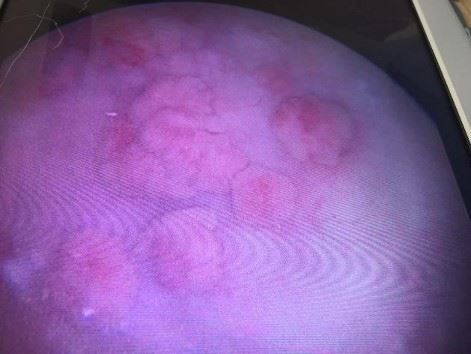
A woman in her early 50s presented with multiple broad based bladder mucosal lesions seen on cystoscopic examination.
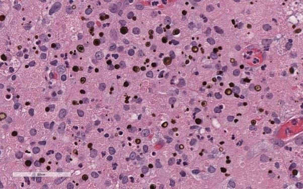
Clinical and histopathology images:
What is your diagnosis?
Diagnosis: Malakoplakia
Test question (answer at the end):
What is the name of the targetoid bodies, which are pathognomonic for this condition?
A. Michaelis-Gutman bodies
B. Aschoff bodies
C. Asbestos bodies
D. Howell-Jolly bodies
E. Psammoma bodies
Stains:
Discussion:
Malakoplakia is caused by defects in phagocytic or degradative functions of histiocytes in response to bacterial infections (usually by gram negative coliforms). It is a relatively uncommon lesion seen in patients with hematuria or signs of urinary tract infections. It occurs more commonly in women (4:1), older patients and immunosuppressed patients (HIV positive and renal transplant recipients). It can also occur, although less commonly, in the prostate, kidney, testis, lymph nodes, lungs and bone. The typical mucosal lesion of malakoplakia is characterized by multiple yellow-brown, soft (malakos) plaques (plakos). Sometimes malakoplakia can form nodules or polypoid mass lesions, mimicking neoplasms.
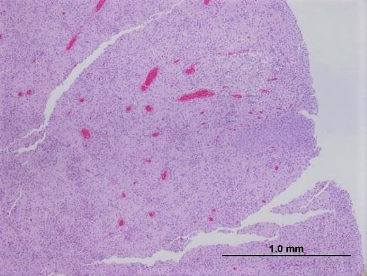
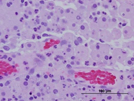
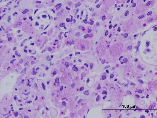
Microscopically, malakoplakia is composed of sheets of von Hansemann histiocytes (macrophages with abundant granular cytoplasm) in the lamina propria. Careful search of Michaelis-Gutman bodies at high power view is key to making the correct diagnosis. The intracellular and extracellular target-like structures (Michaelis-Gutman bodies) are formed by deposition of calcium and iron phosphate on undigested bacterial components (von Kossa stain for calcium, iron stain). The treatment includes using antibiotics that concentrate in macrophages and discontinuation of immunosuppressive drug therapy.
References:
Br J Urol 1987;59:559, Urol Case Rep 2014;3:6, Malakoplakia
Test question answer:
A. Michaelis-Gutman bodies
All cases are archived on our website. To view them sorted by case number, diagnosis or category, visit our main Case of the Month page. To subscribe or unsubscribe to the Case of the Month or our other email lists, click here.
Thanks to Dr. Cheng Wang, Dalhousie University and Nova Scotia Health Authority, Halifax, NS (Canada) and the Genitourinary Pathology Society (GUPS) for contributing this case and discussion. The discussion was reviewed by Dr. Maria Tretiakova, University of Washington.
Advertisement
Case of the Month #496
Clinical history:
A woman in her early 50s presented with multiple broad based bladder mucosal lesions seen on cystoscopic examination.
Clinical and histopathology images:
What is your diagnosis?
Click here for diagnosis, test question and discussion:
Diagnosis: Malakoplakia
Test question (answer at the end):
What is the name of the targetoid bodies, which are pathognomonic for this condition?
A. Michaelis-Gutman bodies
B. Aschoff bodies
C. Asbestos bodies
D. Howell-Jolly bodies
E. Psammoma bodies
Stains:
PAS stain
Von Kossa stain
Discussion:
Malakoplakia is caused by defects in phagocytic or degradative functions of histiocytes in response to bacterial infections (usually by gram negative coliforms). It is a relatively uncommon lesion seen in patients with hematuria or signs of urinary tract infections. It occurs more commonly in women (4:1), older patients and immunosuppressed patients (HIV positive and renal transplant recipients). It can also occur, although less commonly, in the prostate, kidney, testis, lymph nodes, lungs and bone. The typical mucosal lesion of malakoplakia is characterized by multiple yellow-brown, soft (malakos) plaques (plakos). Sometimes malakoplakia can form nodules or polypoid mass lesions, mimicking neoplasms.
Microscopically, malakoplakia is composed of sheets of von Hansemann histiocytes (macrophages with abundant granular cytoplasm) in the lamina propria. Careful search of Michaelis-Gutman bodies at high power view is key to making the correct diagnosis. The intracellular and extracellular target-like structures (Michaelis-Gutman bodies) are formed by deposition of calcium and iron phosphate on undigested bacterial components (von Kossa stain for calcium, iron stain). The treatment includes using antibiotics that concentrate in macrophages and discontinuation of immunosuppressive drug therapy.
References:
Br J Urol 1987;59:559, Urol Case Rep 2014;3:6, Malakoplakia
Test question answer:
A. Michaelis-Gutman bodies
