
Esophagus
Other nonneoplastic
Epidermoid metaplasia
Author: Yukihiro Nakanishi, M.D., Ph.D.
Editorial Board Members: Maryam Kherad Pezhouh, M.D., M.Sc., Raul S. Gonzalez, M.D.
Editor-in-Chief: Debra L. Zynger, M.D.

Last author update: 6 July 2022
Last staff update: 6 July 2022
Copyright: 2018-2024, PathologyOutlines.com, Inc.
PubMed Search: Esophagus epidermoid metaplasia

Table of Contents
Definition / general | Essential features | Terminology | ICD coding | Epidemiology | Sites | Etiology | Clinical features | Diagnosis | Case reports | Treatment | Clinical images | Gross description | Microscopic (histologic) description | Microscopic (histologic) images | Positive stains | Molecular / cytogenetics description | Sample pathology report | Differential diagnosis | Additional references | Board review style question #1 | Board review style answer #1 | Board review style question #2 | Board review style answer #2Cite this page: Nakanishi Y. Epidermoid metaplasia. PathologyOutlines.com website. https://www.pathologyoutlines.com/topic/esophagusepidermization.html. Accessed May 13th, 2024.
Definition / general
- Esophageal squamous epithelium with a prominent granular layer and orthokeratosis / hyperorthokeratosis, resembling the epidermis of the skin
Essential features
- Affects middle aged to elderly individuals with a history of smoking and alcohol intake
- Well demarcated white plaque in the mid to distal esophagus
- Esophageal squamous epithelium with a prominent granular layer and orthokeratosis / hyperorthokeratosis
- Possible association with esophageal squamous cell carcinoma and dysplasia
- Follow up, unless a patient has a concurrent squamous cell carcinoma
Terminology
- Also known as epidermization, esophageal leukoplakia, orthokeratosis and hyperkeratosis
ICD coding
- ICD-10: K22.9 - Disease of esophagus, unspecified
Epidemiology
- Incidence:
- 2 cases (0.2%) of epidermoid metaplasia out of 1,048 consecutive esophageal biopsies; 50x less common than parakeratosis (11%) (Histopathology 2016;68:988)
- 37 cases (2%) of epidermoid metaplasia and hyperkeratosis without a prominent granular layer out of 1,845 consecutive esophageal biopsies (Histopathology 2013;63:463)
- Mean age: 61.5 years with a slight female predominance (Mod Pathol 2014;27:38)
- Possible association with squamous cell carcinoma (Am J Gastroenterol 2021;116:1533, Histopathology 2016;68:988, Histopathology 2013;63:463, Mod Pathol 2014;27:38)
Sites
- Mid to distal esophagus (Mod Pathol 2014;27:38)
- Most common in the mid esophagus (51%) in non-Barrett setting (Histopathology 2013;63:463)
- Most common in the distal esophagus (98%) in setting of Barrett esophagus (Histopathology 2013;63:463)
Etiology
- Remains unknown
- Associated with smoking and alcohol intake (Histopathology 2016;68:988, Histopathology 2013;63:463, Mod Pathol 2014;27:38)
Clinical features
- Dysphagia or asymptomatic
- Endoscopy findings
- Scaly or shaggy white plaque with clear borders
- Mimics crackleware china
- Lugol voiding lesion with clear demarcation, resembling superficial esophageal cancer (Hepatogastroenterology 2011;58:809)
Diagnosis
- Diagnosis is based on esophageal biopsy histologic findings
Case reports
- 53 year old man with epidermoid metaplasia and mucosal bridge formation (Endoscopy 2016;48:E325)
- 57 year old Japanese man with superficial esophageal cancer arising from epidermization (Clin J Gastroenterol 2018;11:29)
- 58 year old Japanese man with epidermization and a concurrent superficial squamous cell carcinoma (Am J Surg Pathol 1997;21:605)
- 69 year old man with an unusual appearance of epidermoid metaplasia (Endoscopy 2015;47:E100)
- 69 year old man with epidermoid metaplasia in association with esophageal intramural pseudodiverticulosis and candidiasis (Case Rep Gastroenterol 2021;15:709)
Treatment
- Follow up, unless a patient has a concurrent squamous cell carcinoma
Clinical images
Images hosted on other servers:

Scaly white plaque
Gross description
- Scaly or shaggy white plaque with clear borders (mean size: 2.6 cm) (Mod Pathol 2014;27:38)
Microscopic (histologic) description
- Esophageal squamous epithelium with a compact layer of orthokeratosis / hyperorthokeratosis and a prominent granular layer 1 - 4 cells thick with keratohyalin granules, resembling the epidermis of the skin
- Abrupt transition from the adjacent normal squamous epithelium
- Reference: Am J Surg Pathol 1997;21:605
Microscopic (histologic) images
Contributed by Yukihiro Nakanishi, M.D., Ph.D.
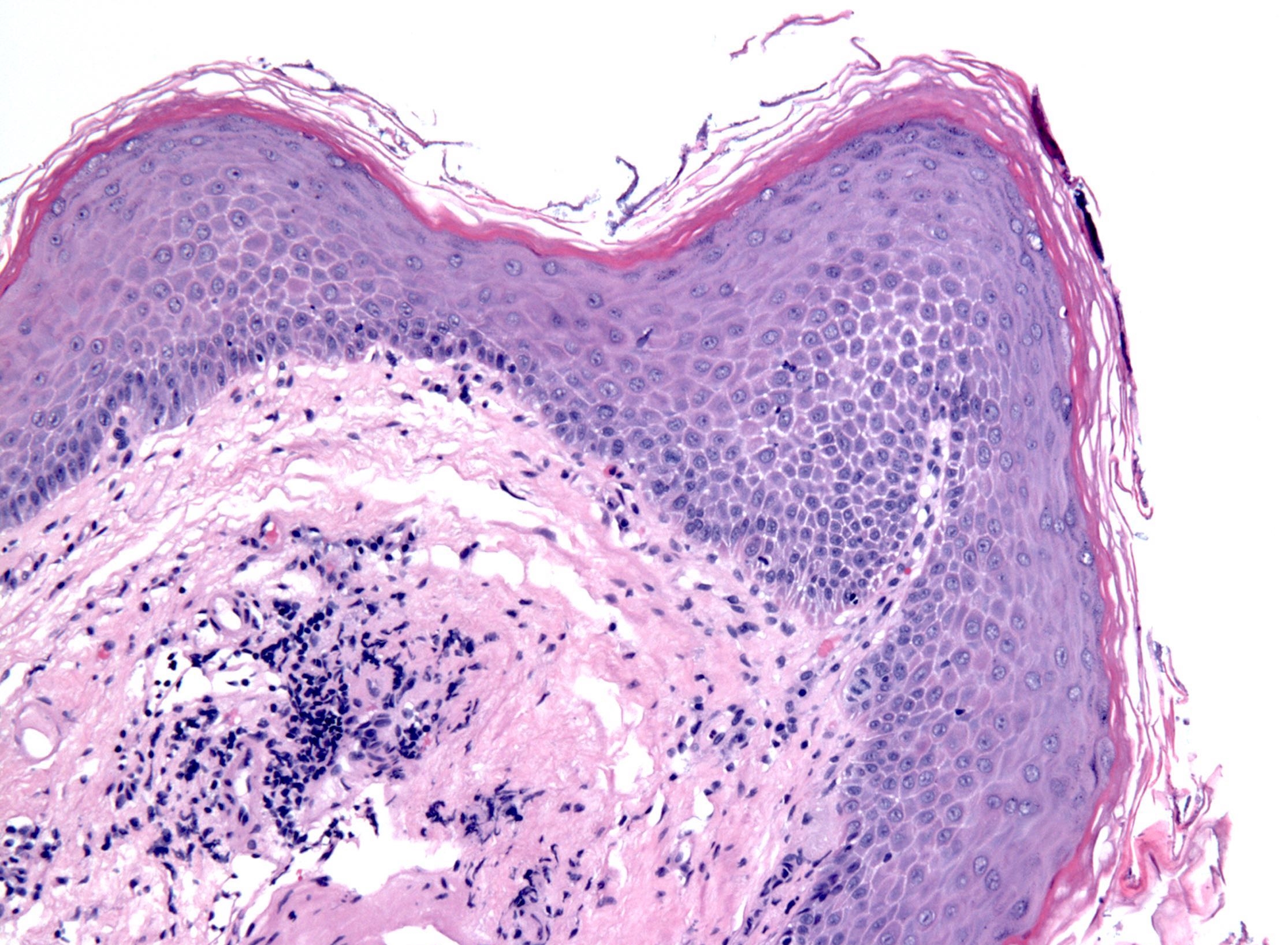
Orthokeratosis /
hyperorthokeratosis
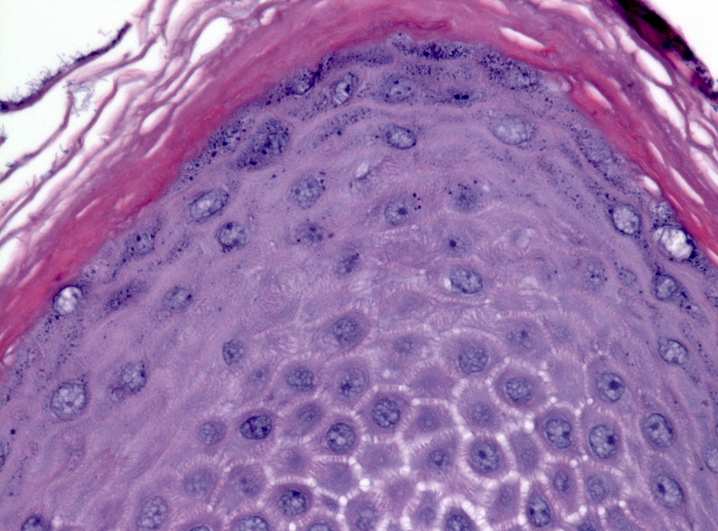
Prominent granular layer
Positive stains
- PAS: reduced expression of PAS+ cells compared with normal esophageal squamous epithelium (Am J Surg Pathol 1997;21:605)
- Antikeratin 1 immunostain labels the suprabasal cells of epidermization (Am J Surg Pathol 1997;21:605)
- Antikeratin 14 immunostain labels all the cell layers of epidermization (Am J Surg Pathol 1997;21:605)
Molecular / cytogenetics description
- Targeted next generation sequencing analysis of epidermoid metaplasia shows frequently mutated genes consisted of TP53, PIK3CA, EGFR, MYCN, HRAS and the TERT promoter (Mod Pathol 2017;30:1613)
Sample pathology report
- Esophagus, mid, plaque, biopsy:
- Epidermoid metaplasia / epidermization / orthokeratosis with prominent granular layer
- Negative for dysplasia or malignancy
Differential diagnosis
- Hyperkeratosis:
- No prominent granular layer
- Parakeratosis:
- Nuclei present in a keratin layer without an associated prominent granular layer
- Dysplasia:
- Variable nuclear atypia present
- Squamous cell carcinoma in situ:
- Full thickness nuclear atypia present
- Atrophic change:
- No orthokeratosis or prominent granular layer
Additional references
Board review style question #1
Which of the following statements about epidermization / epidermoid metaplasia of the esophagus is true?
- Characterized by orthokeratosis / hyperorthokeratosis with a prominent granular layer
- No association with smoking or excessive alcohol consumption
- No association with squamous cell carcinoma and dysplasia
- PAS stain highlights epidermoid metaplasia
Board review style answer #1
A. Epidermization / epidermal metaplasia is characterized by orthokeratosis / hyperorthokeratosis with a prominent granular layer, mimicking epidermis.
Comment Here
Reference: Epidermoid metaplasia
Comment Here
Reference: Epidermoid metaplasia
Board review style question #2

Which of the following statements about histologic features of epidermization / epidermoid metaplasia of the esophagus is true?
- Characterized by the presence of the stratum lucidum beneath the cornified layer
- Finding of increased thickness in the cornified layer is called parakeratosis
- Granular layer is characterized by the presence of basophilic stained granules known as keratohyalin granules
- Orthokeratosis / hyperkeratosis is more frequently seen in the esophagus than parakeratosis
Board review style answer #2
C. Epidermization / epidermoid metaplasia is histopathologically characterized by the presence of the granular layer beneath the cornified layer. The granular layer contains basophilic stained granules known as keratohyalin granules. Parakeratosis is characterized by keratosis with persistence of the cell nuclei. The stratum lucidum is seen only in soles and palms and is not seen in epidermization / epidermoid metaplasia. Parakeratosis is more frequently seen in the esophagus than orthokeratosis / hyperkeratosis.
Comment Here
Reference: Epidermoid metaplasia
Comment Here
Reference: Epidermoid metaplasia
