Gallbladder & extrahepatic bile ducts
Gallbladder nonneoplastic
Papillary hyperplasia
Editorial Board Member: Danielle Hutchings, M.D.
Deputy Editor-in-Chief: Aaron R. Huber, D.O.


Last author update: 17 October 2024
Last staff update: 17 October 2024
Copyright: 2003-2025, PathologyOutlines.com, Inc.
PubMed Search: Papillary hyperplasia
Table of Contents
Definition / general | Essential features | Terminology | Epidemiology | Sites | Pathophysiology | Etiology | Diagnosis | Laboratory | Radiology description | Prognostic factors | Case reports | Treatment | Gross description | Gross images | Microscopic (histologic) description | Microscopic (histologic) images | Sample pathology report | Differential diagnosis | Practice question #1 | Practice answer #1 | Practice question #2 | Practice answer #2Cite this page: Kolch NL, Evason KJ. Papillary hyperplasia. PathologyOutlines.com website. https://www.pathologyoutlines.com/topic/gallbladderpaphyperplasia.html. Accessed August 27th, 2025.
Definition / general
- Benign nonneoplastic change in gallbladder epithelium
- May be primary in cause or secondary to anatomical malformations (pancreaticobiliary maljunction) and inflammatory diseases (including cholecystitis, cholelithiasis)
Essential features
- Can mimic other conditions with diagnostic testing and imaging pointing to gallbladder cancer (Int J Surg Case Rep 2021;88:106542)
- Primary papillary hyperplasia is typically asymptomatic; few cases show right upper quadrant pain (J Pediatr Surg 2001;36:1584)
- Idiopathic or secondary to previous hepatobiliary condition (Int Surg 2014;99:247)
- Typically confined to gallbladder
Terminology
- Adenomatous hyperplasia (J Pediatr Surg 2001;36:1584)
- Primary papillary hyperplasia of the gallbladder (Int Surg 2014;99:247)
Epidemiology
- Unknown at this time
Sites
- Primarily confined to gallbladder
Pathophysiology
- Associated with inflammatory disease (cholecystitis, cholelithiasis, primary sclerosing cholangitis, inflammatory bowel disease) and anatomical malformation (pancreaticobiliary maljunction) (Int J Surg Case Rep 2021;88:106542)
- In pancreaticobiliary maljunction, reflux of pancreatic juice and stagnant gallbladder bile with high concentrations of lysolecithin are thought to drive abnormal proliferation of gallbladder mucosa (Lab Invest 2009;89:1018)
- In cholesterolosis, papillary hyperplasia may be an adaption to increase surface area for enhanced absorption of cholesterol in the gallbladder
- May occur idiopathically without evidence of chronic inflammation or gallstones (Int Surg 2014;99:247)
Etiology
- Potential association with infectious diseases in human immunodeficiency virus (HIV) positive patients (J Glob Infect Dis 2021;13:105)
Diagnosis
- Diagnosed postresection on microscopic evaluation of H&E stained slides
Laboratory
- Nothing conclusive known at this time
Radiology description
- Computed tomography (CT) imaging and ultrasound of the abdomen show gallbladder wall thickening; persistent thickening may be captured on imaging over time
Prognostic factors
- In patients presenting with biliary colic, symptoms resolved after cholecystectomy (J Pediatr Surg 2001;36:1584)
- Due to the potential for misdiagnosis of malignancy, frozen sections during surgery can help prevent unnecessary surgical procedures
Case reports
- 12 year old girl with right upper quadrant pain for 10 months (J Pediatr Surg 2001;36:1584)
- 36 year old woman with upper abdominal discomfort and no prior history of hepatobiliary disease (Pathology 2006;38:591)
- 63 year old man with primary papillary hyperplasia of the gallbladder with widespread erythematous skin rash (Int Surg 2014;99:247)
- 70 year old man diagnosed with gallbladder cancer prior to surgery (Int J Surg Case Rep 2021;88:106542)
Treatment
- Cholecystectomy (J Pediatr Surg 2001;36:1584)
Gross description
- Diffuse thickening of the gallbladder wall
Gross images
Images hosted on other servers:

Thickened gallbladder mucosa
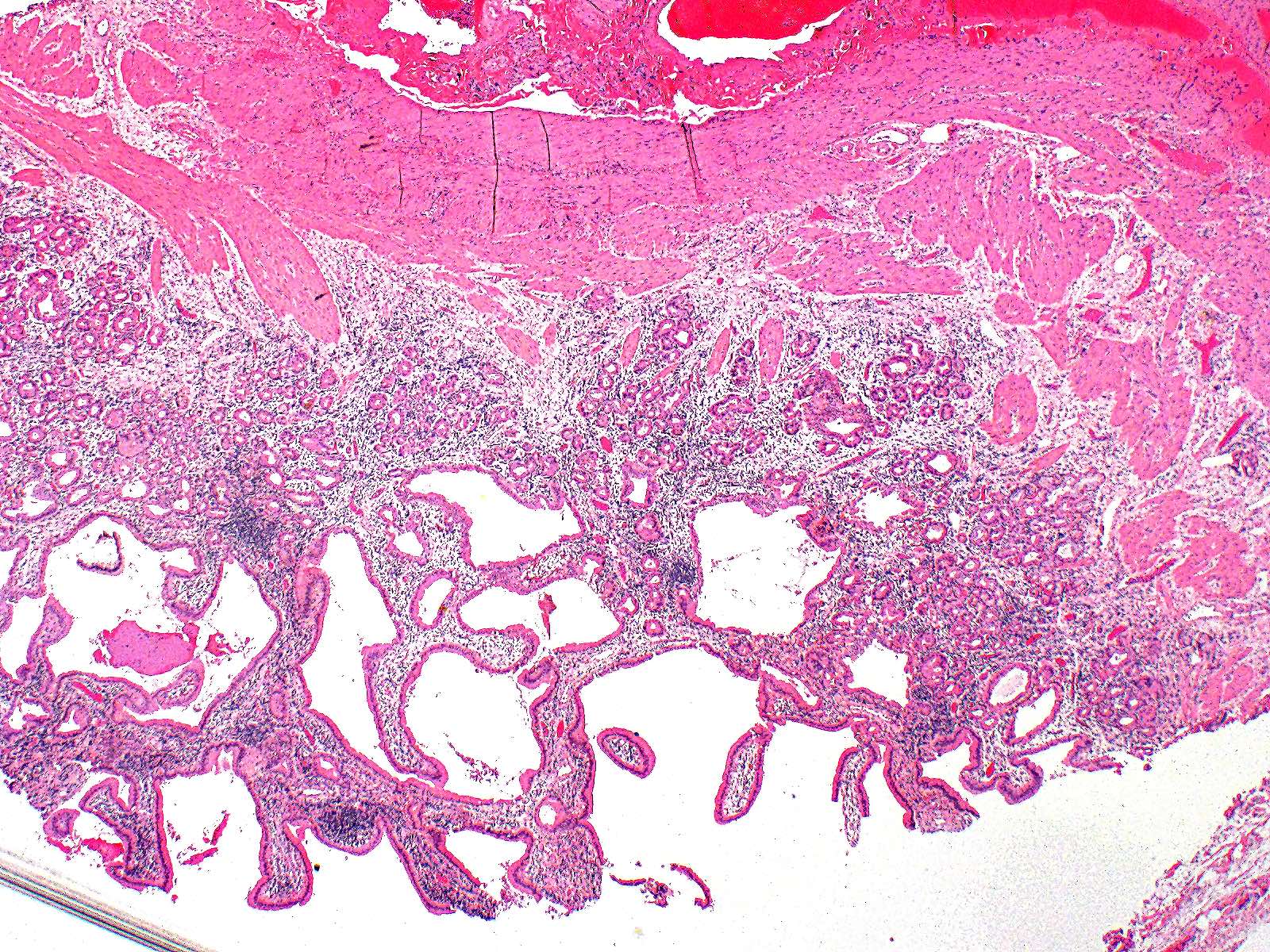
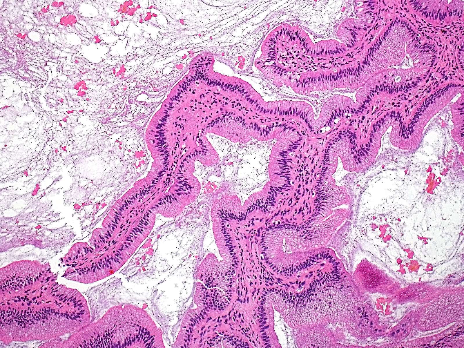
Microscopic (histologic) description
- Diffuse thickened mucosal papillary projections and folds (Int Surg 2014;99:247)
- Papillae are lined by biliary type, columnar cells and have loose fibroblastic stroma (Int Surg 2014;99:247)
- No significant cellular atypia (Int J Surg Case Rep 2021;88:106542)
- Minimal / no mitotic activity (Int J Surg Case Rep 2021;88:106542)
Microscopic (histologic) images
Contributed by Kimberley J. Evason, M.D., Ph.D.
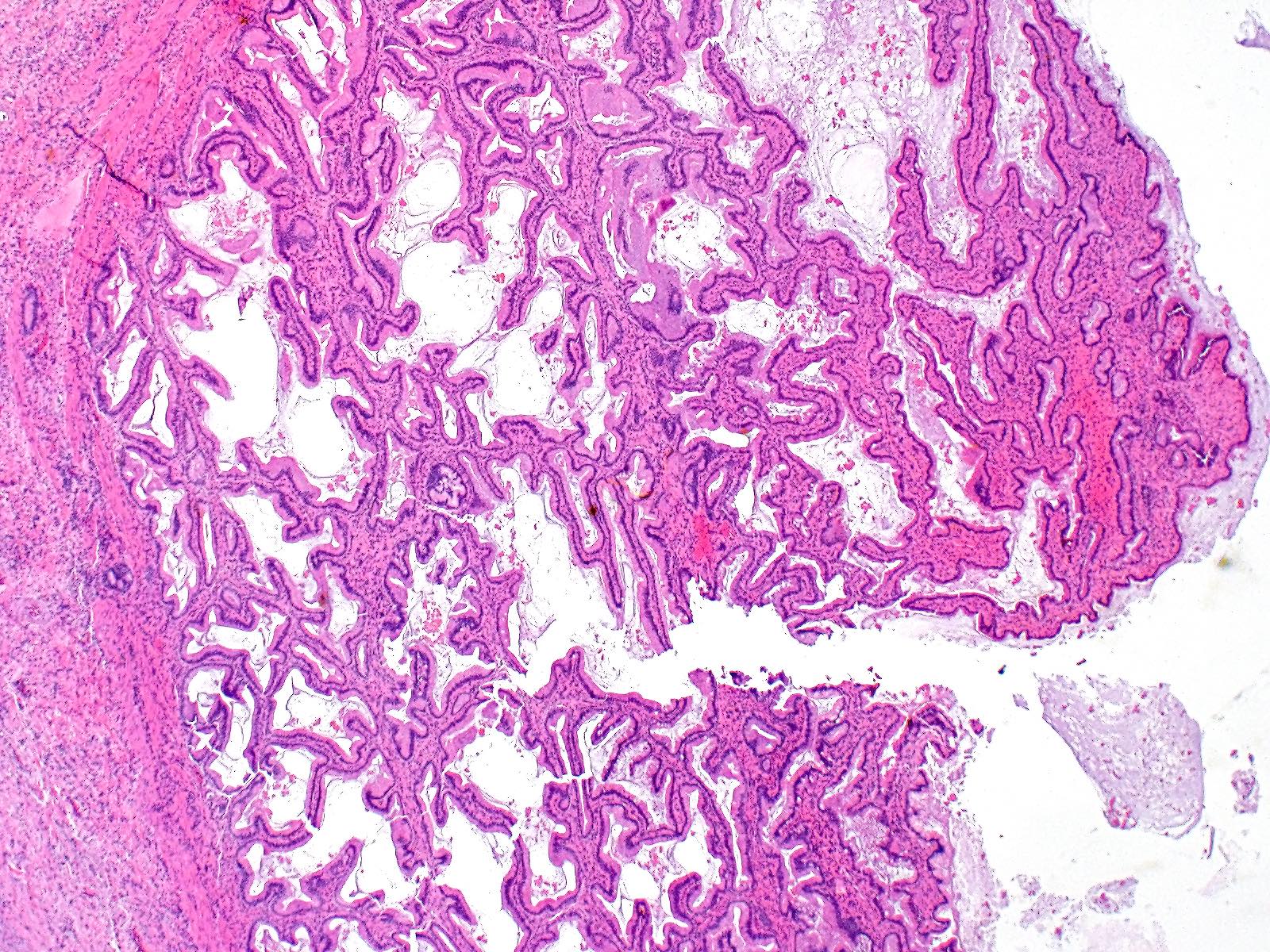
Papillary projections into lumen
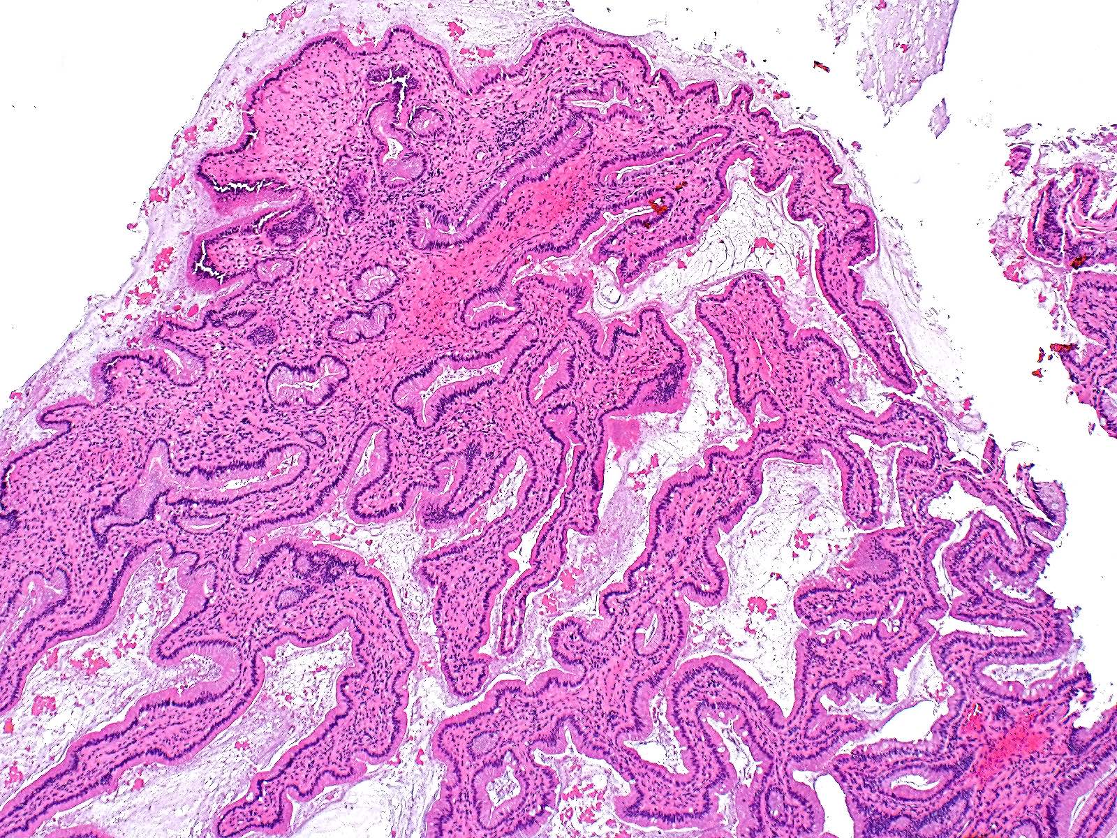
Papillary projections without dysplasia
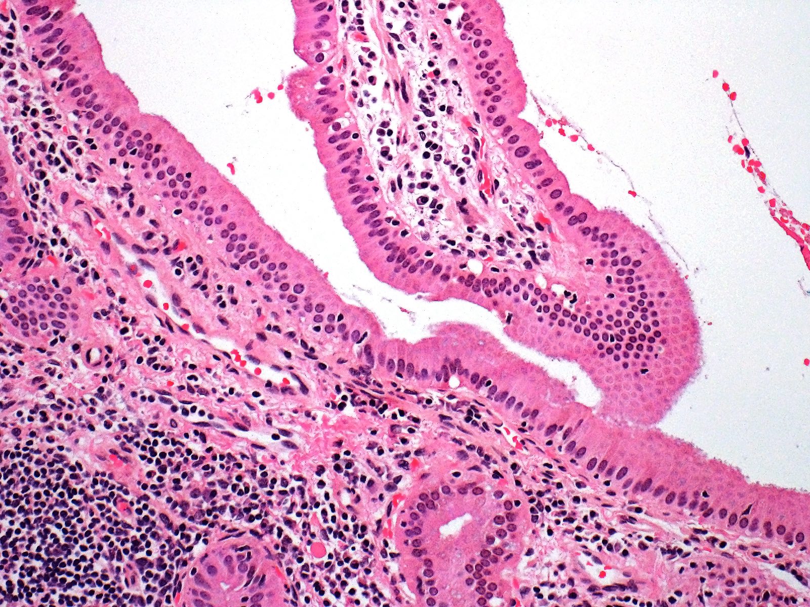
Columnar epithelium without dysplasia
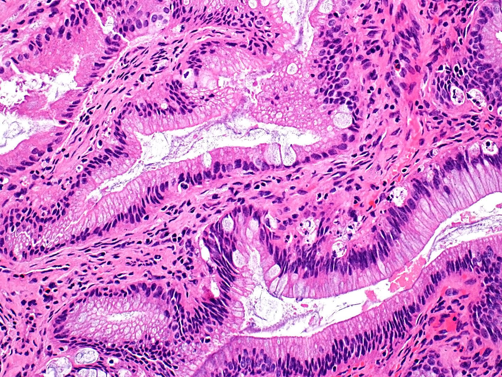
Focal intestinal metaplasia
Sample pathology report
- Gallbladder, cholecystectomy:
- Chronic cholecystitis with papillary hyperplasia
Differential diagnosis
- Gallbladder papillomatosis:
- Cytologic atypia including nuclear enlargement, crowding, hyperchromasia, prominent nucleoli or loss of polarity
- Gallbladder carcinoma:
- Infiltrative glands or single cells surrounded by desmoplastic stroma
- Cytologic atypia including nuclear enlargement, crowding, hyperchromasia, prominent nucleoli or loss of polarity (unless very well differentiated)
- Lymphovascular or perineural invasion
- Pyloric gland adenoma:
- Polypoid mass rather than diffuse thickening of the gallbladder wall
- Glands are pyloric type or Brunner gland-like
- Cytologic atypia (nuclear enlargement, crowding, hyperchromasia, prominent nucleoli or loss of polarity) sufficient for at least low grade dysplasia
- Adenomyomatous hyperplasia:
- Papillary projections not prominent
- Cystically dilated benign biliary glands
- Smooth muscle hypertrophy of gallbladder wall
- Intracholecystic papillary neoplasm:
- Polypoid mass rather than diffuse thickening of the gallbladder wall
- Cytologic atypia (nuclear enlargement, crowding, hyperchromasia, prominent nucleoli or loss of polarity) sufficient for at least low grade dysplasia
Practice question #1
The above slide was taken from a cholecystectomy of a 62 year old woman who presented with biliary colic for the past several months. Presurgical ultrasound revealed diffuse thickening of the gallbladder mucosa. What is the likely diagnosis?
- Adenomyomatous hyperplasia
- Cholesterolosis
- Gallbladder adenocarcinoma
- Papillary hyperplasia
Practice answer #1
D. Papillary hyperplasia. Papillary projections, lined by uniform columnar epithelial cells without nuclear atypia, are seen on histological imaging; these findings are consistent with papillary hyperplasia. Answer A is incorrect because the specimen lacks abundant Rokitansky-Aschoff sinuses and smooth muscle hypertrophy typically seen in adenomyomatous hyperplasia. Answer B is incorrect because lamina propria does not contain lipid laden macrophages. Answer C is incorrect because the lesion lacks nuclear atypia and desmoplastic stroma.
Comment Here
Reference: Papillary hyperplasia
Comment Here
Reference: Papillary hyperplasia
Practice question #2
Based on the provided image, which of the following histopathological findings would be most consistent with papillary hyperplasia of the gallbladder rather than gallbladder adenocarcinoma?
- Irregular glandular architecture
- Loss of nuclear polarity
- No significant cellular atypia
- Stromal invasion
Practice answer #2
C. No significant cellular atypia. Papillary hyperplasia is a benign condition characterized by absence of malignant cellular features. Answer A is incorrect because malignant conditions, such as gallbladder adenocarcinoma, are associated with disruptions in normal cellular architecture, whereas papillary hyperplasia of the gallbladder is a benign condition that preserves normal glandular architecture. Answer B is incorrect because loss of nuclear polarity is associated with malignancy where there is a disruption in the normal arrangement of nuclei. Answer D is incorrect because disruption in the basement membrane and infiltration of associated connective tissue is a characteristic of malignant processes, such as gallbladder adenocarcinoma.
Comment Here
Reference: Papillary hyperplasia
Comment Here
Reference: Papillary hyperplasia
