Larynx, hypopharynx & trachea
Benign tumors / nonneoplastic
Laryngocele
Authors: Jamie Brown, M.D., Ruta Gupta, M.D.
Editorial Board Member: Elizabeth Ann Bilodeau, D.M.D., M.D., M.S.Ed.
Deputy Editor-in-Chief: Kelly Magliocca, D.D.S., M.P.H.


Last author update: 2 June 2025
Last staff update: 2 June 2025
Copyright: 2019-2025, PathologyOutlines.com, Inc.
PubMed Search: Laryngocele
Table of Contents
Definition / general | Essential features | ICD coding | Epidemiology | Sites | Pathophysiology | Etiology | Clinical features | Diagnosis | Radiology description | Radiology images | Case reports | Treatment | Gross description | Microscopic (histologic) description | Microscopic (histologic) images | Positive stains | Sample pathology report | Differential diagnosis | Practice question #1 | Practice answer #1Cite this page: Brown J, Gupta R. Laryngocele. PathologyOutlines.com website. https://www.pathologyoutlines.com/topic/larynxlaryngocele.html. Accessed August 27th, 2025.
Definition / general
- Air filled dilation of the laryngeal saccule that communicates with the laryngeal lumen
- Subclassified as internal, external or combined based on relation to the thyrohyoid membrane
Essential features
- Usually unilateral
- Congenital or acquired
- Lined by respiratory epithelium with squamous or mucinous metaplasia
ICD coding
- ICD-10: Q31.3 - laryngocele
Epidemiology
- Rare; epidemiological data is limited
- A literature review in 1975 estimated an incidence of 1 per 2.5 million, with a ratio of M:F = 5:1 (J Laryngol Otol 1975;89:915)
- Few studies into incidence of asymptomatic cases, so true incidence may be much higher (see Etiology)
Sites
- By definition, a laryngocele involves the saccule of the laryngeal ventricle and communicates with the laryngeal lumen
- Usually unilateral, less often bilateral
- Subclassified as
- Internal: confined to the space between the larynx and thyrohyoid membrane
- External: extending through the thyrohyoid membrane
- Combined: dilated internal and external components
- References: Goldblum: Rosai and Ackerman's Surgical Pathology, 11th Edition, 2017, Longacre: Mills and Sternberg's Diagnostic Surgical Pathology, 7th Edition, 2022
Pathophysiology
- Saccule (appendix) of the laryngeal ventricle is surrounded by loose connective tissue
- Increased intralaryngeal pressure may lead to laryngocele formation
- Congenital laryngocele has been reported (J Otolaryngol Head Neck Surg 2020;49:34)
Etiology
- Most laryngoceles are thought to be acquired; however, congenital cases have been reported (J Otolaryngol Head Neck Surg 2020;49:34)
- A study in 1966 evaluated 94 members of the Royal Canadian Air Force who played wind instruments and found 52 (56%) to have conclusive radiological evidence of asymptomatic laryngocele, most of which were of a mixed bilateral type (Arch Otolaryngol 1966;83:270)
- Other reported causes include chronic cough, glass blowing or activities / conditions that lead to increased intraluminal laryngeal pressure
- Obstruction of the ventricular orifice by a tumor may lead to a secondary laryngocele
Clinical features
- Internal or combined laryngoceles may present with hoarseness, cough, dysphagia or (rarely) stridor or airway compromise
- External and combined laryngoceles can also present as a soft, fluctuant neck mass
- May be asymptomatic
- Infection (laryngopyocele) is a rare complication that may increase the risk of airway compromise (J Clin Imaging Sci 2018;8:42)
Diagnosis
- Typically diagnosed on computed tomography (CT) or plain radiograph (Radiopaedia: Laryngocele [Accessed 20 May 2025])
- Direct visualization with nasopharyngoscopy or laryngoscope (Ear Nose Throat J 2017;96:133)
Radiology description
- Plain radiograph: low density air pocket adjacent to larynx
- CT: well defined, air filled paraglottic lesion in continuity with laryngeal ventricle (Radiopaedia: Laryngocele [Accessed 20 May 2025])
Radiology images
Images hosted on other servers:


Right sided combined laryngocele, CT scan
Case reports
- 5 day old boy diagnosed with congenital laryngocele (J Otolaryngol Head Neck Surg 2020;49:34)
- Early 40s man presented with difficulty in breathing, change in voice and left neck swelling (BMJ Case Rep 2022;15:e248126)
- 72 year old man with laryngeal dyspnea for about 1 year (Ann Med Surg (Lond) 2020;60:356)
- 75 year old man with fever, right neck swelling and shortness of breath (J Clin Imaging Sci 2018;8:42)
Treatment
- Symptomatic lesions are surgically removed with an external or endoscopic approach, with or without the use of CO2 laser (Ear Nose Throat J 2017;96:133)
Gross description
- Often submitted as fragmented pieces of cyst wall
- Intact specimens consist of a thin walled unilocular cyst containing air (by definition) or collapsed
Microscopic (histologic) description
- Clinical or radiological correlation is essential; connection with lumen of larynx is required for diagnosis
- Simple cysts lined by ciliated columnar (respiratory) epithelium
- May have mucinous, oncocytic or squamous metaplasia
- May have a cuff of chronic inflammation
- (Rarely) may have acute suppurative inflammation (laryngopyocele)
- References: Goldblum: Rosai and Ackerman's Surgical Pathology, 11th Edition, 2017, Longacre: Mills and Sternberg's Diagnostic Surgical Pathology, 7th Edition, 2022, Thompson: Diagnostic Pathology - Head and Neck, 2nd Edition, 2016
Microscopic (histologic) images
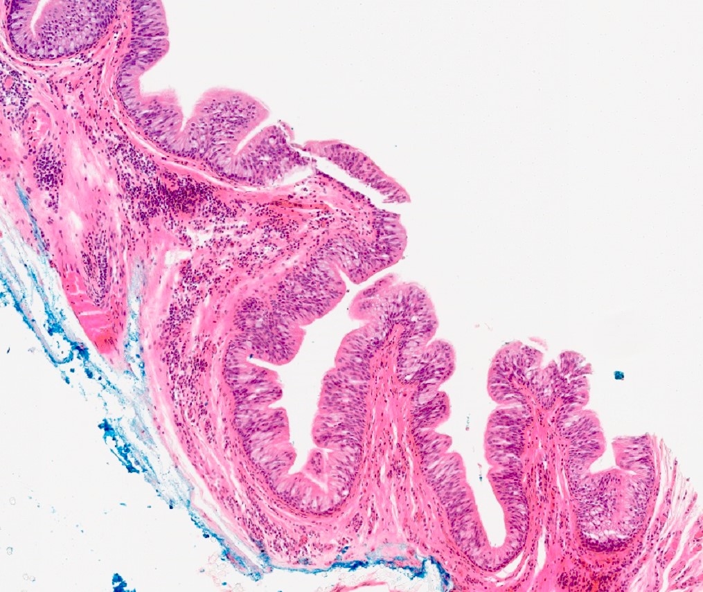
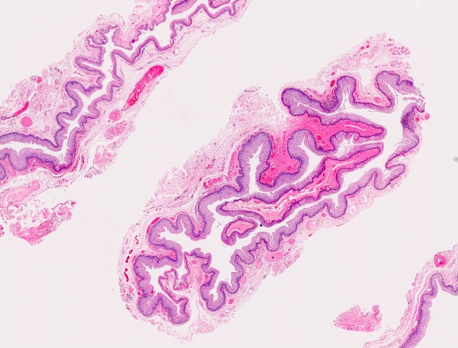
Contributed by Jamie Brown, M.D.
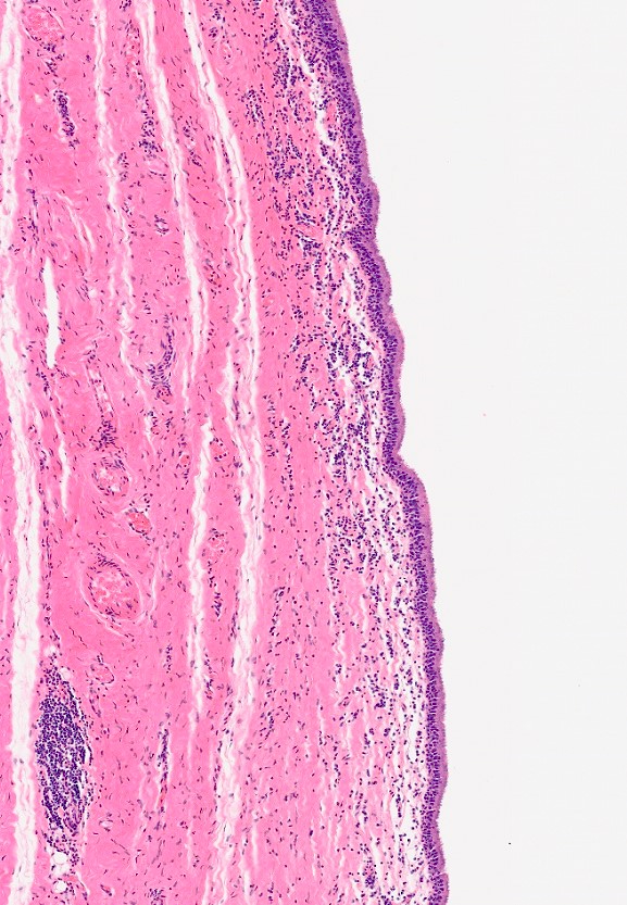
Respiratory epithelium
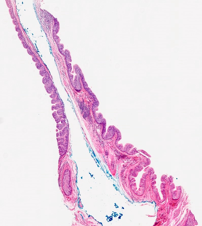
Mucinous metaplasia
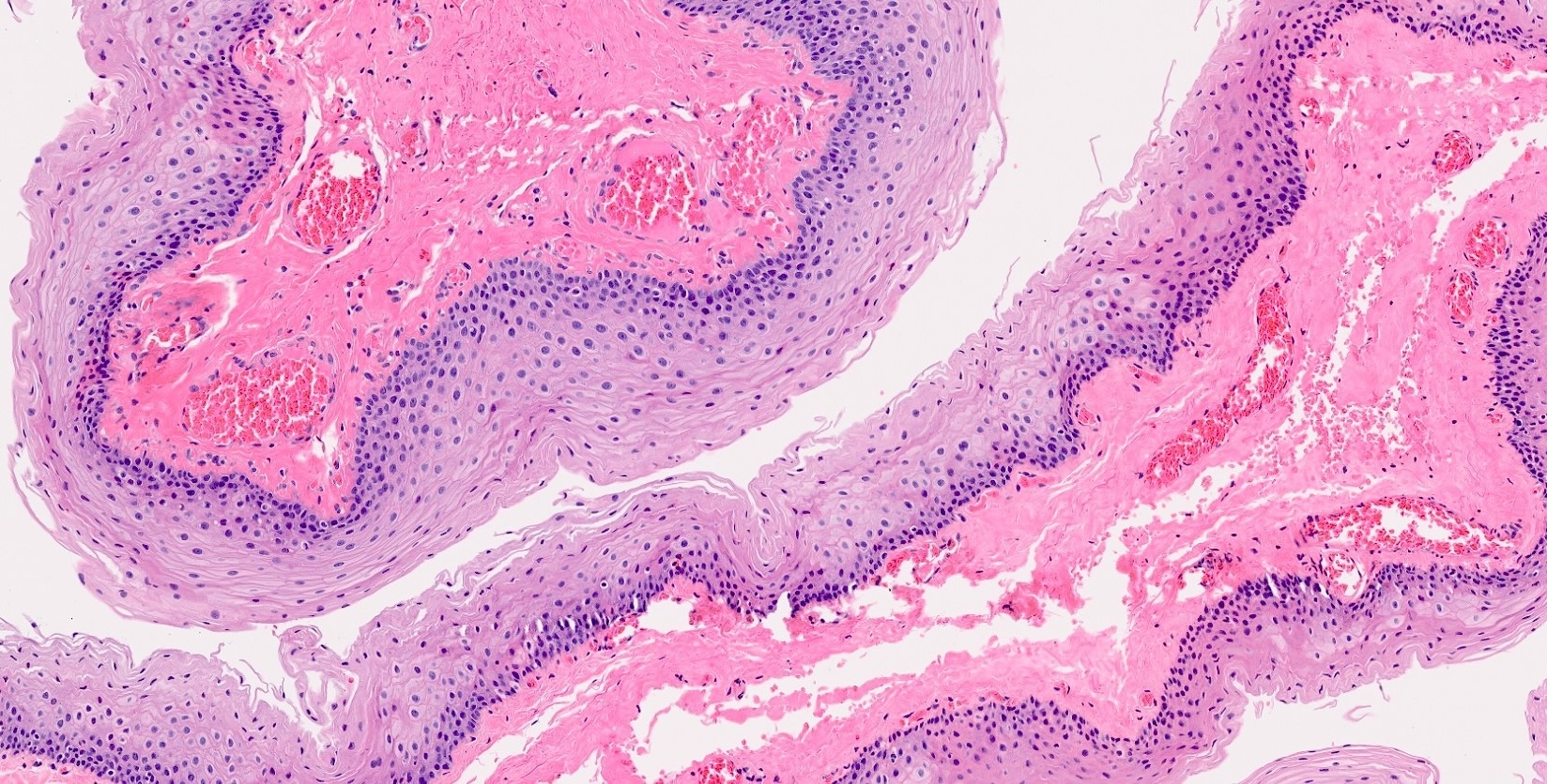
Squamous metaplasia
Chronic inflammation
Positive stains
- Immunohistochemistry generally not used in diagnosis of laryngocele
Sample pathology report
- Right neck, cyst resection:
- Combined laryngocele (see comment)
- Comment: Microscopic examination reveals a simple cyst lined by respiratory type epithelium with focal mucinous metaplasia. There is associated chronic inflammation without evidence of acute inflammation. Microscopic features are consistent with laryngocele; however, this diagnosis requires clinical or radiological correlation to distinguish this entity from saccular or ductal cysts.
Differential diagnosis
- Saccular cyst:
- Also present at appendix of laryngeal ventricle
- Does not communicate with laryngeal lumen
- Contains mucin rather than air
- Ductal cyst:
- Secondary to obstructed mucinous gland
- Sites other than the appendix of the ventricle
- Contains mucin rather than air
Practice question #1
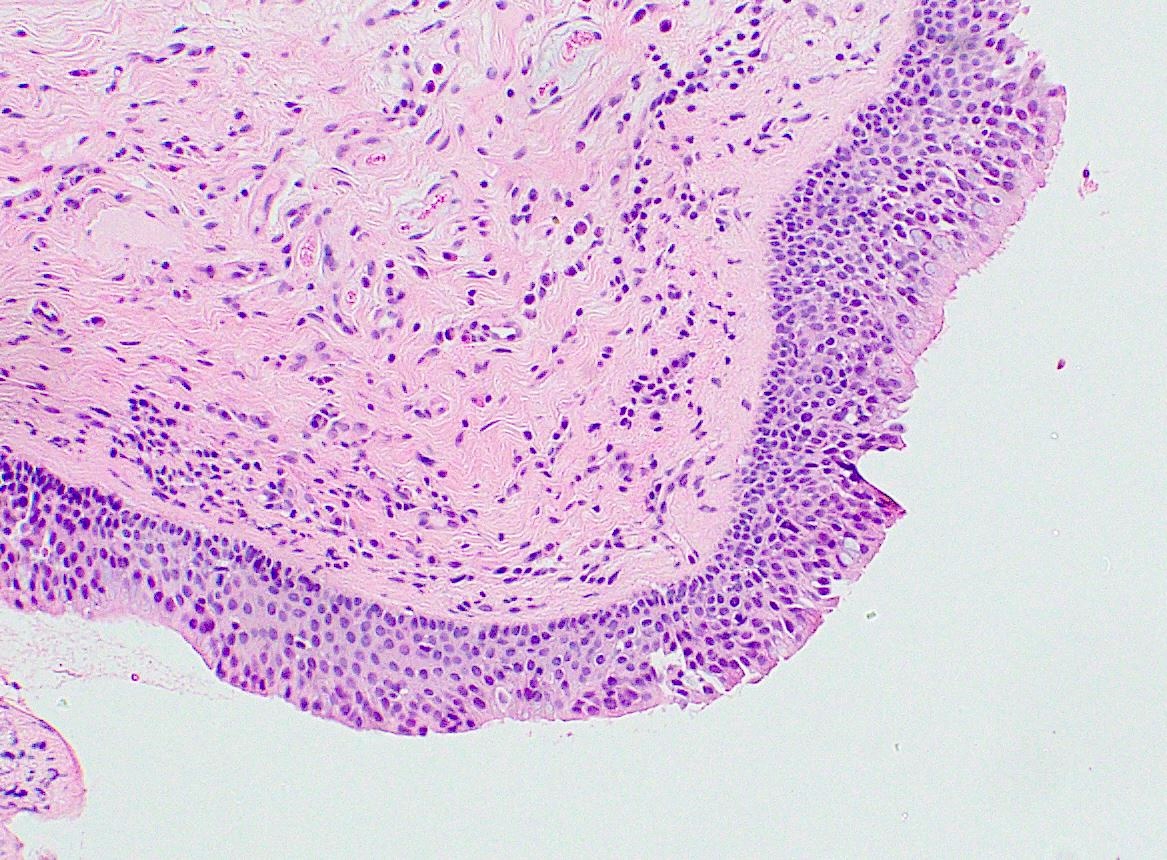
The image above is from an H&E stained section from a cyst wall removed from the lateral neck of a 45 year old man. The cyst was filled with air and connected with the laryngeal lumen at the ventricle. Based on the microscopic and clinical features provided, what type is cyst is shown?
- Ductal cyst
- Laryngocele
- Saccular cyst
- Thyroglossal duct cyst
Practice answer #1
B. Laryngocele. The micrograph shows a cyst lined by respiratory type epithelium; the clinical history provided was that the cyst connects to the laryngeal lumen and contains air, which are diagnostic features of a laryngocele. Answer C is incorrect because saccular cysts do not connect to the laryngeal lumen and contain mucin, rather than air. Answer A is incorrect because ductal cysts contain mucin rather than air and are found at sites other than the laryngeal ventricle. Answer D is incorrect because thyroglossal duct cysts do not connect with the laryngeal lumen and typically contain thyroid tissue within the wall.
Comment Here
Reference: Laryngocele
Comment Here
Reference: Laryngocele
