Lung
Infectious
Fungal
Other fungi
Author: Elliot Weisenberg, M.D.

Last author update: 1 September 2011
Last staff update: 4 April 2024
Copyright: 2003-2025, PathologyOutlines.com, Inc.
PubMed search: blastomyces [title] lungs, candida [title] lungs, cryptococcus neoformans [title] pulmonary, mucor [title] pulmonary
Cite this page: Weisenberg E. Other fungi. PathologyOutlines.com website. https://www.pathologyoutlines.com/topic/lungnontumorotherfungi.html. Accessed September 3rd, 2025.
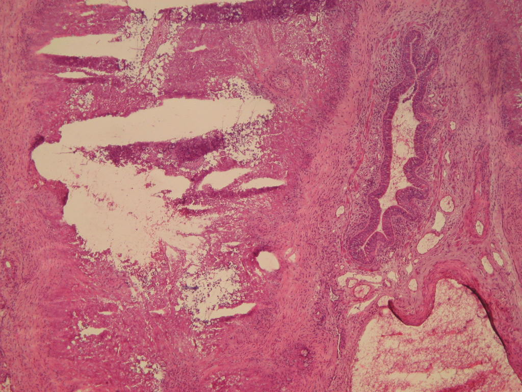
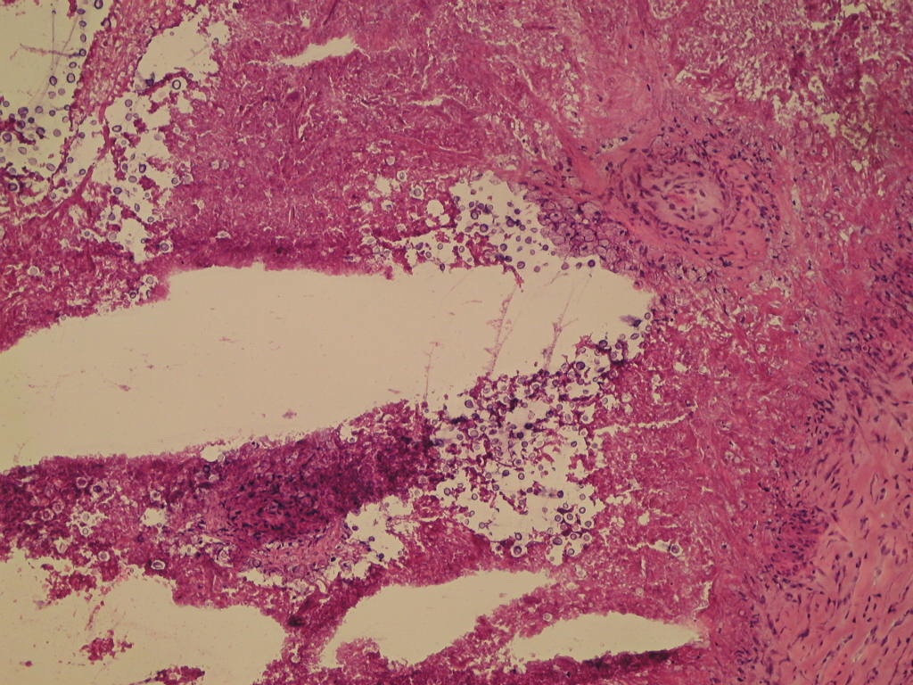
Blastomyces
Clinical features
- See also Skin - nontumor chapter
- Dimorphic fungus found in soil in Central and Southeastern United States and Canada (bordering Great Lakes, St. Lawrence River, Mississippi River and Ohio River basins), Mexico, Africa, India, Middle East
- More common in males
- Due to inhalation of soil containing microfoci of mycelia (Clin Microbiol Rev 2010;23:367)
- Difficult to isolate in clinical microbiology laboratory
- May present as consolidative pneumonia, ARDS or nodules resembling carcinoma; prefers upper lobes
- May also infect skin and bone
- Grossly often resembles tuberculosis
Microscopic (histologic) description
- Mixed acute and granulomatous inflammation caused by large budding yeasts (15 - 10 μm) with broad based buds and refractile walls
- Easily seen with H&E
Microscopic (histologic) images
Images hosted on other servers:

Typical appearance of B. dermatitidis

Microabscess with a nonbudding yeast cell


Budding yeast of B. dermatitidis
Candida
Definition / general
- See also Skin - nontumor chapter
- Normal flora of mouth, GI tract, vagina
- May be commensal or pathogen
- Commonly present in upper airways
- Often present in aspiration pneumonia or pulmonary abscess due to colonization but invasive disease due to candida is rare in lung except in setting of candidal sepsis
- Candida pneumonia associated with malignancy or immunosuppression (West J Med 1979;131:196)
Microscopic (histologic) description
- Pseudohyphae, occasionally true hyphae and budding yeasts
- In some cases, only yeast may be present
Microscopic (histologic) images
Contributed by Yale Rosen, M.D.
Lung: GMS staining
Images hosted on other servers:

Candida
Positive stains
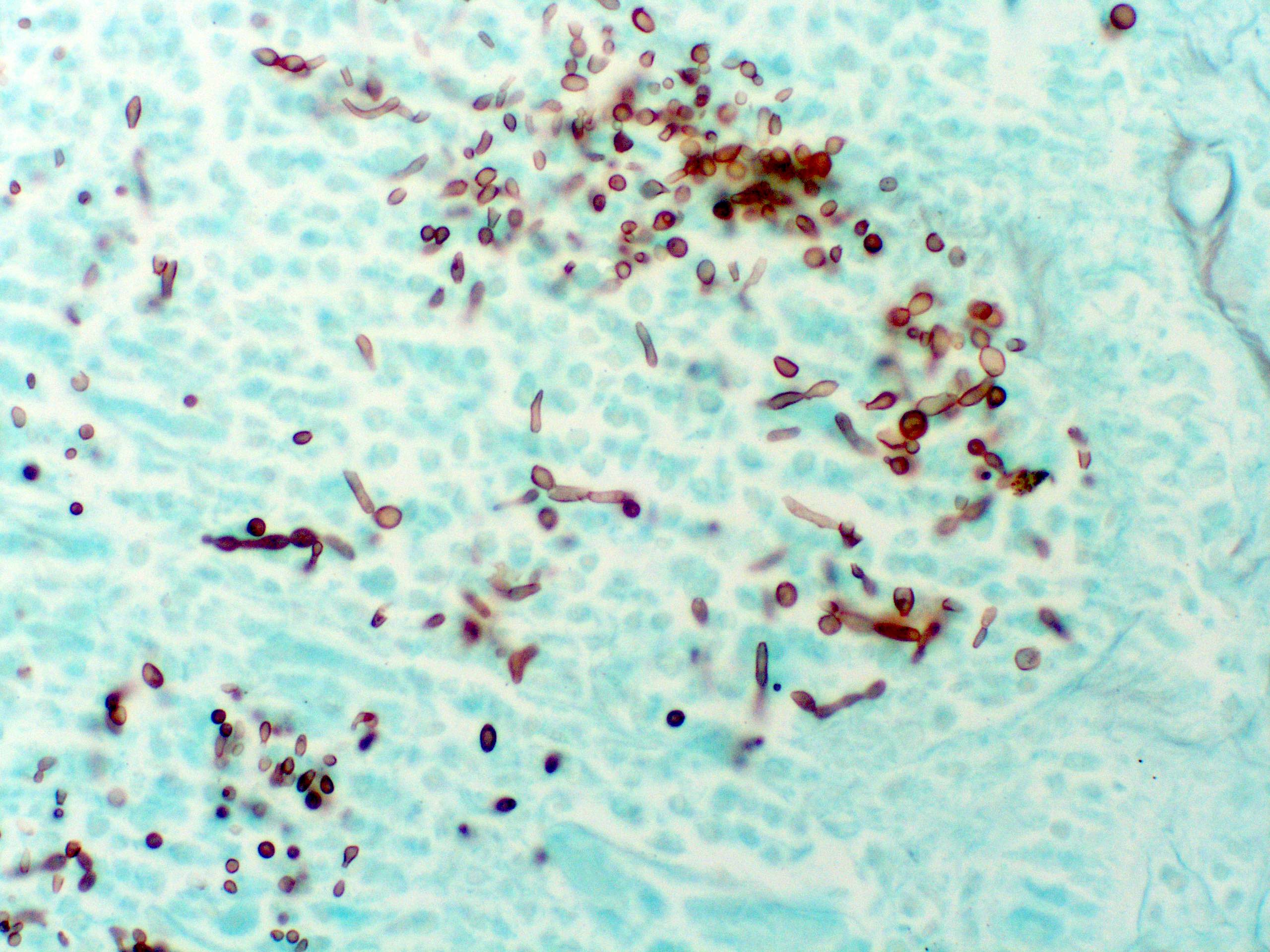
Cryptococcus
Clinical features
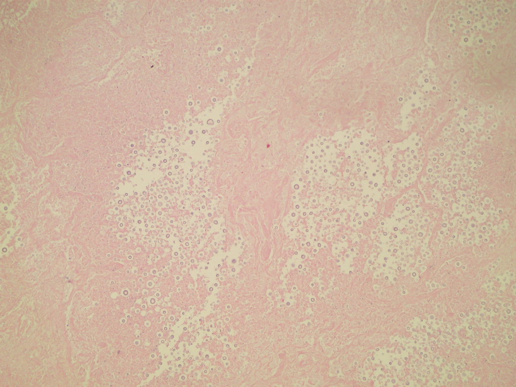
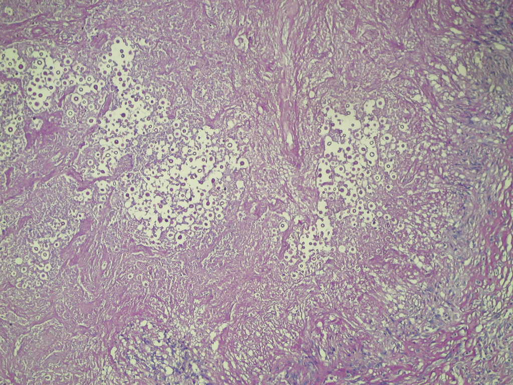
- Yeast, mostly encapsulated, found in pigeon droppings, that may cause mild infection after inhalation; usually confluent bronchopneumonia with "yeast lakes" of microorganisms and possibly coin lesions but no evident host response (eMedicine: Cryptococcosis [Accessed 22 March 2021])
- May also cause meningitis
- Latent infections can reactivate in immunosuppressed
- CNS disease is a major concern in immunocompromised
- Most commonly an opportunistic infection but disease may occur in immunocompetent patients
- Major virulence factor is capsular polysaccharide glucuronoxylomannin, which hinders phagocytosis by alveolar histiocytes and inflammatory cell recruitment and migration
- Other virulence factors include melanin production (Fontana-Masson stain may be positive; melanin may have antioxidant properties) and enzymes that increase invasiveness
Case reports
- 65 year old woman with arthritis and lung mass (Case of the Week #298)
Microscopic (histologic) description
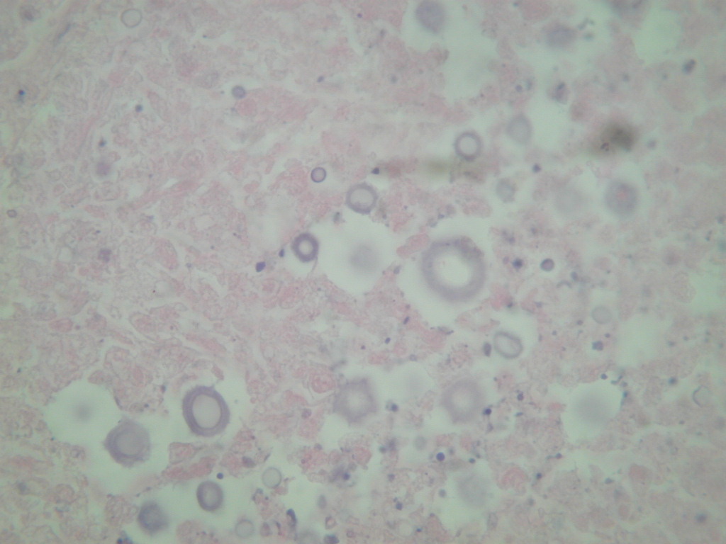
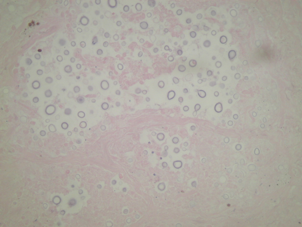
- Somewhat pleomorphic, round / oval yeast, 4 - 10 microns
- Thick, mucinous capsule stains bright red with mucicarmine; some are unencapsulated
- Narrow necked budding takes place
- Smaller, unencapsulated forms resemble Histoplasma capsulatum
Microscopic (histologic) images
Case #298
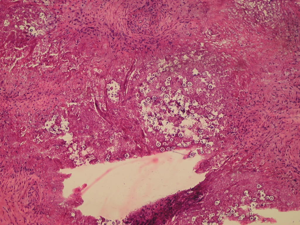
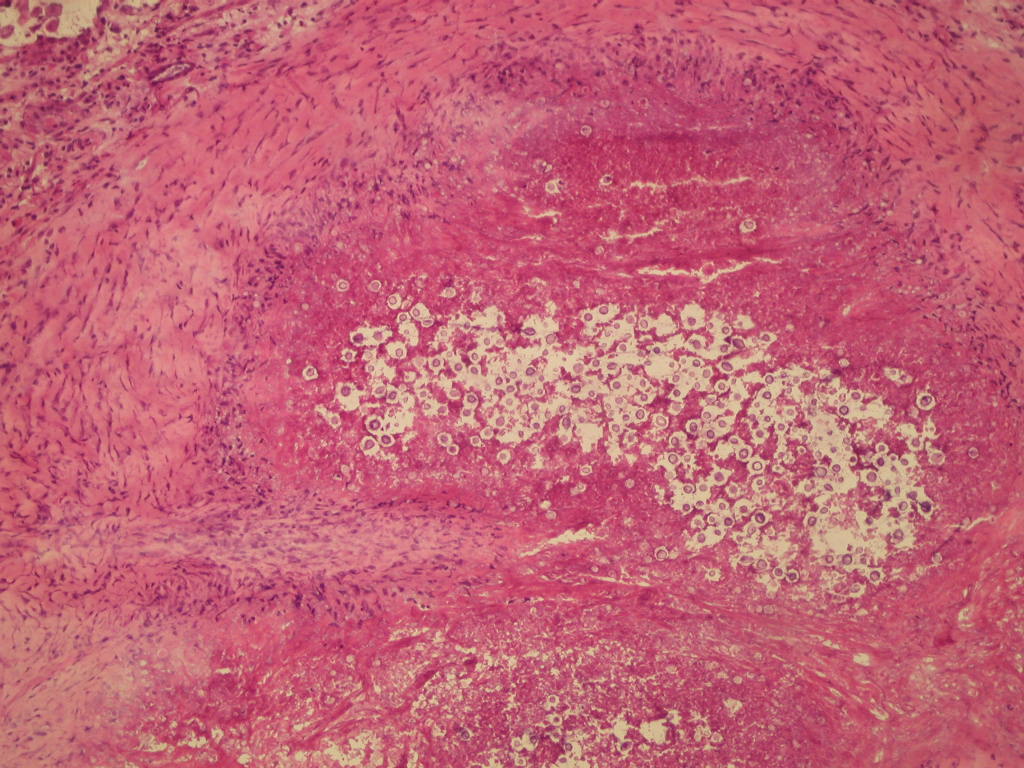
Frozen section
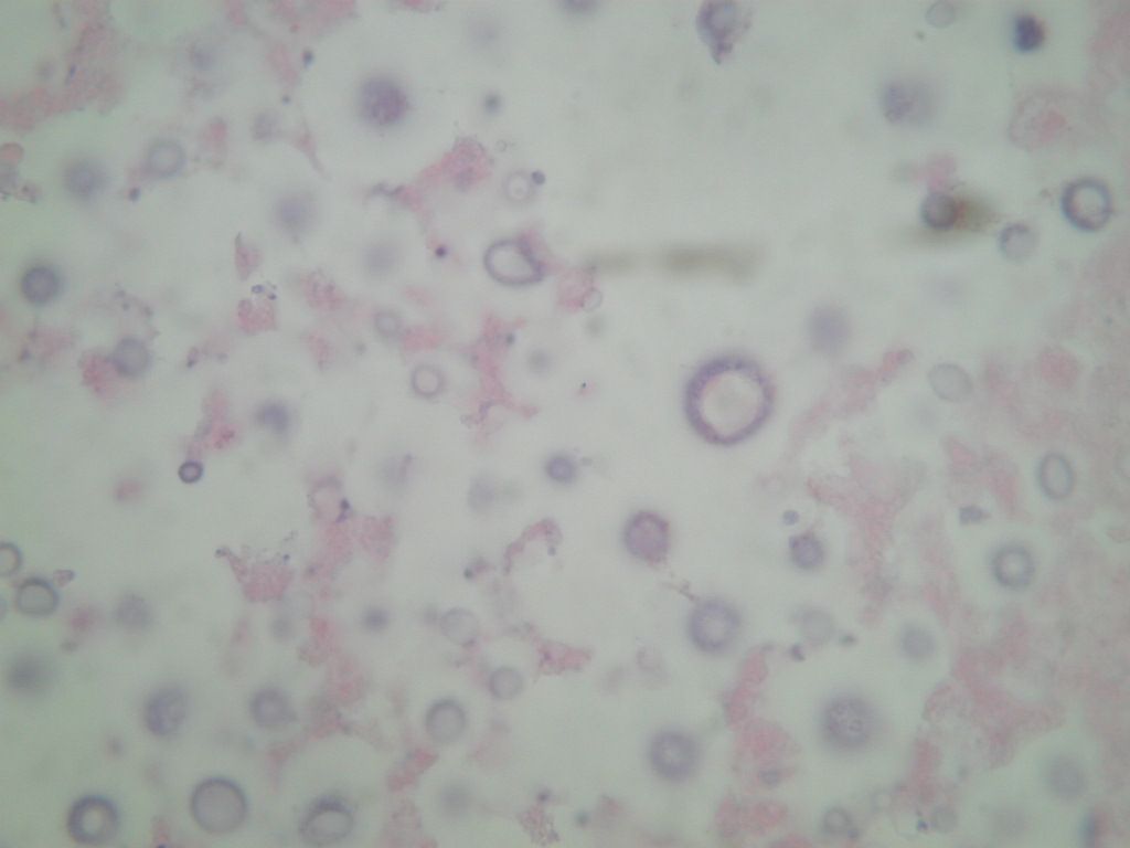
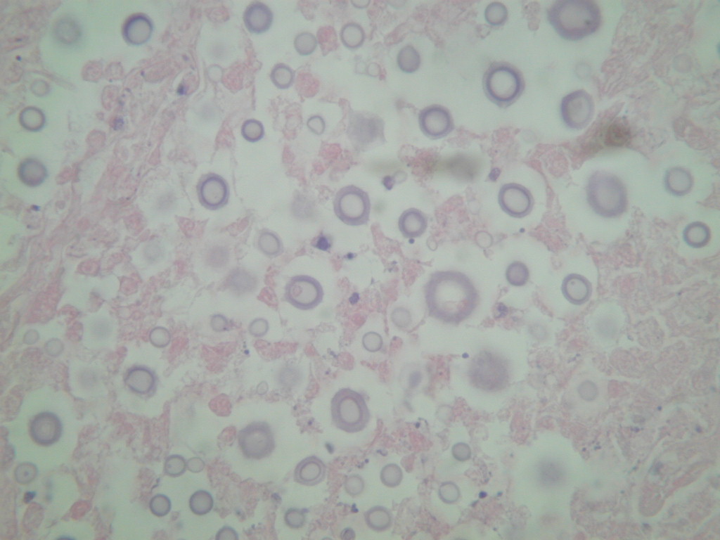
Oil immersion images
Oil immersion images

H&E
PAS stain
Images hosted on other servers:

Large mucoid capsule

Cryptococcosis in patient with AIDS
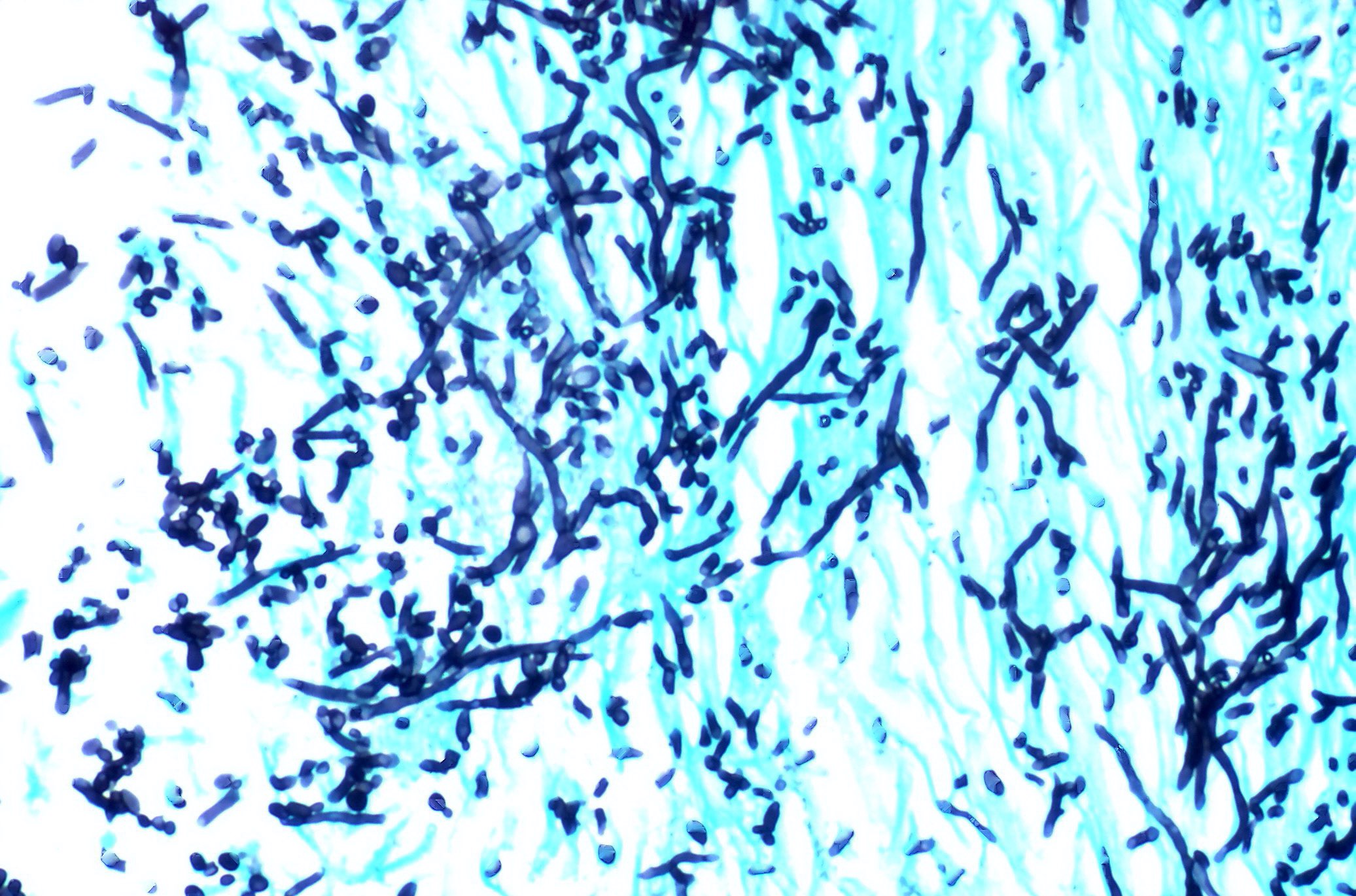
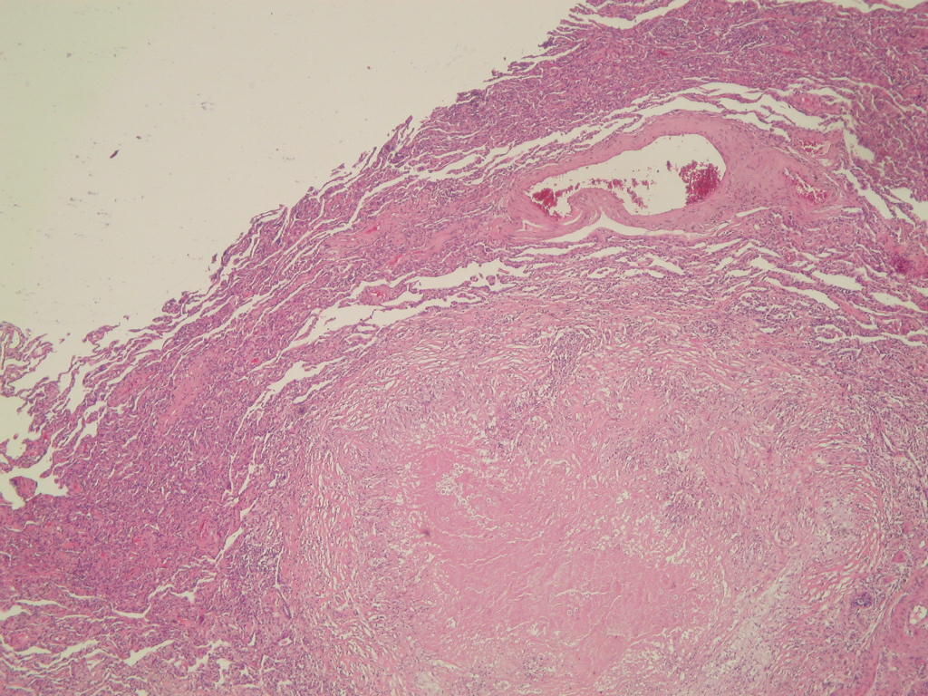
Mucor
Definition / general
- Also known as zygomycosis or mucormycosis
- Ubiquitous fungi of class Zygomycetes, includes Mucor, Rhizopus, Absidia and Cunninghamella
- Opportunistic infection especially associated with diabetes; other predisposing factors are neutropenia, corticosteroid therapy, iron overload and mucocutaneous trauma
- Pulmonary and sinusoidal infection caused by inhaled spores or secondary to rhinocerebral mucormycosis
Case reports
- 39 year old man with necrotizing pneumonia (University of Pittsburgh: Case 181 [Accessed 22 March 2021])
Microscopic (histologic) description
- Large, nonsepta hyphae with 90 degree angle branching and nonparallel walls, angioinvasive causing tissue necrosis and hemorrhage
Microscopic (histologic) images
Images hosted on other servers:

Culture shows young sporangia of Mucor species
