

Mediastinum
Nonneoplastic
Other nonneoplastic
Author: Hanni Gulwani, M.B.B.S.

Last author update: 1 December 2012
Last staff update: 15 March 2021
Copyright: 2003-2024, PathologyOutlines.com, Inc.
PubMed Search: Acute thymic involution, Diffuse thymic fibrosis, Ectopic thymus[TI], Ectopic tissue in thymus[TI], Acute mediastinitis[TI], Chronic mediastinitis[TI], Thymic dysplasia, Thymic follicular hyperplasia, True thymic hyperplasia

Table of Contents
Acute thymic involution | Diffuse thymic fibrosis | Ectopic thymus | Ectopic tissue in thymus | Mediastinitis, acute | Mediastinitis, chronic | Thymic dysplasia | Thymic follicular hyperplasia | True thymic hyperplasiaCite this page: Gulwani H. Other nonneoplastic. PathologyOutlines.com website. https://www.pathologyoutlines.com/topic/mediastinumothernonneoplastic.html. Accessed April 26th, 2024.
Acute thymic involution
Definition / general
- Due to stress (chronic debilitating disease), HIV or other infections, prolonged protein malnutrition and immunosuppressive or cytotoxic drugs, graft versus host reaction
- Seen in newborn infants with chorioamnionitis and sepsis
- Thymus size is significantly reduced in preterm infants born to mothers with subclinical, histologically proven chorioamnionitis (Hum Pathol 2000;31:1121)
Microscopic (histologic) description
- Preservation of lobular architecture and Hassall corpuscles but marked lymphocyte depletion (particularly with HIV)
- Vessels are large compared to size of lobules
- Frequent plasma cells, fibrohyaline changes of basement membrane of vessels and thymic epithelium
- HIV patients also have effacement of corticomedullary junction and inconspicuous Hassall corpuscles
Differential diagnosis
Diffuse thymic fibrosis
Definition / general
- Uncommon disorder with no / limited symptoms; may have dyspnea, cough or hemoptysis
- Area of diffuse fibrosis varies from 3.5 - 17 cm, confined to anterior mediastinum
- Unknown etiology; altered immunity or infection may play a role
- Males and females, mean age 48 years
Microscopic (histologic) description
- Diffuse fibrosis with variable collagen deposition, lymphoplasmacytic infiltrates and involution / atrophy of thymus
- May show IgG4+ plasma cells and focal obliterative phlebitis (Am J Surg Pathol 2010;34:211)
Ectopic thymus
Definition / general
- Remnants, implants or accessory nodules that may appear from angle of mandible to thyroid gland, most commonly at level of thyroid gland
- Rarely becomes hyperplastic or neoplastic
Epidemiology
- Usually an incidental finding during thyroid surgery in preteens, very rare in adults due to thymic involution
Case reports
- 12 month old boy with neck mass (Arch Pathol Lab Med 2001;125:278)
- 22 year old woman with anterior neck mass (Arch Pathol Lab Med 2001;125:842)
- With micronodular epithelial hyperplasia (Int J Surg Pathol 2006;14:73)
Microscopic (histologic) description
- Normal appearing thymic tissue
Microscopic (histologic) images
Images hosted on other servers:
Central cervical epithelial rest

CK5 / 6, p63, CD10
Differential diagnosis
- Ectopic cervical thymoma:
- "The great mimic"
- Considered in differential diagnosis of neck masses in elderly (Indian J Pathol Microbiol 2007;50:553)
- Other heterotopic epithelial elements:
- Including misplaced cutaneous structures and salivary gland tissue (Am J Clin Pathol 2009;132:707)
Ectopic tissue in thymus
Definition / general
- Usually parathyroid tissue or sebaceous glands, rarely thyroid tissue
Case reports
- Woman with heterotopic intrathymic thyroid tissue (Pathol Int 2006;56:629)
Mediastinitis, acute
Definition / general
- Usually in posterior mediastinum, due to traumatic perforation of esophagus or descending infection along prevertebral fascia
- Initial lesion may be a neck abscess
- Often causes mediastinal abscess which requires surgical drainage
- Other causes: chest wall infection or postcardiac surgery, often due to CMV
Case reports
- Sternum osteolysis following staphylococcus mediastinitis (Cardiovasc Pathol 2006;15:297)
- Descending necrotizing mediastinitis: rare spreading of cervical infection to mediastinum (Clin Imaging 2005;29:138)
Microscopic (histologic) images
Images hosted on other servers:

Extensive acute inflammation

Fibrinoinflammatory
exudate
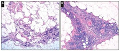
Mediastinitis, chronic
Definition / general
- May compress superior vena cava and simulate malignancy
- Usually anterior to tracheal bifurcation
- Some cases may represent fibrosing mediastinitis
Microscopic (histologic) description
- Granulomas, fibrosis; may be fungus, Histoplasma (with thick fibrous capsule), mycobacteria (thin fibrous capsule)
Thymic dysplasia
Definition / general
- Congenital thymic alteration due to developmental arrest
- Lack of differentiation of thymic epithelium, responsible for absence of Hassall corpuscles, is main feature (Histopathology 1992;21:499)
Clinical features
- Associated with severe combined immunodeficiency syndrome, ataxia telangiectasia, chromosomal instability syndromes, Nezelof syndrome (Arch Pathol Lab Med 1987;111:1118)
- Incomplete form of DiGeorge syndrome is congenital anomaly with a constellation of findings that includes thymic hypoplasia (J Cutan Pathol 2008;35:380); complete form has absent thymus
Gross description
- Small thymus (< 5 g)
Microscopic (histologic) description
- Tubules and rosettes of primitive appearing epithelium without segregation into cortical and medullary regions
- No Hassall corpuscles, no / rare lymphocytes
Differential diagnosis
Thymic follicular hyperplasia
Definition / general
- Defined as substantial numbers of lymphoid follicles in thymus of adults
- Thymus usually has normal size / weight
Clinical features
- Present in 65% with myasthenia gravis
- Also associated with hyperthyroidism, Addison disease, SLE, early HIV, multilocular cysts, other immune related diseases
- Often different clinical history than true thymic hyperplasia (Pathologica 2009;101:175)
Microscopic (histologic) description
- Follicles with germinal centers, medullary epithelial cells may be disordered or hypertrophied
Differential diagnosis
- Normal lymphoid follicles of infants / children (few present)
True thymic hyperplasia
Definition / general
- Thymus larger than normal limits for age, based on tables
- Otherwise histologically unremarkable
Epidemiology
- Often in infants or children or in adults after cancer chemotherapy
Case reports
- 5 week old boy with severe thymic cyst bleeding (Ann Diagn Pathol 2007;11:358)
- 22 year old man with thymic hemorrhage (Pathol Res Pract 2010;206:331)
- 24 year old woman who was treated for T cell lymphoma (J Clin Oncol 2004;22:953)
- 26 year old woman with thymolipoma, suggesting an origin from thymic true hyperplasia (Int J Surg Pathol 2010;18:526)
- 53 year old woman with unilocular thymic cyst (Ann Diagn Pathol 2006;10:32)
Immunohistochemistry
- p63+, XIAP- (Am J Clin Pathol 2009;131:689)
