
Microbiology & infectious diseases
Gram negative bacteria
Ehrlichia
Author: Dragos C. Luca, M.D.

Last author update: 1 March 2015
Last staff update: 18 October 2021
Copyright: 2002-2024, PathologyOutlines.com, Inc.
PubMed Search: Ehrlichia
Table of Contents
Definition / general | Clinical presentation and diagnosis | Case reports | Treatment | Microscopic (histologic) description | Microscopic (histologic) images | Peripheral smear images | Electron microscopy images | Additional referencesCite this page: Luca DC. Ehrlichia. PathologyOutlines.com website. https://www.pathologyoutlines.com/topic/microbiologyehrlichia.html. Accessed April 26th, 2024.
Definition / general
- Ehrlichia: obligate intracellular gram negative bacteria belonging to recently reorganized family Anaplasmataceae
- Species producing disease in humans (3):
- E. chaffeensis: human monocytic ehrlichiosis (HME)
- Anaplasma phagocytophilia: human granulocytotropic anaplasmosis (HGA, formally termed human granulocytic ehrlichiosis)
- E. ewingii: rare granulocytic disease (increased frequency in transplant patients)
- Vector borne disease transmitted through bite of Ixodes ticks
- Bacteria is obligate intracellular pathogen that binds to P selectin glycoprotein ligand 1 (PSGL1 / CD162)
- Susceptibility also associated with expression of CD15s (J Clin Invest 1999;103:407)
- First described in USA in 1994
- Geographic distribution of E. chaffeensis (HME) reflects regions of US where their hard tick vectors reside: south central, southeastern and mid Atlantic states
Clinical presentation and diagnosis
- HME is more often serious with hospitalization in 50% of patients; septic shock-like syndrome possible, especially if immunosuppressed
- Presents with fever, leukopenia, thrombocytopenia (70 - 90%) and elevated liver enzymes
- Mortality rate is 2 - 3% for HME
- Particularly severe infections occur in elderly / immunocompromised
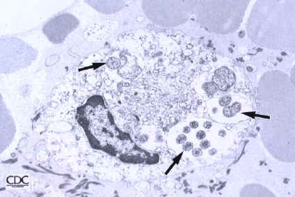
- Characteristic intracytoplasmic morulae (morula is Latin for mulberry): cytoplasmic membrane bound vacuoles with irregular edges containing hundreds to thousands of clustered gram negative bacteria
- Infected cells typically contain only 1 or 2 morulae although as many as 15 may be seen in immunosuppressed individuals
- Morulae are present in less than 0.2% of circulating WBCs in HME infection; examination of buffy coats facilitates detection
- Greatly variable percentage of peripheral blood films with detectable morulae in the literature (3 - 80%) with a higher number seen with immunosuppressed individuals
Case reports
- 3 pancreas transplant recipients with HGA / human granulocytic ehrlichiosis (Transpl Infect Dis 2001;3:34)
Treatment
- Most patients are seronegative during first few weeks of acute infection (60 - 97%), so therapeutic decisions must be based on clinical suspicion, peripheral blood findings and PCR (sensitivity is 60 - 85%, high degree of false positive results)
- Became a nationally reportable disease to US Centers for Disease Control in 1999
- Organisms are susceptible to tetracyclines and their derivatives, particularly doxycycline
Microscopic (histologic) description
- Peripheral blood: buffy coat examination may reveal intracytoplasmic inclusions (morulae - spherical structures with irregular edges) within neutrophils or monocytes
- Bone marrow: epithelioid granulomas; usually normo or hypercellular with intact trilineage maturation; rare hypoplasia; possible increased megakaryocytes
- Histopathologic bone marrow findings: inconsistent and likely to change during the course of the disease
Microscopic (histologic) images
Images hosted on other servers:

Left: HGA (HGE)

Rhodamine labeled HGE organisms

HGE antigens
Peripheral smear images
Images hosted on other servers:

HME
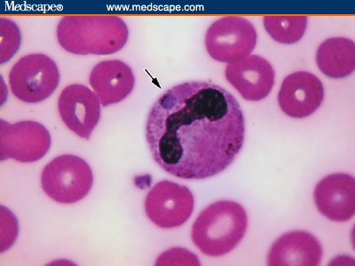
Intracytoplasmic inclusion
Electron microscopy images
Images hosted on other servers:
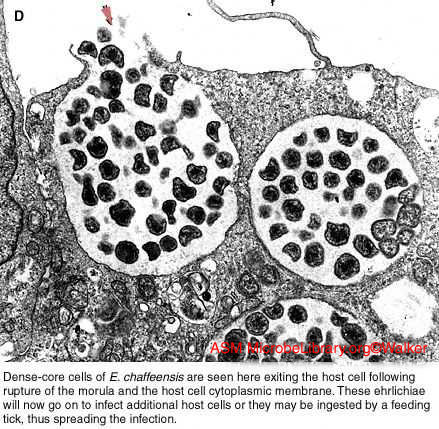
Dense core cells of E. chaffeensis
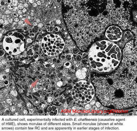
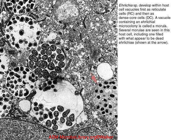
Several morulae
Bone marrow white blood cell
