Table of Contents
Definition / general | Essential features | Terminology | ICD coding | Epidemiology | Sites | Clinical features | Diagnosis | Prognostic factors | Case reports | Treatment | Clinical images | Microscopic (histologic) description | Microscopic (histologic) images | Sample pathology report | Differential diagnosis | Additional references | Practice question #1 | Practice answer #1 | Practice question #2 | Practice answer #2Cite this page: Elbendary A. Behçet disease. PathologyOutlines.com website. https://www.pathologyoutlines.com/topic/skinnontumorbehcetdisease.html. Accessed August 27th, 2025.
Definition / general
- Autoinflammatory multisystemic disease
- Involvement includes mucocutaneous, ocular, cardiovascular, renal, gastrointestinal, pulmonary, urologic, central nervous system, joints, blood vessels and lungs
Essential features
- Recurrent oral and genital ulcers in 90% of cases
- Variable vessel vasculitis and thrombosis
- Affects mainly Mediterranean basin and far east Asia
- Variable cutaneous manifestations and pathology
Terminology
- Behçet disease, Behçet syndrome, Adamantiades-Behçet syndrome
Epidemiology
- Distinct geographic distribution pattern along the ancient Silk Road
- Prevalence of 0.64 - 660/100,000 (Clin Rheumatol 2020;39:3449)
- M:F = 0.36 - 4.9
- Lowest male ratios reported in Western Europe; significant male predominance in Arab populations (Clin Rheumatol 2020;39:3449)
- Erythema nodosum, arthritis / arthralgias and central nervous system symptoms more frequent in females
- Males show a lower mean age at onset, a higher frequency of pseudofollicular lesions and panuveitis (Clin Rheumatol 2020;39:3449)
Sites
- Mucocutaneous, ocular, cardiovascular, renal, gastrointestinal, pulmonary, urologic, central nervous system, joint, blood vessel and lung involvement
Clinical features
- Oral ulcerations (95% of cases) on lips, gingival, buccal mucosa and tongue, leaves no scars
- Genital ulcers (90% of cases) on scrotum, major and minor labia, forms scars when healed
- Acne-like papulopustular lesions on face, upper trunk and extremities
- Erythema nodosum-like lesions, healing with hyperpigmentation
- Recurrent asymmetric mono or oligoarthritis, arthralgia, usually involving lower extremities, resolves with no deformity or erosion
- Ocular involvement (50% of cases), mainly posterior or panuveitis and retinal vasculitis
- Superficial and deep venous thrombosis
- Gastrointestinal involvement ranging from mild abdominal pain to emergency complication such as hemorrhage or perforation
- Neurologic involvement (5% of cases) such as severe headache, cranial nerve palsy, ataxia and hemiparesis
- Variable vessel vasculitis with contemporary involvement of arteries and veins and tendency for aneurysm formation
- Reference: Rheumatology (Oxford) 2020;59:iii101
Diagnosis
- International criteria for Behçet disease - point score system: scoring ≥ 4 indicates Behçet diagnosis (J Eur Acad Dermatol Venereol 2014;28:338)
- Ocular lesions: 2 points
- Genital aphthosis: 2 points
- Oral aphthosis: 2 points
- Skin lesions: 1 point
- Neurological manifestations: 1 point
- Vascular manifestations: 1 point
- Positive pathergy test: 1 point
Prognostic factors
- Worst prognosis reported in patients with retinal vasculitis, where blindness can occur (Indian J Ophthalmol 2011;59:240)
- Patients with vascular aneurysm formation have a possibility to rupture (Korean J Thorac Cardiovasc Surg 2020;53:381)
- Neuro Behçet syndrome may lead to dementia (Lancet Neurol 2009;8:192)
Case reports
- 24 year old man with recurrent oral and genital ulcers and cerebral venous thrombosis (J Ayub Med Coll Abbottabad 2020;32:124)
- 32 year old man with oral ulcerations and myocarditis (Can J Cardiol 2020;36:1554.e9)
- 45 year old woman with acneform eruption, genital ulcers, chest pain and esophageal and gastric ulcers (J Clin Rheumatol 2020;26:e33)
Treatment
- Treatment is tailored to each patient's clinical manifestations
- Corticosteroids are useful in controlling acute manifestations
- Cytotoxic medications are indicated in patients with ocular, central nervous system and vascular disease
- European League Against Rheumatism (EULAR) has recently released guidelines for the management of Behçet disease (Ann Rheum Dis 2008;67:1656)
- Otezla® (apremilast) is the only FDA approved treatment for Behçet
- Colchicine is also commonly used
- Reference: (Clin Exp Rheumatol 2018;36:13)
Clinical images
Images hosted on other servers:

Erythematous papules and pustules resembling acne

Erythematous plantar maculae
Microscopic (histologic) description
- Oral and genital ulcers: not specific; mixed dermal inflammatory infiltrate perivascular and at the base of the ulcer
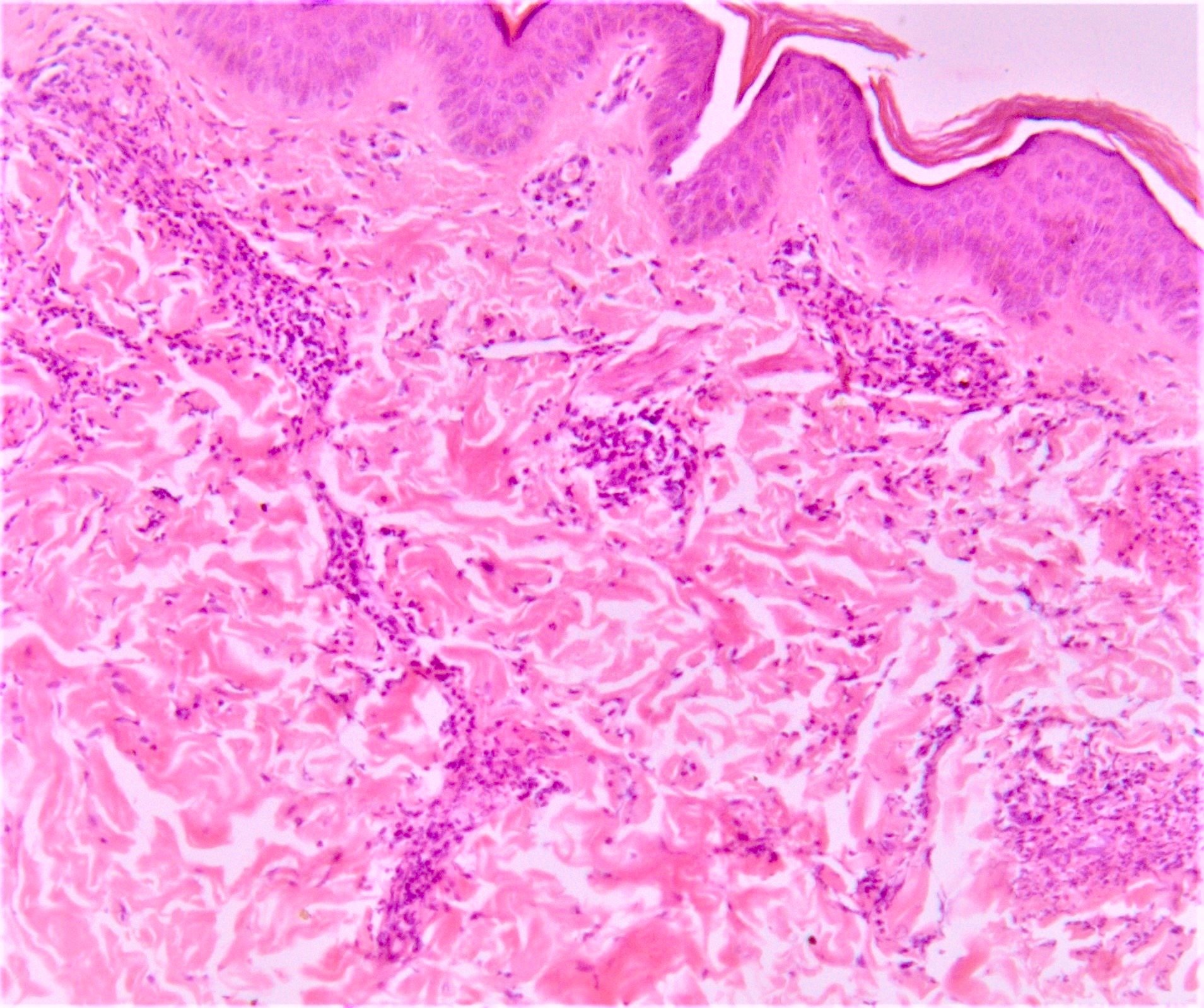
- Erythema nodosum-like lesions: septal panniculitis; vasculitis (lymphocytic or neutrophilic) and necrobiosis may be found
- Pathergy reaction: perivascular infiltrate of mononuclear cells; mast cell infiltrate and neutrophilic vasculitis may be found
- Papulopustular lesions: spongiosis, basal keratinocyte vacuolization, intraepidermal pustules, suppurative folliculitis
- Thrombophlebitis: thrombi in vessel lumens, perivascular inflammatory infiltrate
- Leukocytoclastic vasculitis
- Reference: Patholog Res Int 2012;2012:209316
Microscopic (histologic) images
Contributed by Amira Elbendary, M.B.B.Ch., M.Sc.
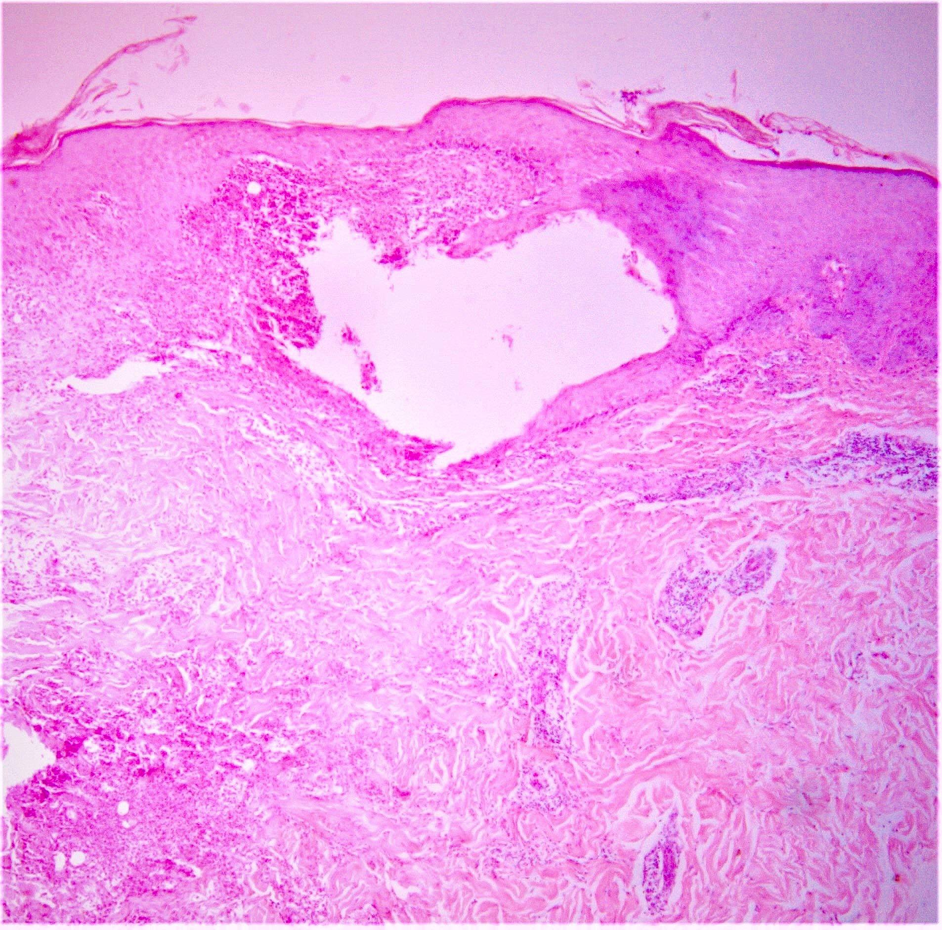
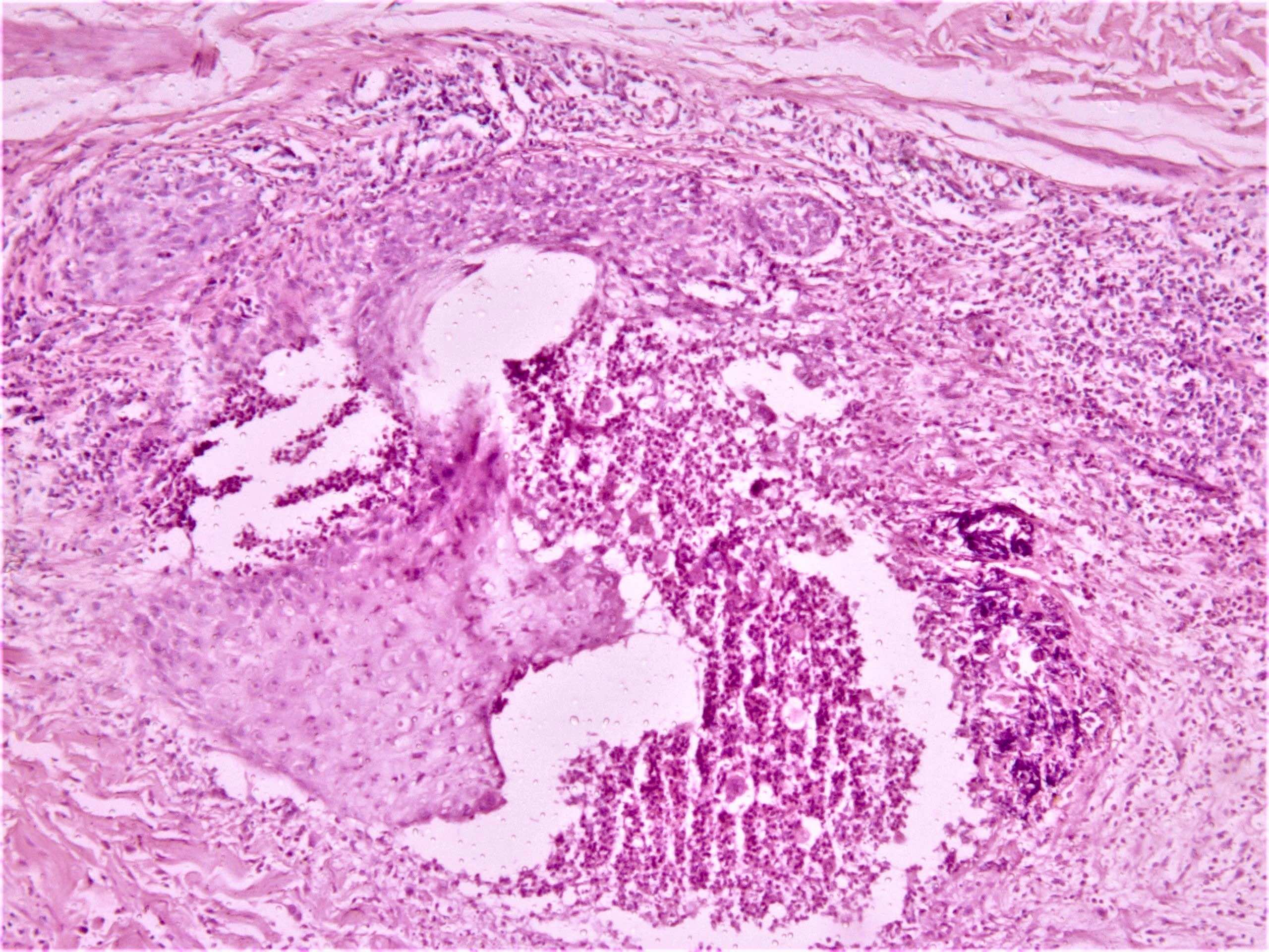
Acne-like lesion
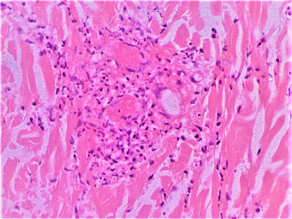
Vasculitis
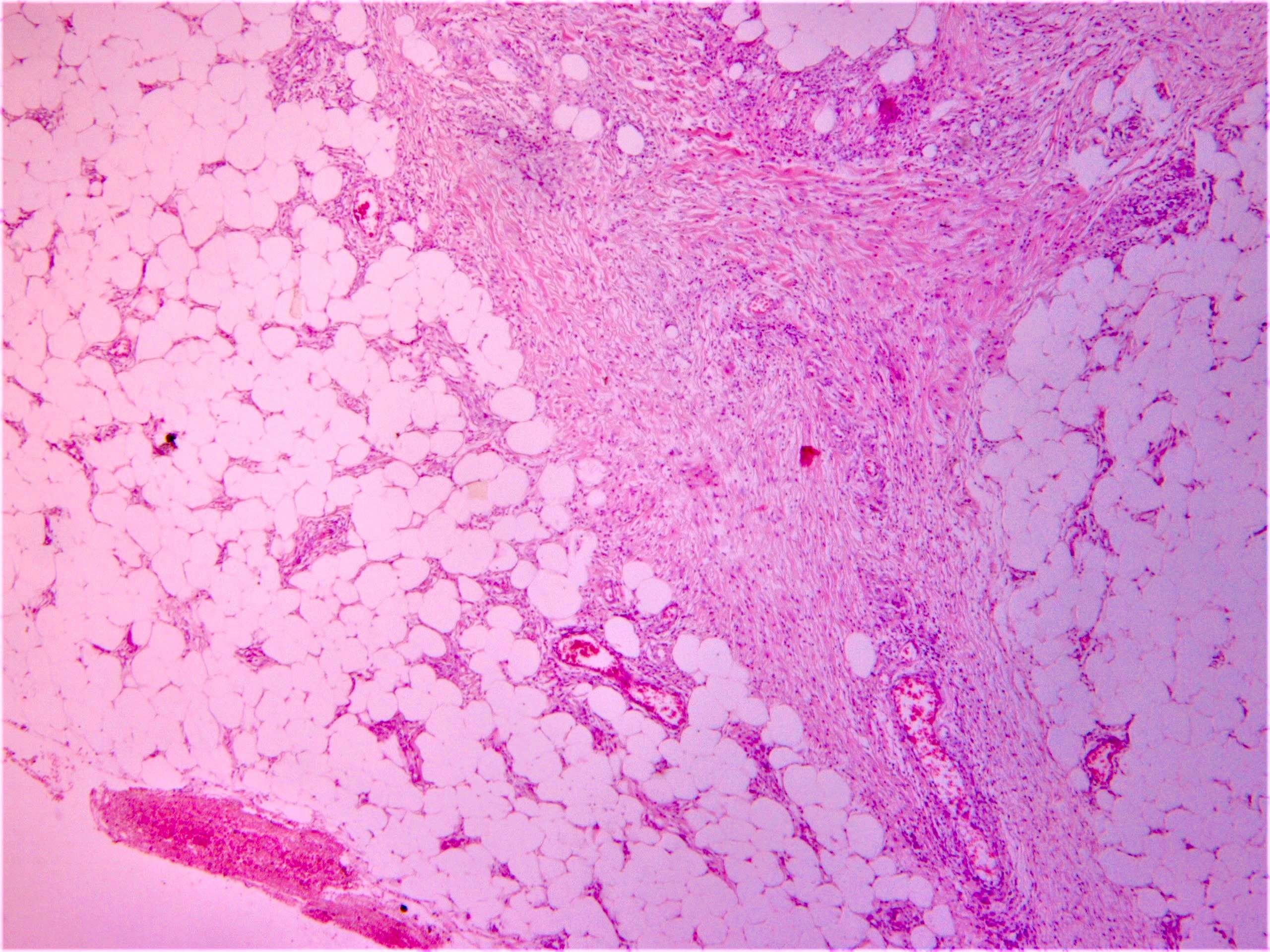
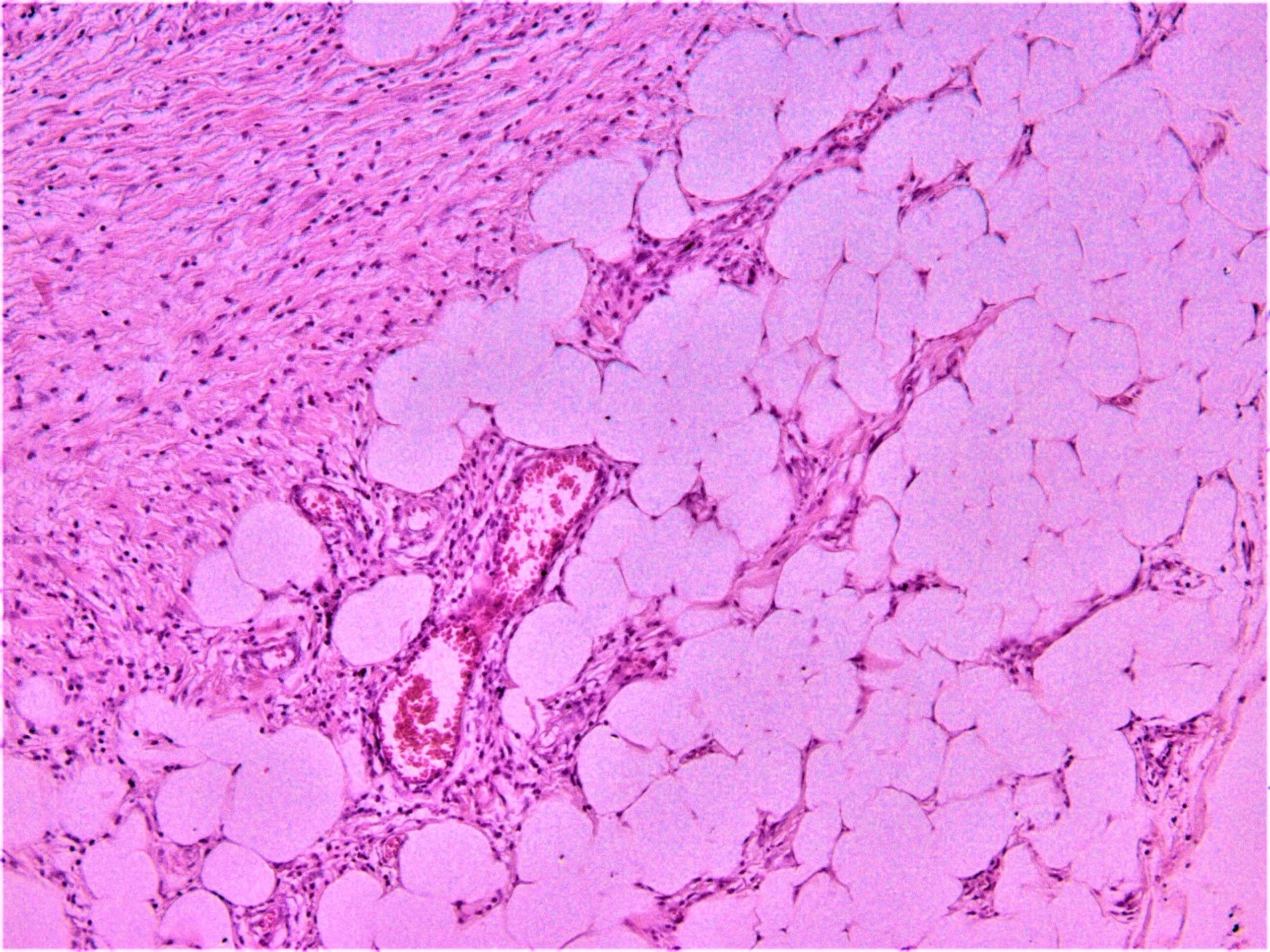
Erythema nodosum-like lesion
Sample pathology report
- Right leg, erythematous subcutaneous nodule, incisional biopsy:
- Erythema nodosum-like lesion (given the history, compatible with Behçet disease) (see comment)
- Comment: Septal panniculitis with unremarkable epidermis. Dermis shows moderate perivascular lymphocytic infiltrate. The subcutaneous fat demonstrates widening of the fibrous septa with edema and infiltration of lymphocytes and histiocytes. Spillover of infiltrate to the adjacent fat lobules is present. Small focus of lymphocytic vasculitis is noted.
Differential diagnosis
- Localized chronic fibrosing vasculitis:
- Histologically resembles late stage erythema elevatum diutinum (EED) but has a different clinical presentation
- Erythemaotous nodules on lower limbs
- Dense concentric and lamellar fibrosis, foci of neutrophilic dust, sparse infiltrate of histiocytes, neutrophils, eosinophils and lymphocytes
- No granulomas present; no involvement of subcutaneous fat
- Tissue cultures for micro-organisms are negative
- Evaluations for systemic vasculitides and connective tissue disorders are negative
- Chronic granulomatous disease (CGD) (Clin Immunol 2020;214:108381):
- A primary immunodeficiency
- Genetic mutations in any of 4 genes within the nicotinamide adenine dinucleotide phosphate oxidase complex
- Impaired oxidative burst after phagocytosis
- Clinical features: abscess formation and granulomatous lesions in skin and mucous membranes, lungs, liver and lymph nodes
- Recurrent oral ulceration without genital or skin lesions has been reported
- Positive nitroblue tetrazolium (NBT) test
- Periodic fever, aphthous stomatitis, pharyngitis and cervical adenitis (PFAPA) (Clin Immunol 2020;214:108381):
- A noninherited periodic fever syndrome
- Abnormal IL1β signaling
- Intermittent episodes of high fever associated with at least 1 of the features mentioned in its name
- Fever lasts 3 - 7 days with periods of health in between attacks
- None will fulfill the criteria for Behçet disease
- Inflammatory bowel disease:
- Marked overlap between the clinical features of inflammatory bowel disease and Behçet syndrome, including oral ulceration, similar skin manifestations, arthritis and uveitis
- Lower lip thickening, tags and cobblestoning within the oral mucosa would suggest Crohn’s disease rather than Behçet disease
- Perianal abscesses, fistula, strictures occur more commonly in Crohn’s disease
- Presence of noncaseating granulomas and absence of vasculitis in small veins and venules
Additional references
Practice question #1
A 25 year old male with a history of recurrent orogenital ulcers presented with erythematous nodules that ulcerate and blurred vision and pain in the right eye. What is the best next step?
- ANA, pANCA, cANCA
- Biopsy from an oral ulcer
- Perform skin biopsy from the edge of the ulcer
- Upper and lower endoscopy
- Urgent ophthalmology consultation
Practice answer #1
E. Urgent ophthalmology consultation. The patient has Behçet disease.
Comment Here
Reference: Behçet disease
Comment Here
Reference: Behçet disease
Practice question #2
A 19 year old male patient presented with recalcitrant papulopustular lesions on the face, shoulders, back and occasionally on lower limbs. He started to suffer from recurrent orogenital ulcers. What pathology finding in a skin biopsy from a pustule on the back would suggest further evaluation for Behçet disease?
- Follicular plug
- Folliculitis
- Leukocytoclastic vasculitis
- Perifolliculitis
- Superficial perivascular lymphocytic infiltrate
Practice answer #2
