Skin nontumor
Lichenoid and interface reaction patterns
Lichen nitidus
Editor-in-Chief: Debra L. Zynger, M.D.


Last author update: 5 May 2020
Last staff update: 20 June 2025
Copyright: 2002-2025, PathologyOutlines.com, Inc.
PubMed Search: Lichen nitidus
Table of Contents
Definition / general | Essential features | ICD coding | Epidemiology | Sites | Pathophysiology | Etiology | Clinical features | Diagnosis | Prognostic factors | Case reports | Treatment | Clinical images | Microscopic (histologic) description | Microscopic (histologic) images | Immunofluorescence description | Positive stains | Sample pathology report | Differential diagnosis | Additional references | Practice question #1 | Practice answer #1 | Practice question #2 | Practice answer #2Cite this page: Collie CJ, Ho JD. Lichen nitidus. PathologyOutlines.com website. https://www.pathologyoutlines.com/topic/skinnontumorlichennitidus.html. Accessed September 15th, 2025.
Definition / general
- Uncommon, idiopathic, lichenoid disorder of the skin with characteristic clinical and histopathologic features
Essential features
- Papular eruption most commonly seen in the young
- Discrete lymphohistiocytic infiltrate expanding 1 - 4 dermal papillae
- Epidermal collarette gives ball and claw appearance
- Vacuolar interface change may be prominent
- Multinucleated giant cells often present
- Atypical clinical variants have classical histopathologic findings
Epidemiology
- More common in children and young adults (Clin Dermatol 2015;33:631, J Am Acad Dermatol 2004;51:606)
- No consistent sex or ethnic predilection (Cutis 1978;21:634)
Sites
- Skin of upper limbs, dorsal hands, trunk and genitalia (Pediatr Dermatol 2019;36:189)
- Less commonly involves oral mucosa, nails and skin of the lower limbs, palms / soles or face (Indian J Dermatol 2019;64:62, Pediatr Dermatol 2013;30:e100)
Pathophysiology
- Largely unknown at this time
- CD4+ T helper cell driven disease (Clin Exp Dermatol 1990;15:273)
Etiology
- Generally unknown at this time
- Rarely drug and vaccination associated (JAMA Dermatol 2018;154:367, Int J Dermatol 2004;43:956)
Clinical features
- Localized > generalized disease (J Dermatol 2012;39:185)
- 1 - 2 mm flesh colored or hypopigmented, pinpoint papules
- Koebnerization common
- Typically asymptomatic but occasionally pruritic
- White / gray / yellow papules and plaques if oral mucosa involved
- Sometimes onychodystrophy (Pediatr Dermatol 2015;32:386)
- Unusual presentations: purpuric lesions, palmar involvement, vesicles, hypertrophic / hyperkeratotic lesions, perifollicular papules and perforating lesions (Acta Derm Venereol 2011;91:108, Pediatr Dermatol 2013;30:e100, Arch Dermatol 1972;105:430, Dermatol Online J 2017;23:15, Am J Dermatopathol 2018;40:694, Am J Dermatopathol 2015;37:406)
- Rare familial cases and associations with trisomy 21 and Crohn's disease reported (Case Rep Dermatol Med 2012;2012:982084, Pediatr Dermatol 2009;26:109, J Am Acad Dermatol 2012;67:e218)
Diagnosis
- Classical clinical presentation
- Histopathologic findings are diagnostic even in atypical variants
Prognostic factors
- Majority resolve spontaneously
Case reports
- 8 and 12 year old boys with papules on skin and nail dystrophy (Skin Appendage Disord 2019;5:158)
- 11 year old boy with linear papules on the thumb and nail involvement (Indian J Dermatol 2019;64:62)
- 24 year old woman with white plaques on the tongue (Ear Nose Throat J 2016;95:143)
- 35 year old man with a 10 year history of hyperkeratotic plaques on the hands (Dermatol Online J 2017;23:15)
- 42 year old man with right sided blaschkolinear papules (JAAD Case Rep 2016;2:102)
Treatment
- Benign neglect
- Topical steroids
- Topical calcineurin inhibitors (J Dermatol 2013;40:499)
- Phototherapy (Photodermatol Photoimmunol Photomed 2013;29:215)
Clinical images
Contributed by Jonathan D. Ho, M.B.B.S., D.Sc.
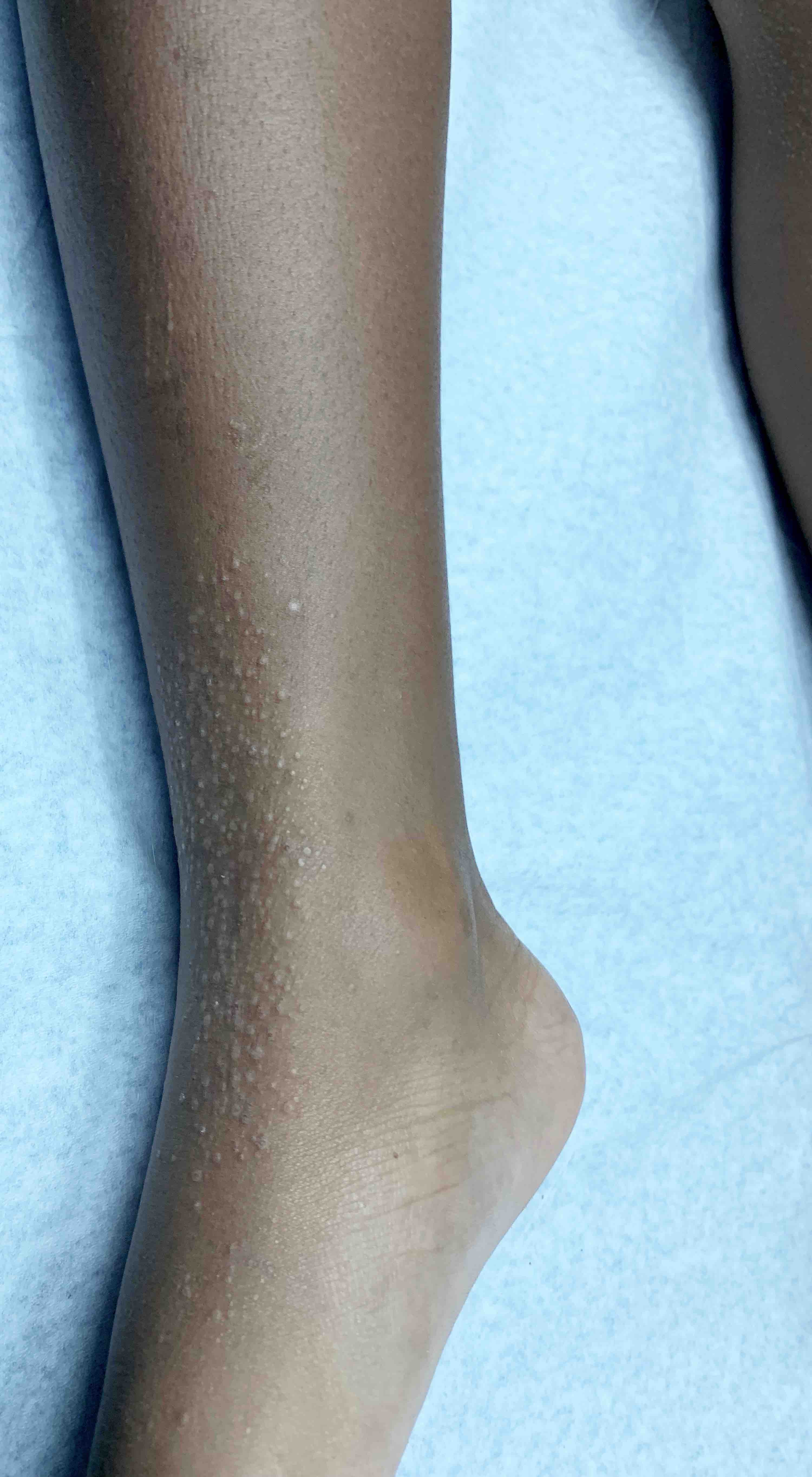
Pinpoint koebnerized papules

Subtle papules
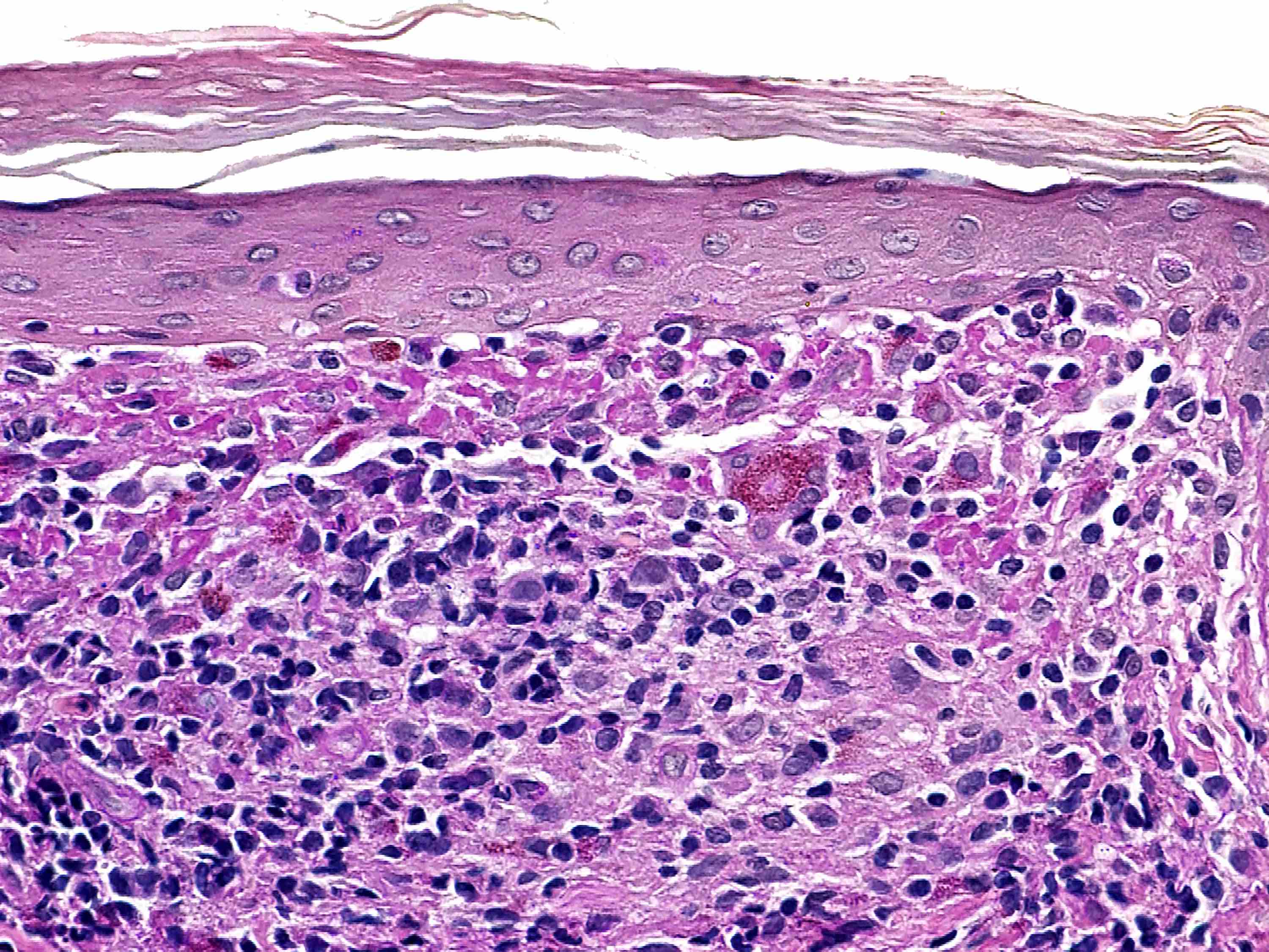
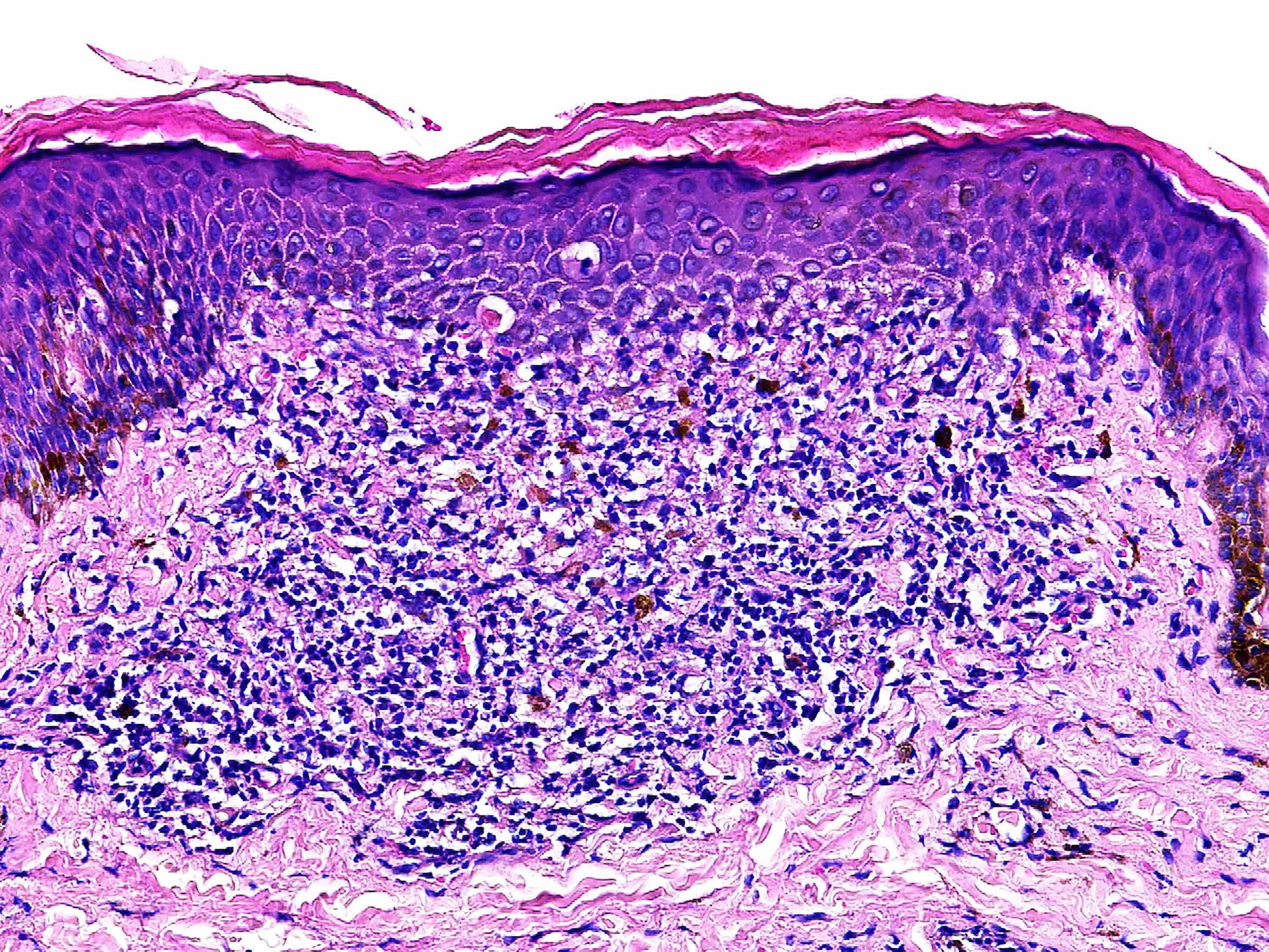
Microscopic (histologic) description
- Discrete inflammatory cell infiltrate obscuring the dermoepidermal junction
- Lichenoid infiltrate expands 1 - 4 dermal papillae
- Epidermal collarette gives a ball and claw appearance
- Variable admixture of lymphocytes and histiocytes with occasional giant cells
- Basal layer vacuolation and colloid bodies may be prominent
- Overlying hyperkeratosis with or without parakeratosis frequent
- Epidermal atrophy may be noted
- Melanophages prominent in richly pigmented individuals
- Extravasated erythrocytes in purpuric variant (Dermatol Online J 2007;13:5)
- Rarely, may have perifollicular distribution (Pediatr Dermatol 2013;30:e20)
Microscopic (histologic) images
Contributed by Jonathan D. Ho, M.B.B.S., D.Sc.
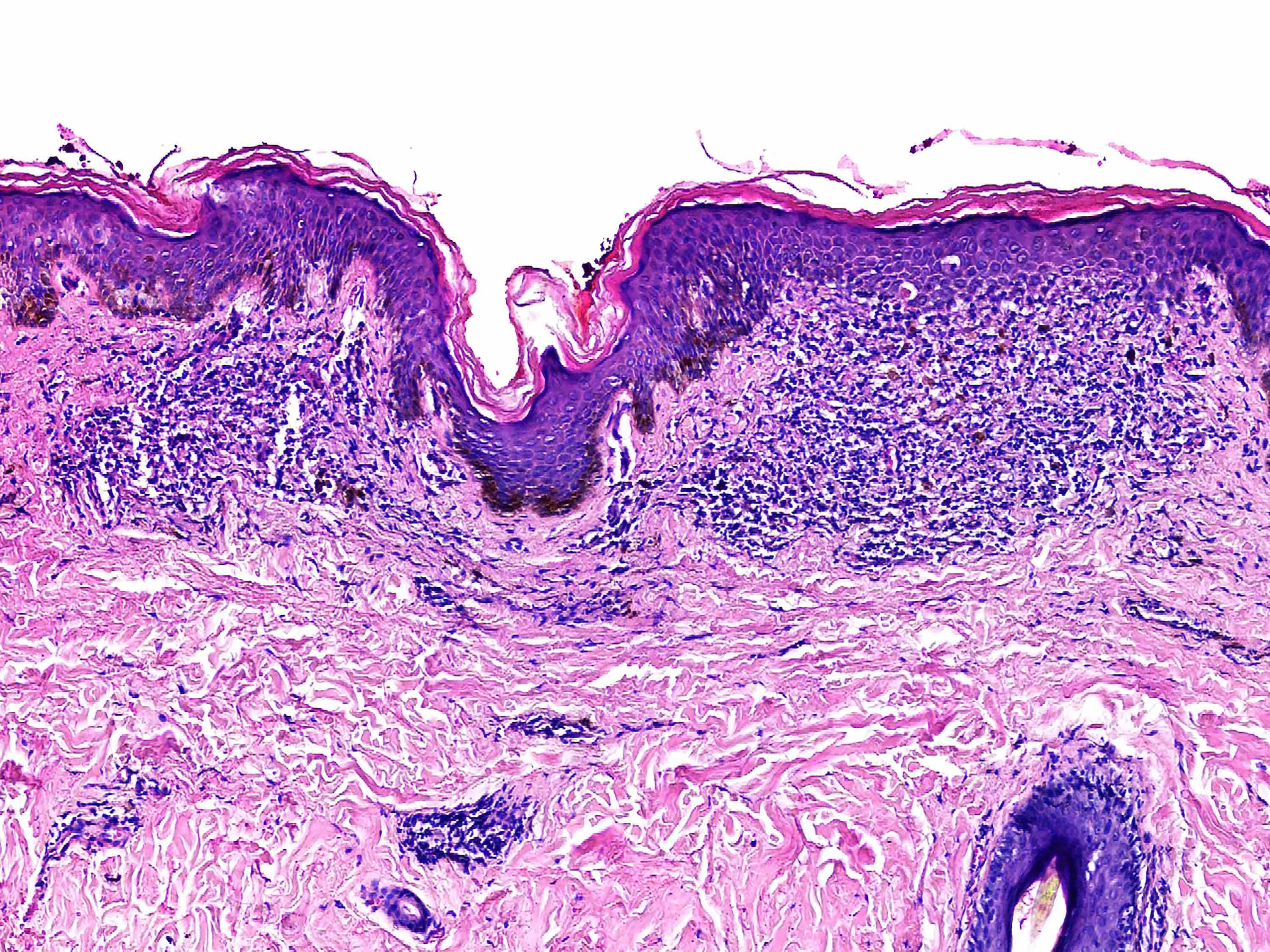
Expanded dermal papillae

Ball and claw appearance
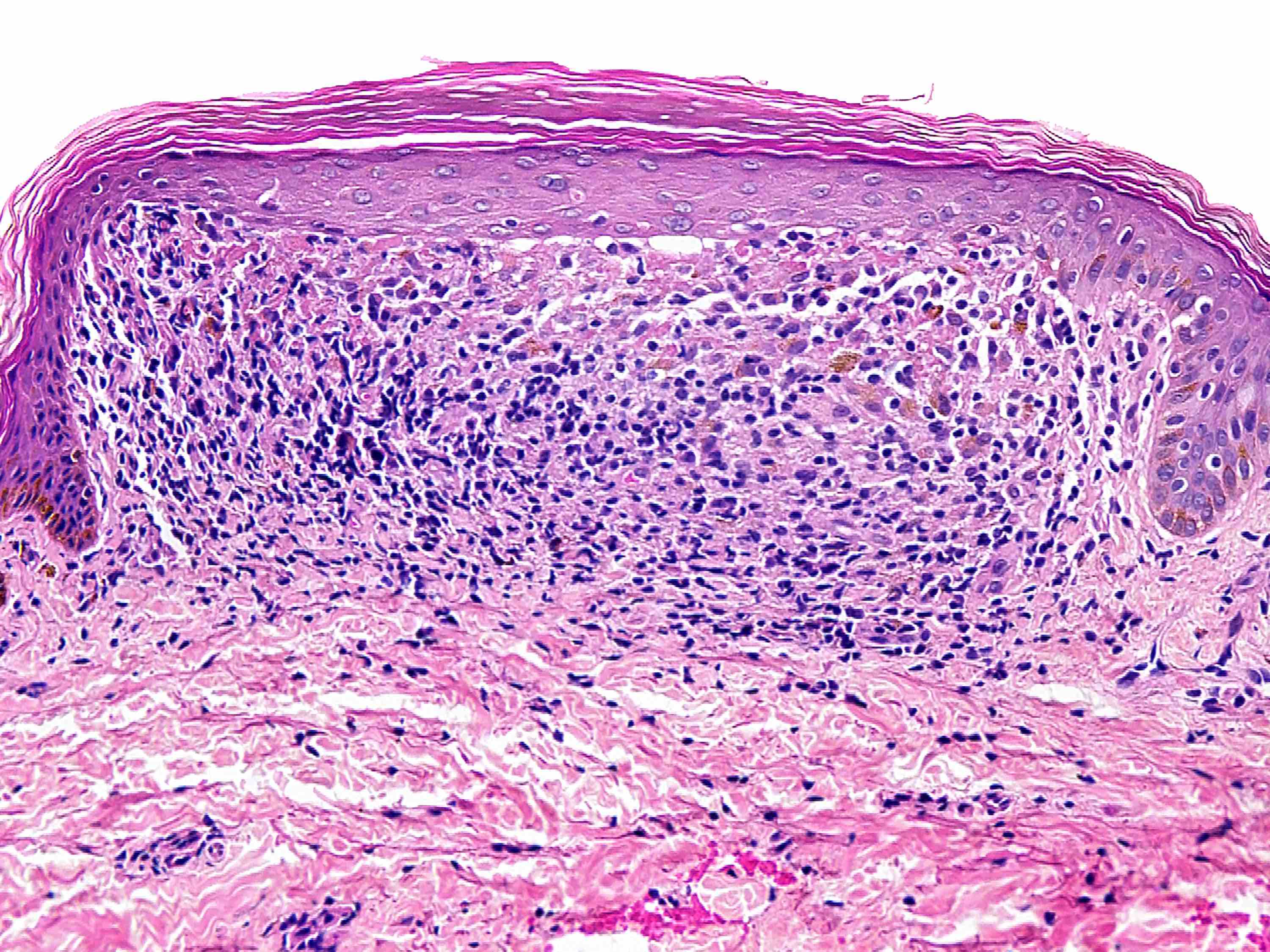
Lymphohistiocytic infiltrate
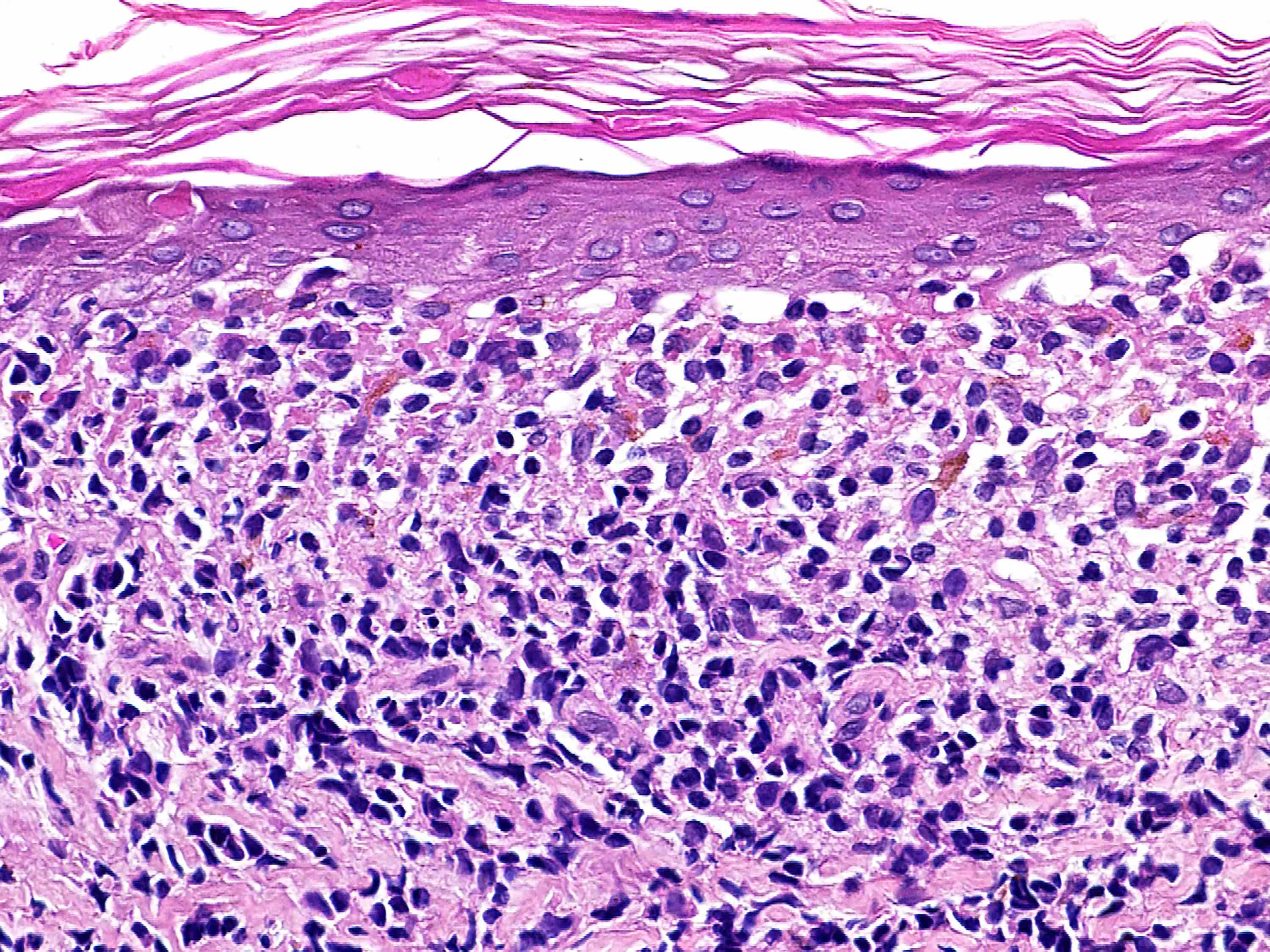
Epidermal changes
PAS stain
Immunofluorescence description
- Immunofluorescence not used for diagnosis
- Immunoglobulins typically absent
- May have fibrinogen in a linear pattern at the dermoepidermal junction (Arch Dermatol 1973;107:200)
Positive stains
- PAS highlights papillary dermal colloid bodies
Sample pathology report
- Left knee, punch biopsy:
- Lichen nitidus (see comment)
- Comment: The specimen exhibits focal parakeratosis, focal epidermal atrophy, basal layer vacuolation with associated apoptotic keratinocytes and a subjacent lichenoid lymphohistiocytic infiltrate expanding 1 - 2 dermal papillae. There are scattered multinucleated giant cells, colloid bodies and occasional melanophages. These features are diagnostic of lichen nitidus.
Differential diagnosis
- Lichen planus:
- Wedge shaped hypergranulosis
- Sawtooth appearance of rete ridges
- Broader lichenoid infiltrate
- Fewer histiocytes in infiltrate
- Lichen scrofulosorum:
- Tuberculid
- Prominent epithelioid granulomas centered around adnexal structures (Dermatology 2000;201:272)
Additional references
Practice question #1
A 9 year old boy presents with an asymptomatic eruption of hypopigmented pinpoint papules involving the skin of his elbows, knees and penile shaft. The child is otherwise well. A punch biopsy reveals focal hyperkeratosis, epidermal atrophy, basal layer vacuolation and a discrete lymphohistiocytic infiltrate expanding a single dermal papilla and obscuring the dermoepidermal junction. What is the most likely diagnosis?
- Lichen nitidus
- Lichen planus
- Lichen scrofulosorum
- Lichen simplex chronicus
- Lichen striatus
Practice answer #1
Practice question #2
Which of the following histopathologic features favors a diagnosis of lichen nitidus over lichen planus?
- Basal layer vacuolar change
- Broad band-like lymphocytic infiltrate with scattered colloid bodies
- Multinucleated giant cells within a focal lichenoid infiltrate
- Scattered melanophages
- Wedge shaped hypergranulosis
Practice answer #2
C. Multinucleated giant cells within a focal lichenoid infiltrate
Comment Here
Reference: Lichen nitidus
Comment Here
Reference: Lichen nitidus
