Stains & CD markers
Synaptophysin

Copyright: 2002-2025, PathologyOutlines.com, Inc.
PubMed Search: Synaptophysin
Synaptophysin
Author: Y. Albert Yeh, M.D., Ph.D.
Editorial Board Members: Christian M. Schürch, M.D., Ph.D., Debra L. Zynger, M.D.
Last author update: 6 January 2022
Last staff update: 13 June 2022
Copyright: 2002-2025, PathologyOutlines.com, Inc.
PubMed Search: Synaptophysin
Table of Contents
Definition / general | Essential features | Terminology | Pathophysiology | Diagrams / tables | Clinical features | Interpretation | Uses by pathologists | Prognostic factors | Microscopic (histologic) description | Microscopic (histologic) images | Cytology images | Positive staining - normal | Positive staining - disease | Negative staining | Sample pathology report | Additional references | Practice question #1 | Practice answer #1Cite this page: Yeh YA. Synaptophysin. PathologyOutlines.com website. https://www.pathologyoutlines.com/topic/stainssynaptophysin.html. Accessed September 28th, 2025.
Definition / general
- A protein (38 kD), encoded by the SYP gene; is an integral membrane glycoprotein localized to presynaptic neurosecretory vesicles (Am J Hum Genet 1990;47:551)
Essential features
- An integral membrane protein of small synaptic vesicles in brain and endocrine cells (Am J Hum Genet 1990;47:551)
- Positive in well differentiated neuroendocrine tumor (carcinoid tumor), neuroendocrine carcinoma, neuroblastoma, adrenal cortical tumors, paraganglioma, pheochromocytoma, Merkel cell carcinoma, parathyroid tumors and medullary thyroid carcinoma
- Used in conjunction with chromogranin A, INSM1 and CD56 for diagnosing neuroendocrine tumors (Am J Surg Pathol 2020;44:757)
Terminology
- Major synaptic vesicle protein p38
- MRX96
- MRXSYP
- XLID96
Pathophysiology
- SYP gene is at Xp11.23 (Am J Hum Genet 1990;47:551)
- Subcellular locations:
- Synaptic vesicle membrane, cytoplasmic vesicles, secretory vesicles, synaptosome
- SYP gene encodes an integral membrane protein of small synaptic vesicles in brain and endocrine cells (Am J Hum Genet 1990;47:551)
- Involved in cholesterol binding and biogenesis and maturation of synaptic vesicles (Nat Cell Biol 2000;2:42)
- Regulates endocytosis of synaptic vesicles
- SYP mutations are associated with X linked intellectual developmental disorder (Nat Genet 2009;41:535)
Diagrams / tables
Images hosted on other servers:

Synaptophysin in synaptic vesicle endocytosis
Clinical features
- SYP mutations cause X linked intellectual developmental disorder (Nat Genet 2009;41:535)
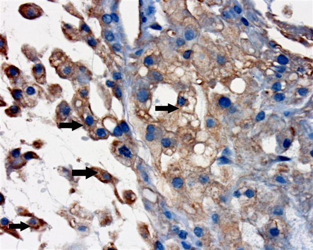
Interpretation
- Cytoplasmic staining
Uses by pathologists
- Common marker of neuroendocrine differentiation
- Used in combination with chromogranin A, INSM1 and CD56 for diagnosing neuroendocrine tumors (Am J Surg Pathol 2020;44:757, Am J Surg Pathol 2017;41:1561)
Prognostic factors
- Expression correlates with resistance to abiraterone and enzalutamide treatment in patients with castration resistance prostate cancer (Urol Oncol 2018;36:162.e1)
- Synaptophysin positive, BRAF mutated colorectal cancers characterized by worse progression free survival and overall survival (Eur J Cancer 2021;146:145)
- Expression associated with a longer median overall survival in patients with advanced nonsmall cell lung cancer (Respiration 2013;85:289)
- Synaptophysin positive stage I squamous cell carcinoma and adenocarcinoma of the lung associated with worse prognosis (Cancer 2007;110:1776)
- Aberrant expression in a small subset of epithelioid hemangioendothelioma associated with aggressive clinical course (Am J Surg Pathol 2021;45:616)
- Associated with worse prognosis in extrahepatic cholangiocarcinoma (Hum Pathol 2005;36:732)
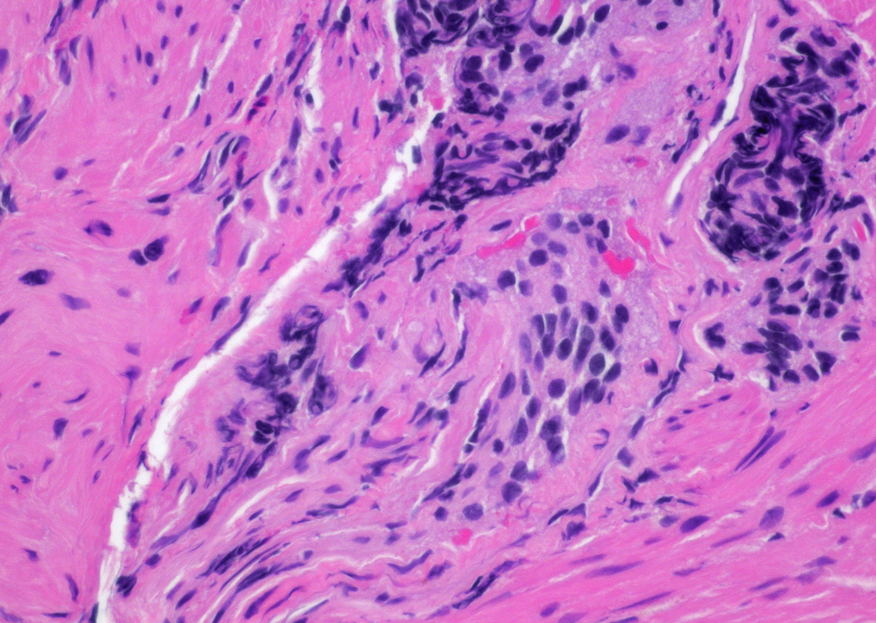
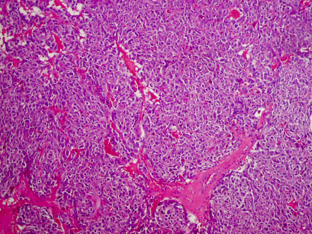
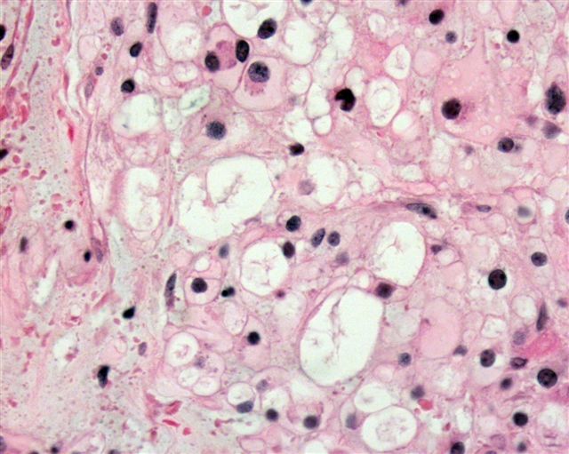
Microscopic (histologic) description
- Diffuse cytoplasmic staining is regarded as positive
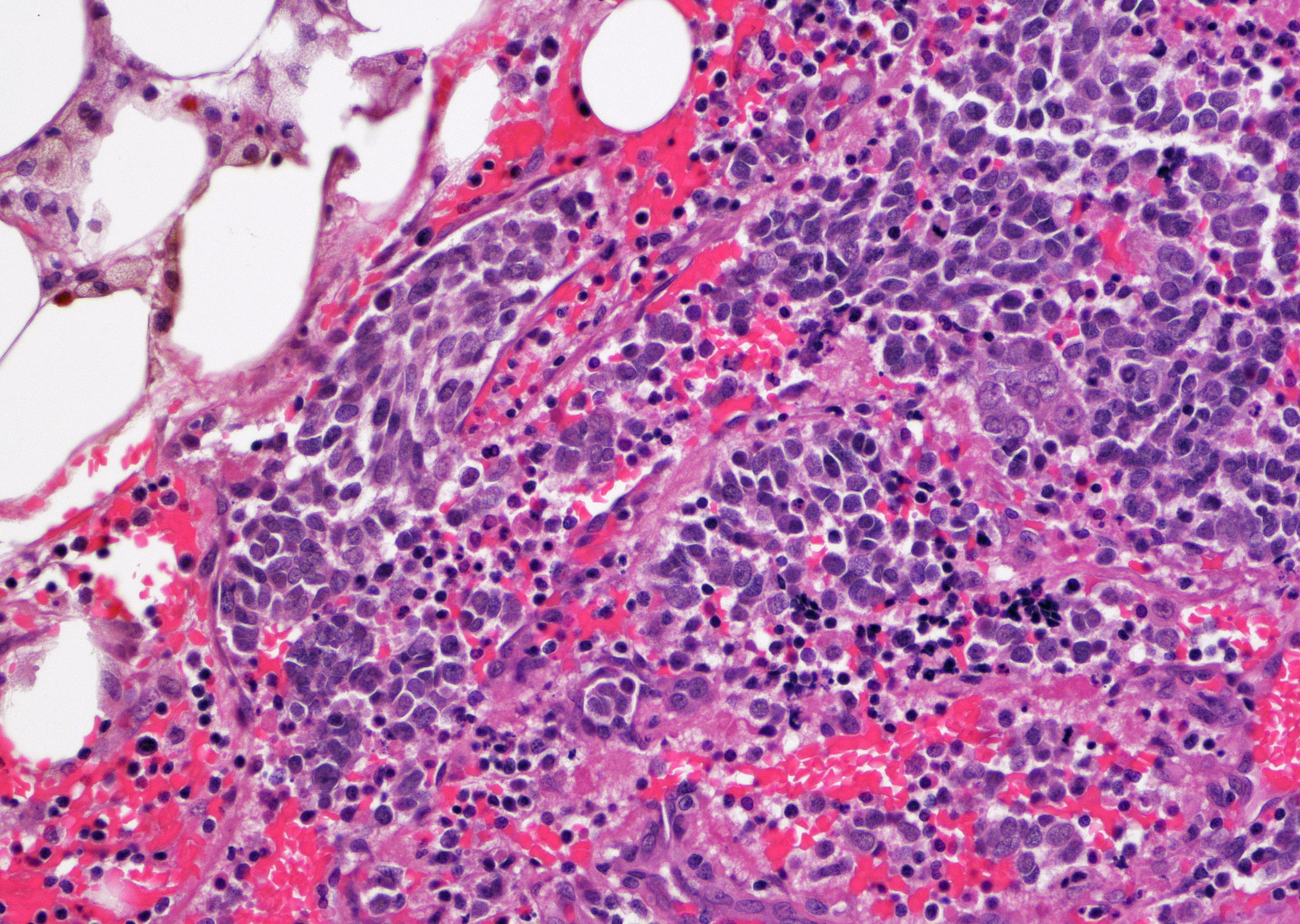
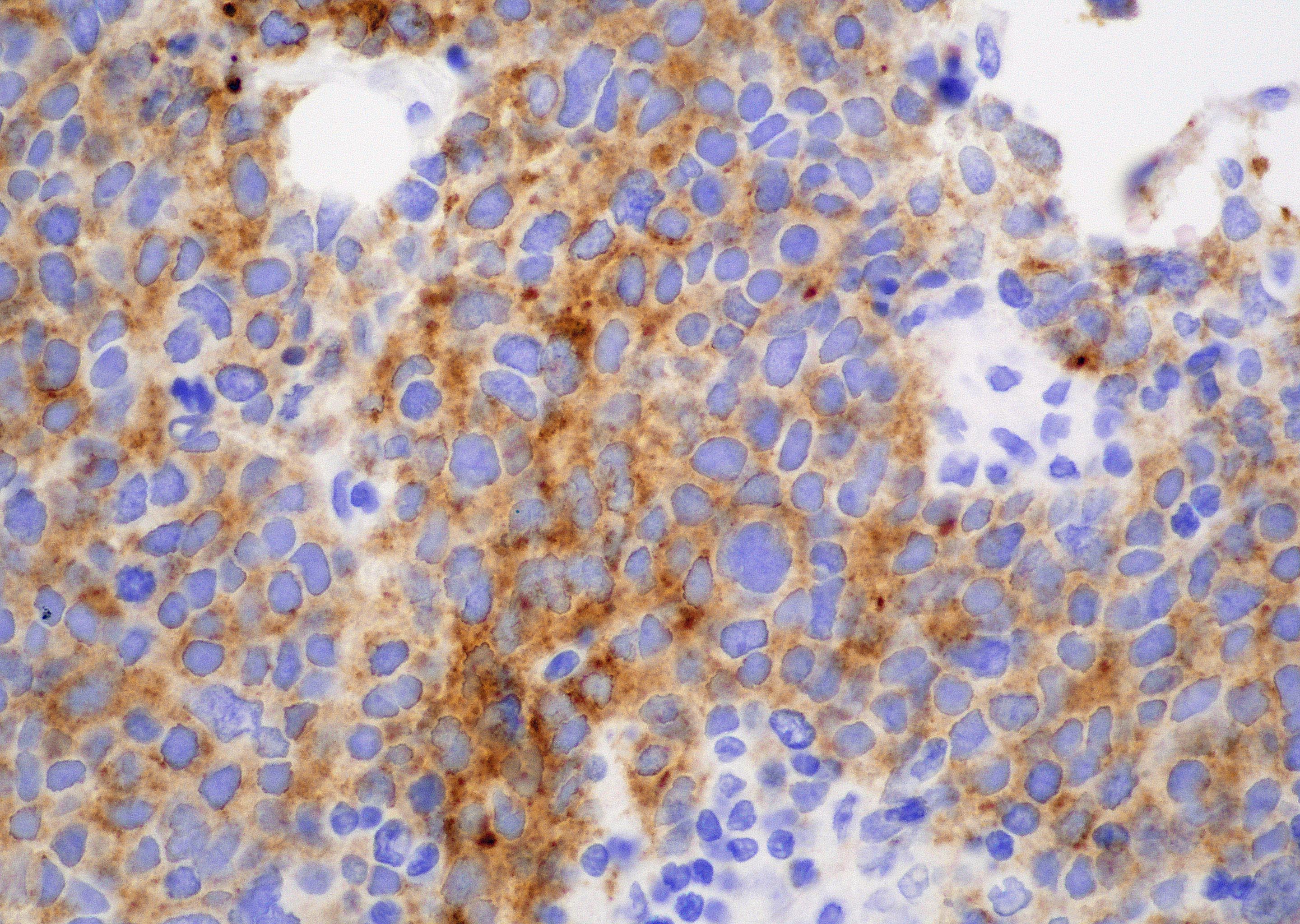
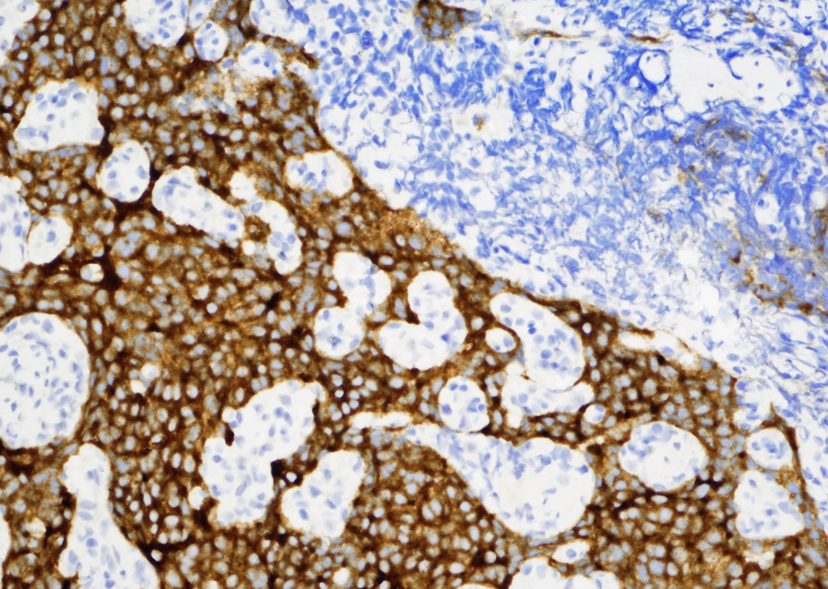
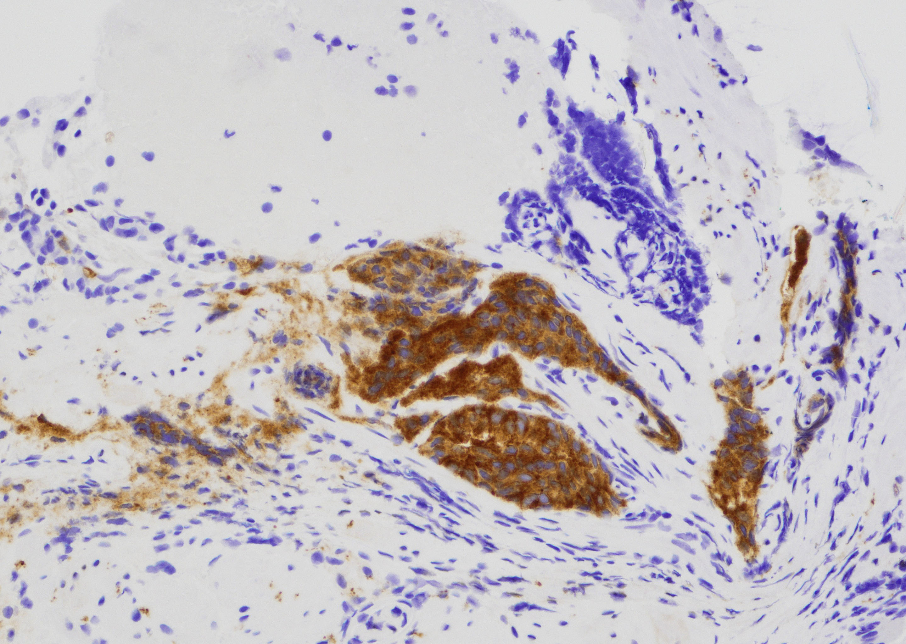
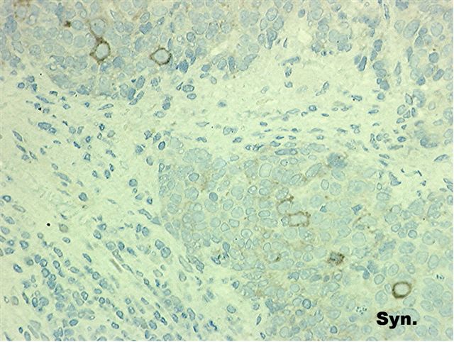
Microscopic (histologic) images
Contributed by Y. Albert Yeh, M.D., Ph.D.
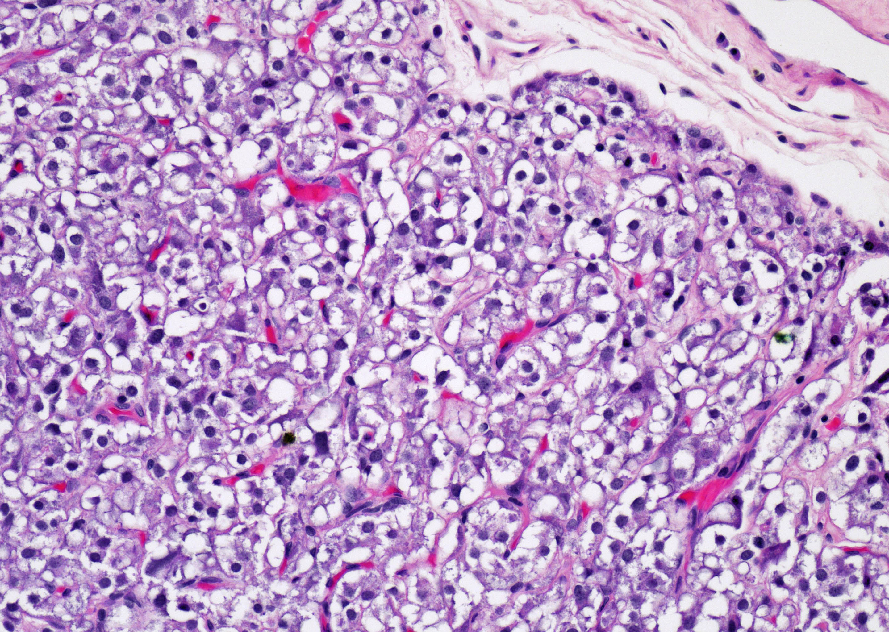
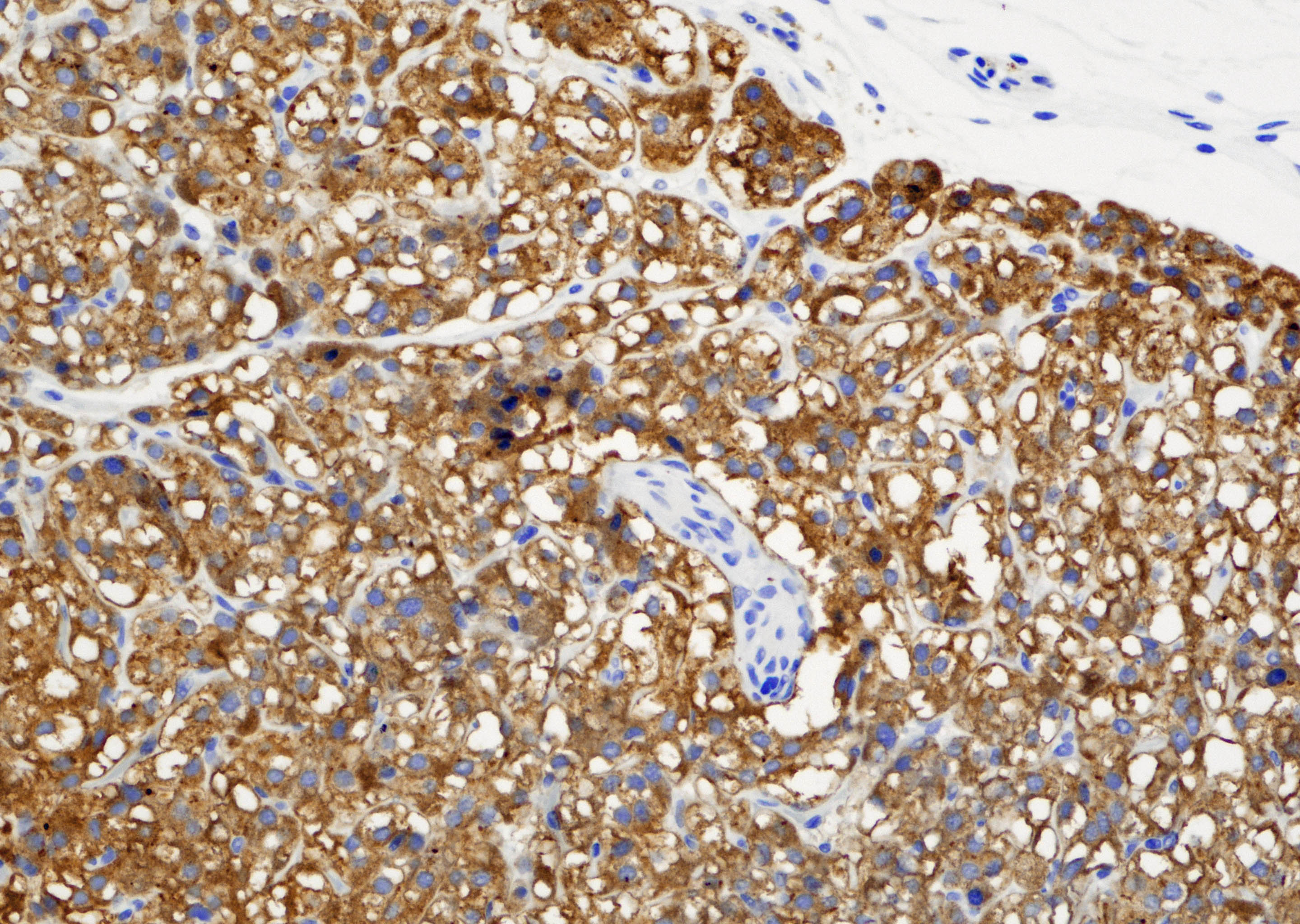
Prostatic paraganglioma
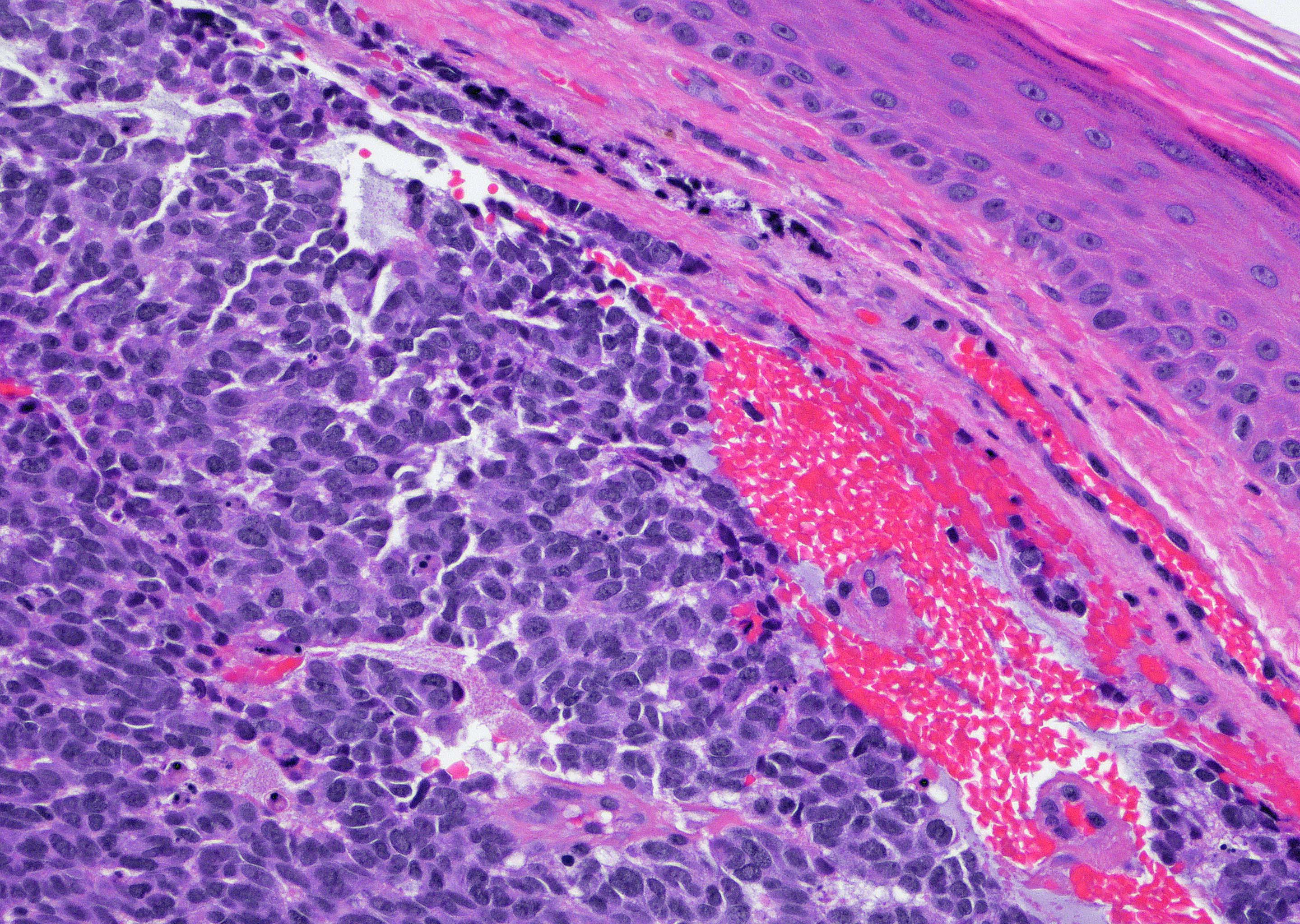
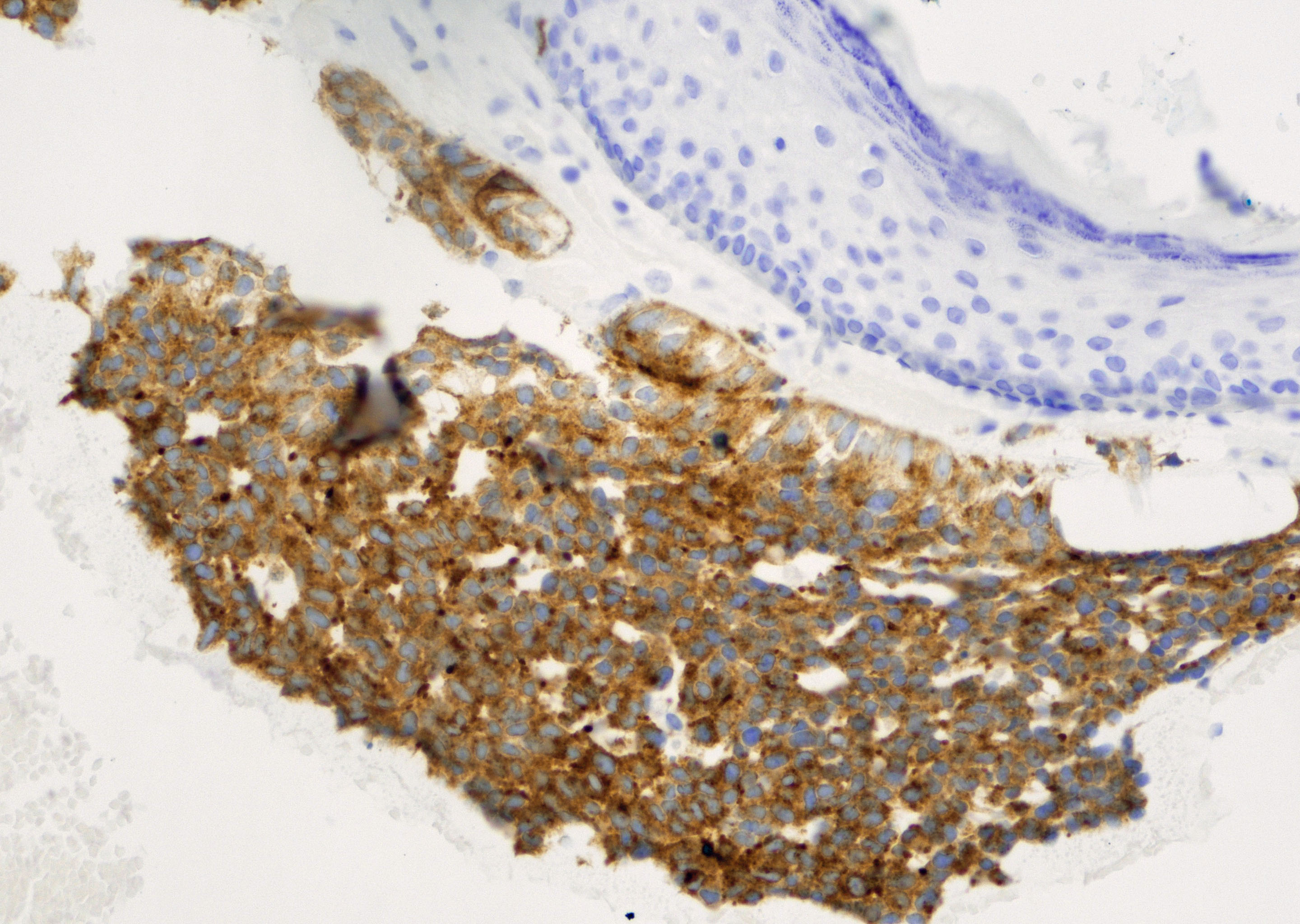
Merkel cell carcinoma
Metastatic Merkel cell carcinoma
Lung small cell carcinoma
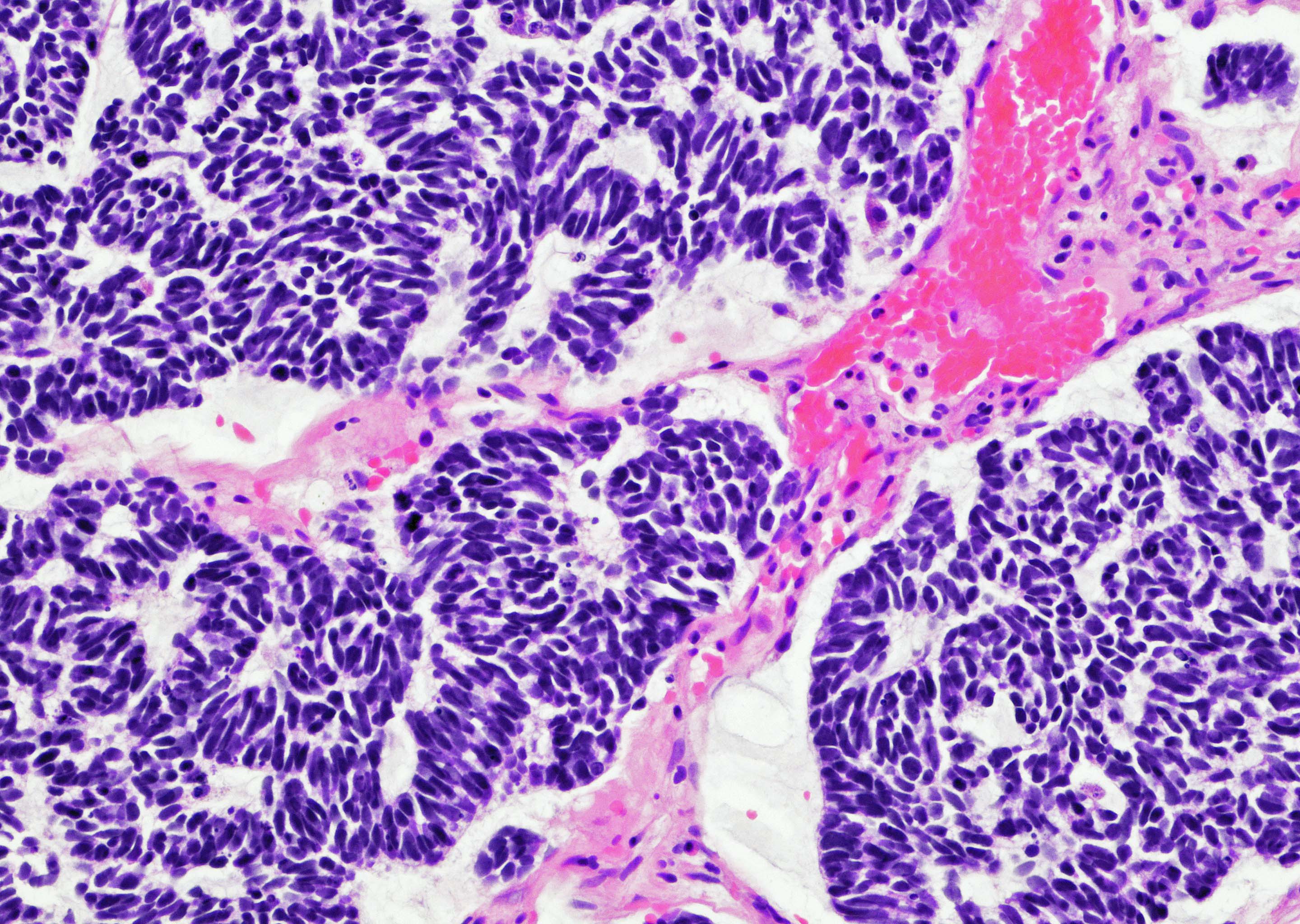

Bladder small cell neuroendocrine carcinoma
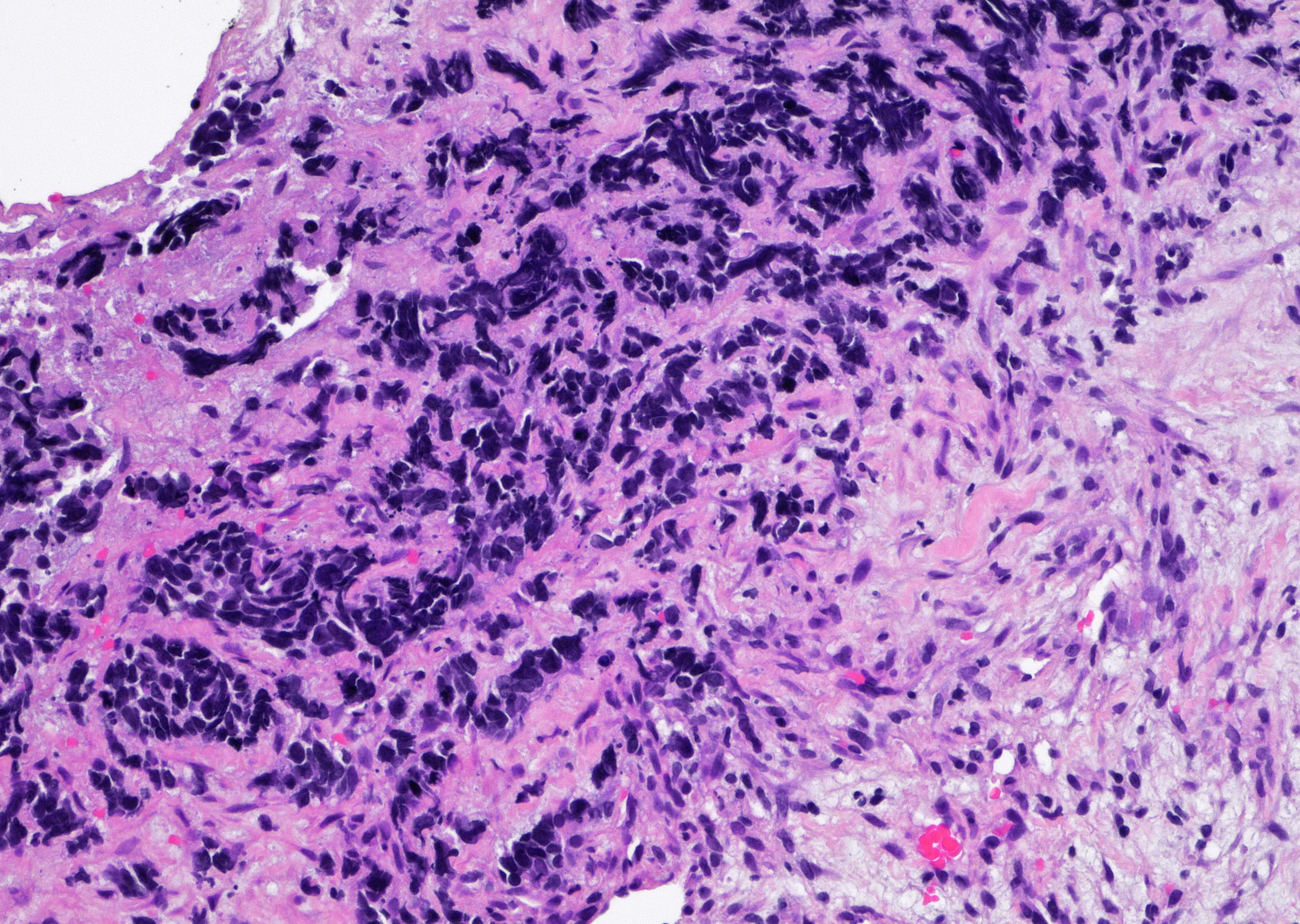
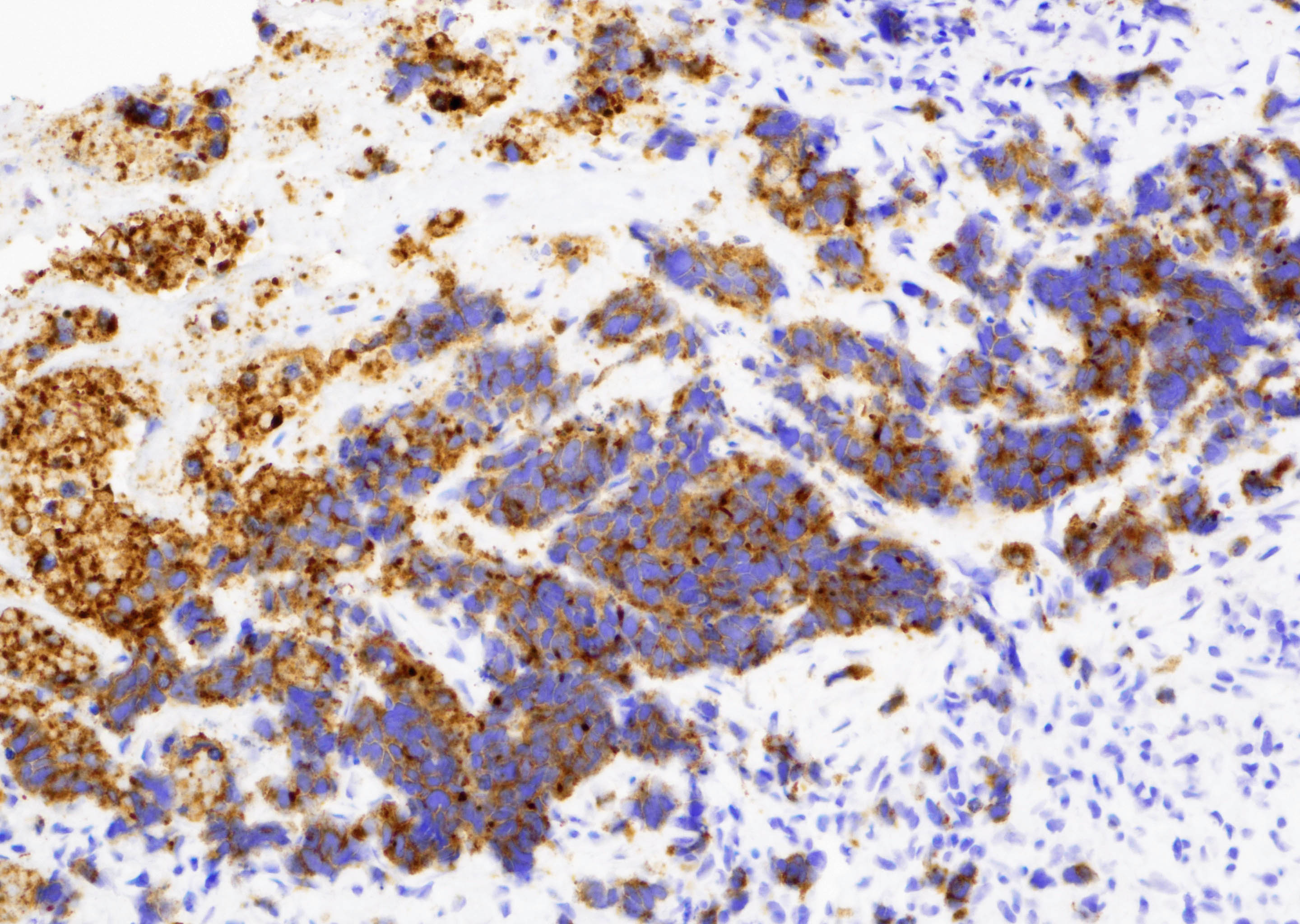
Liver small cell neuroendocrine carcinoma
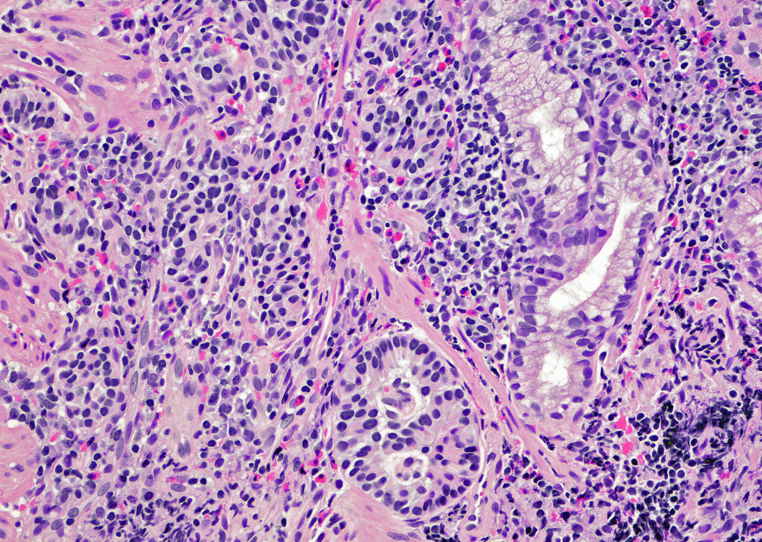
Duodenal well differentiated neuroendocrine tumor
Duodenal neuroendocrine dysplastic nodule
Cases #195, #110, #189, #185 and #17
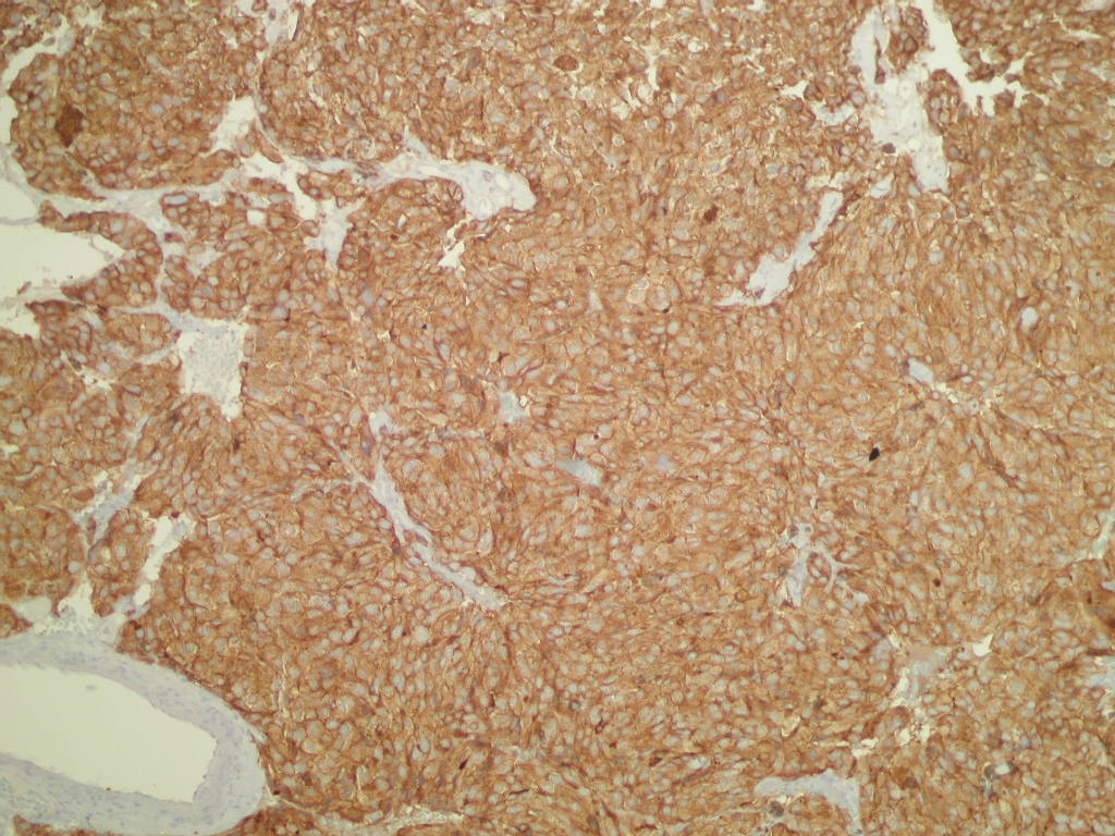
Bladder paraganglioma
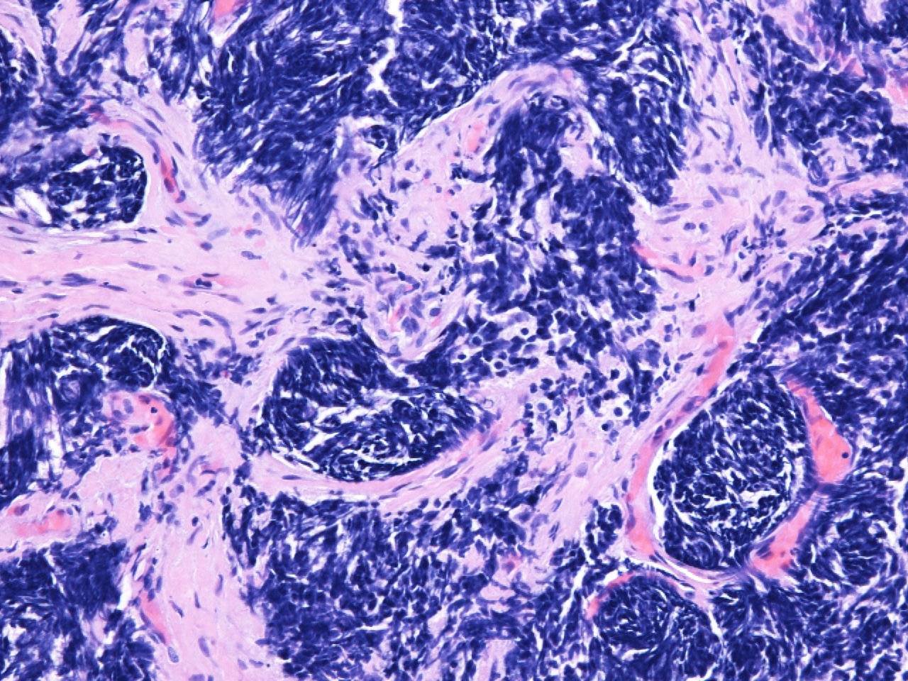
Sacrococcygeal chordoma
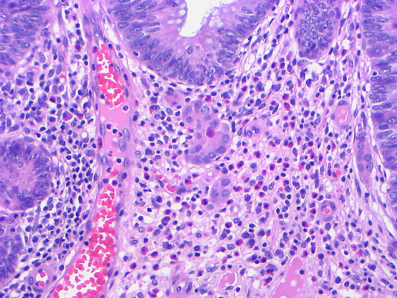
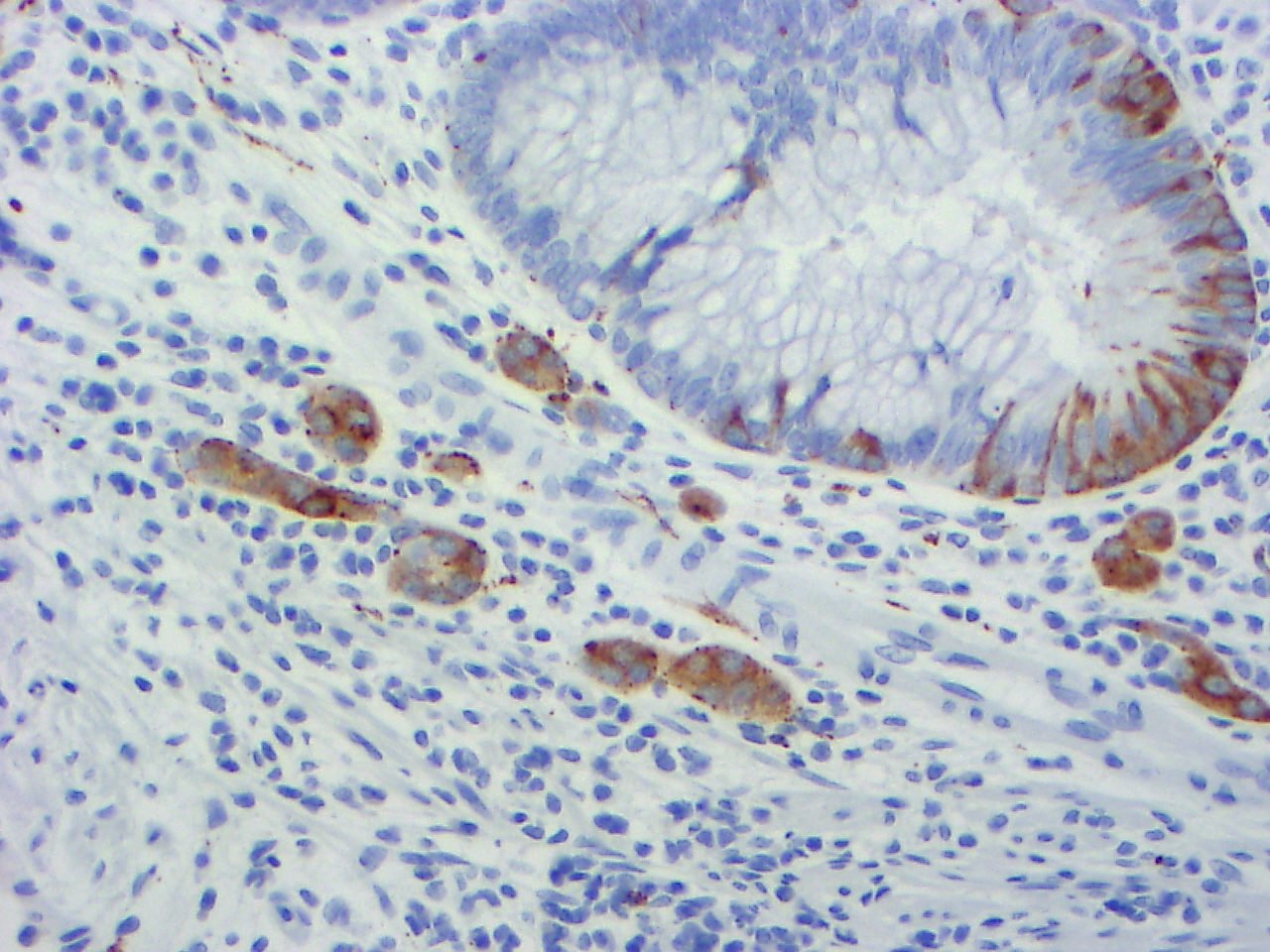
Colonic tubulovillous adenoma with microcarcinoids
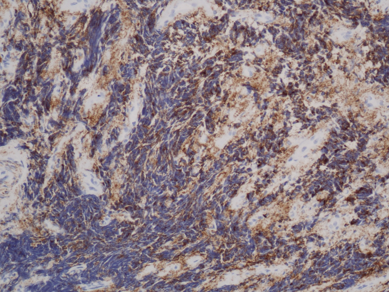
Retropharyngeal neuroblastoma
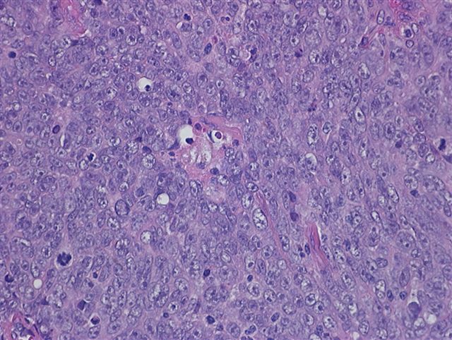
Thymic carcinoma (type C thymoma), nonkeratinizing squamous cell carcinoma type
Contributed by Jijgee Munkhdelger, M.D., Ph.D. and Andrey Bychkov, M.D., Ph.D.
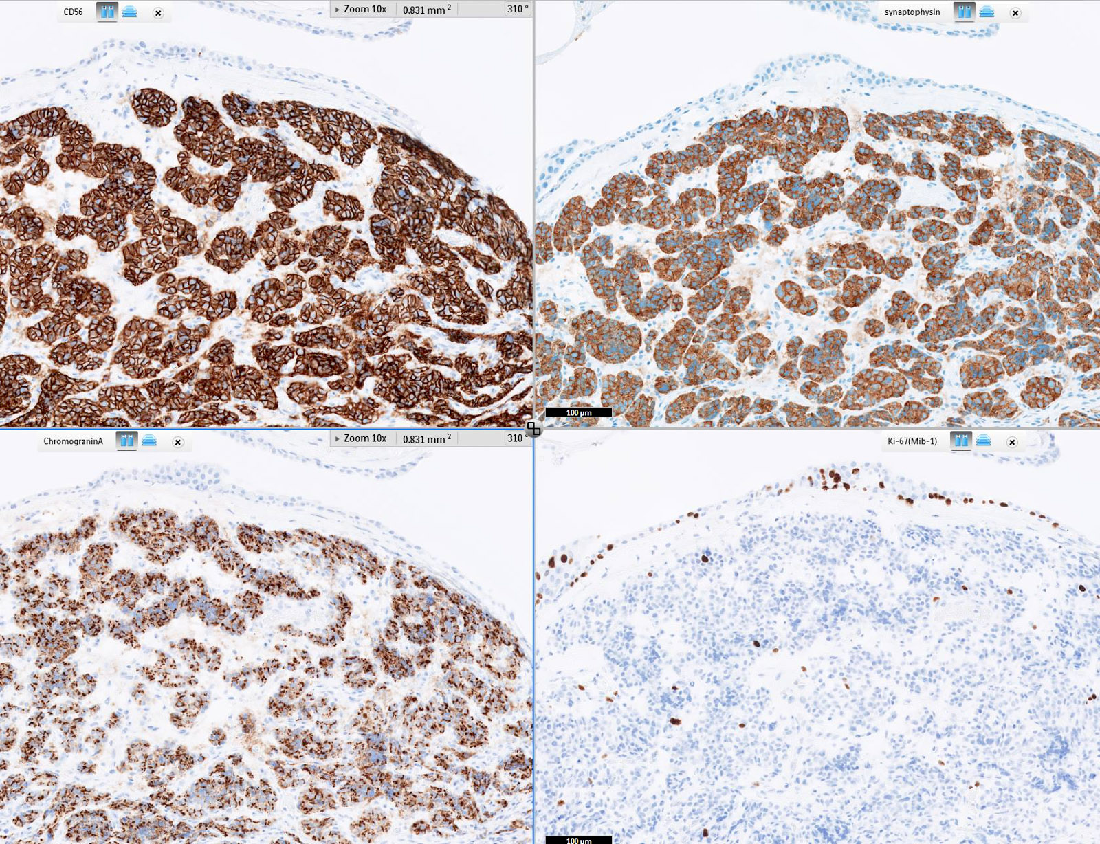
Lung typical carcinoid immunoprofile
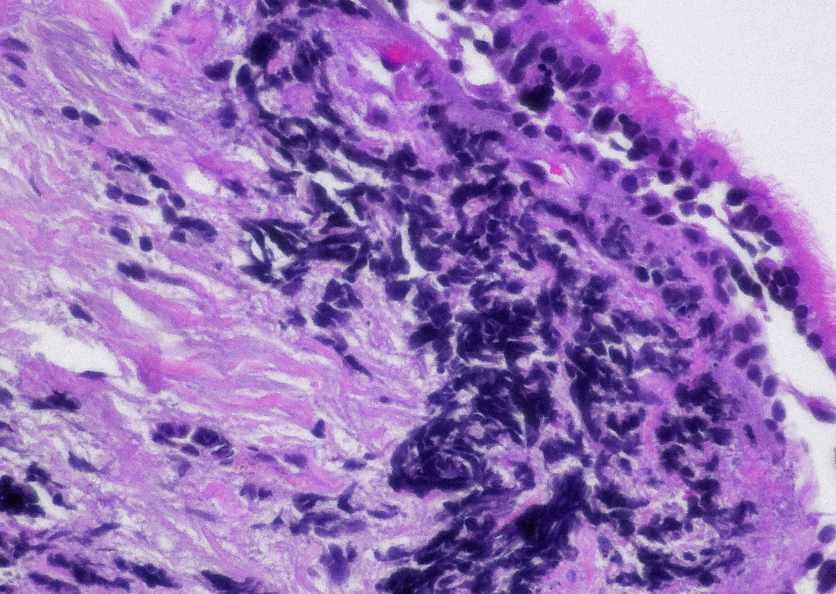
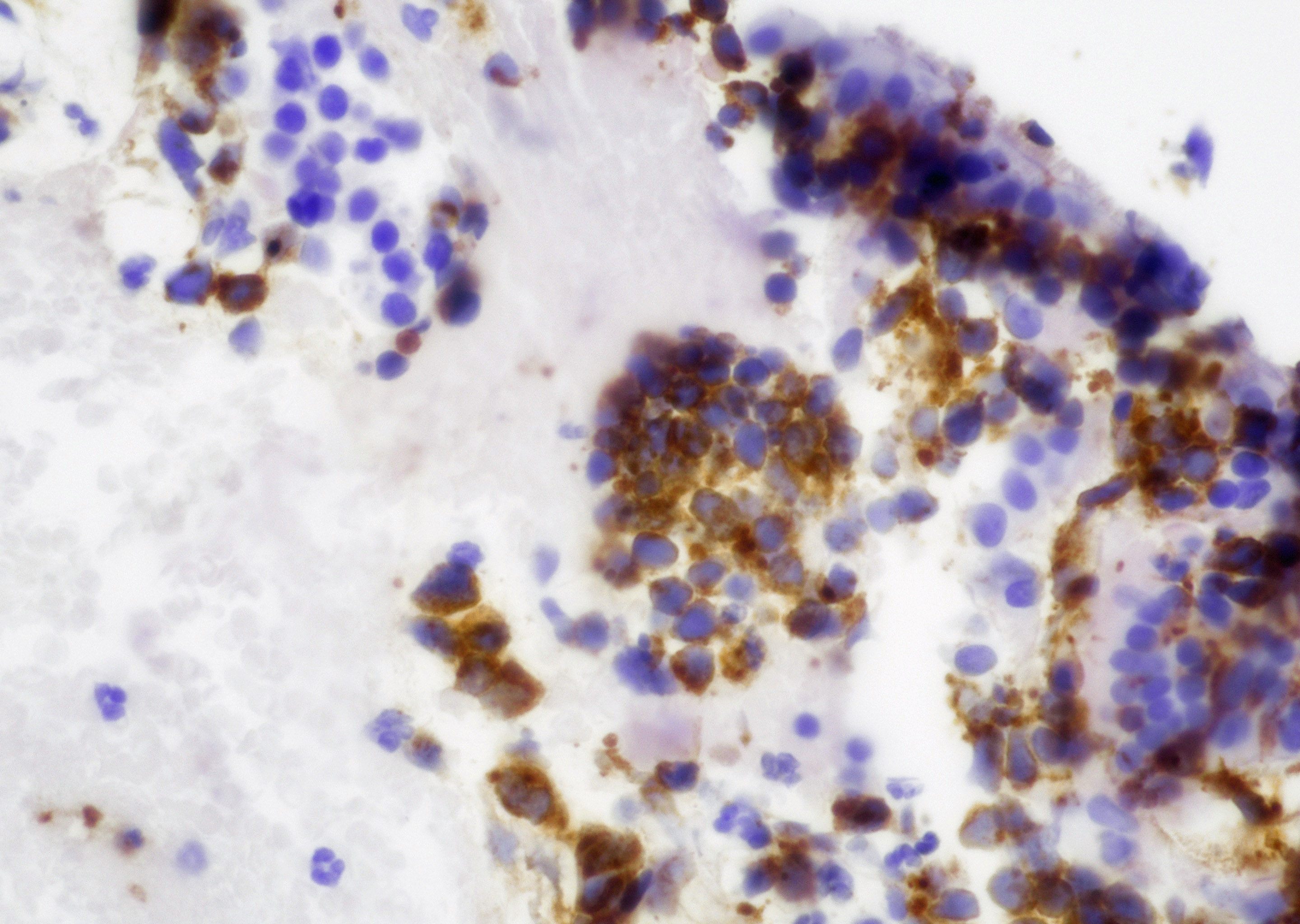
Cytology images
Contributed by Y. Albert Yeh, M.D., Ph.D. and Ashley Moreland, B.S., C.T. (ASCP)
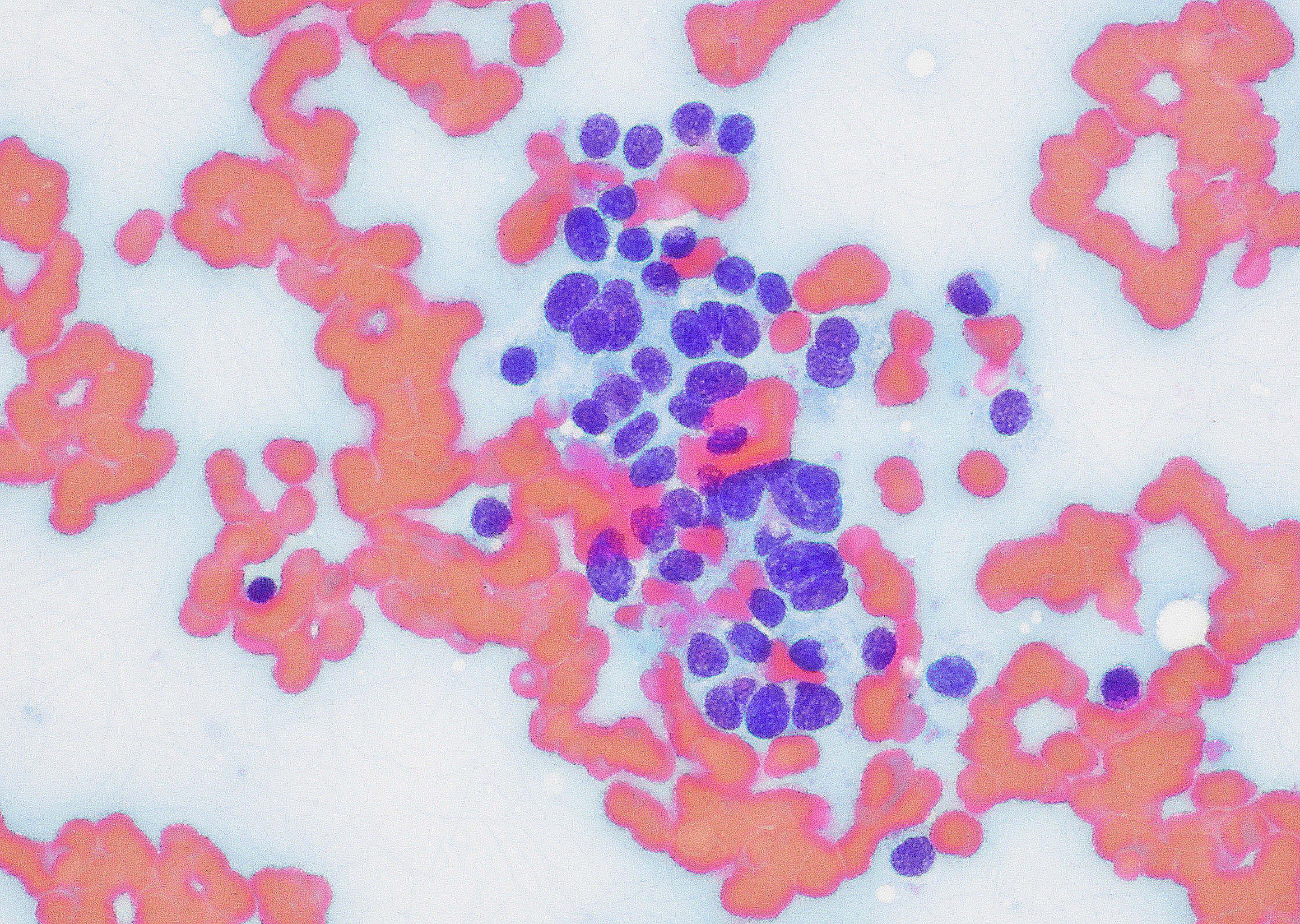
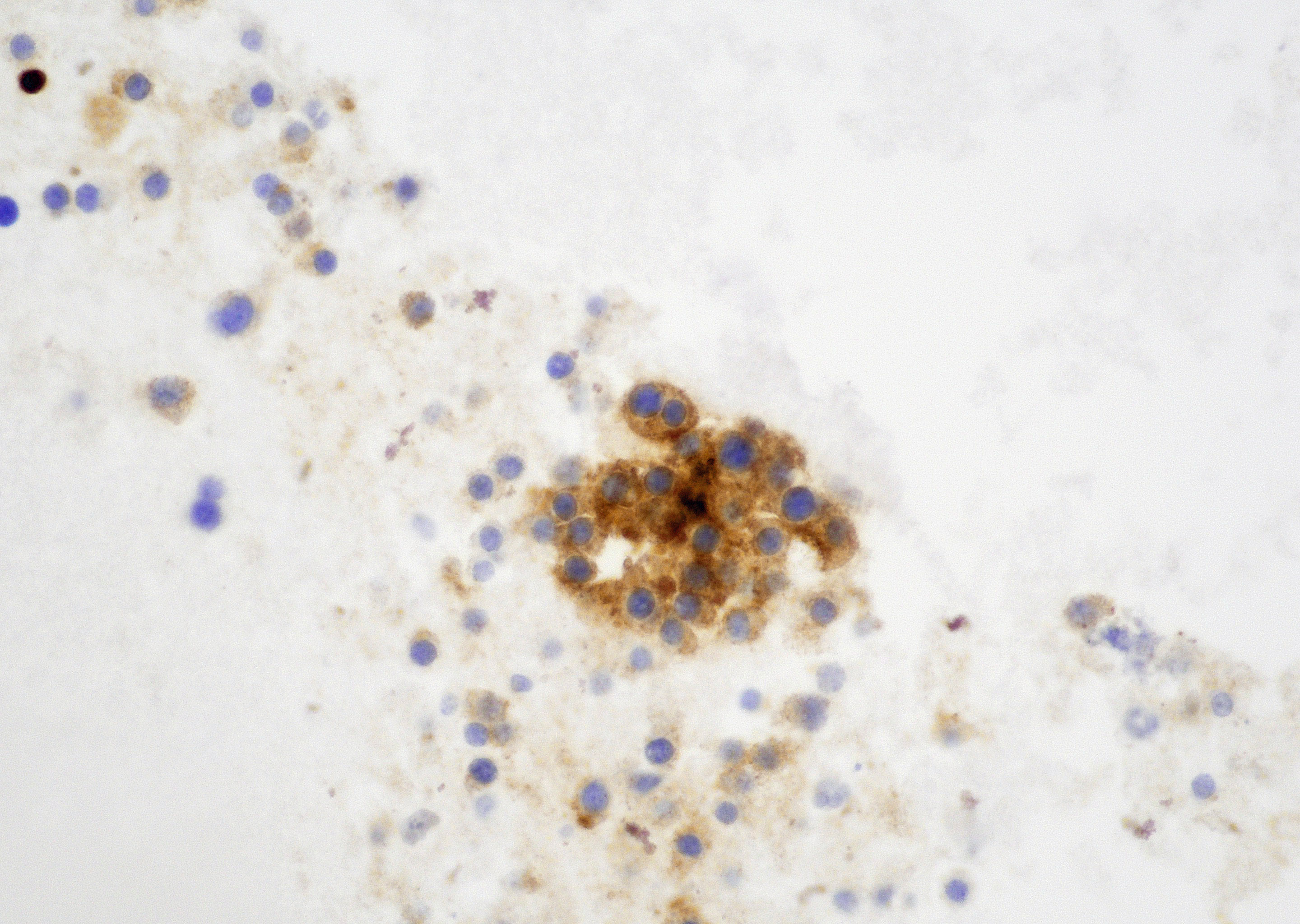
Medullary thyroid carcinoma
Positive staining - normal
- Brain (Mol Cell Proteomics 2014;13:397)
- Neuroendocrine tissues
- Adrenal medulla
- Pancreatic islets
- Pineal gland
- Parathyroid tissue
Positive staining - disease
- Well differentiated neuroendocrine tumor (carcinoid tumor)
- Small cell neuroendocrine carcinoma (Am J Surg Pathol 2020;44:757)
- Large cell neuroendocrine carcinoma
- Adrenal cortical adenoma and carcinoma (Eur J Endocrinol 2009;161:939)
- Adrenal cortical oncocytic tumors
- Pheochromocytoma (Mod Pathol 2011;24:S58)
- Paraganglioma (Arch Pathol Lab Med 2015;139:1062)
- Neuroblastoma, ganglioneuroblastoma (Endocr Pathol 2019;30:173)
- Ganglioneuroma
- Central neurocytoma
- Primitive neuroectodermal tumor (PNET) (Neuropathology 2017;37:35)
- Gangliocytoma, ganglioglioma
- Pilocytic astrocytoma (Am J Surg Pathol 2012;36:43)
- Medulloblastoma, desmoplastic medulloblastoma (Arch Pathol Lab Med 2007;131:234)
- Pineocytoma
- Choroid plexus papilloma, choroid plexus carcinoma
- Retinoblastoma
- Medullary thyroid carcinoma
- Merkel cell carcinoma (J Cutan Pathol 2021;48:411)
- Glomus tumor
- Desmoplastic small round cell tumor (Am J Surg Pathol 1999;23:1408)
- Ewing sarcoma (Am J Clin Pathol 2015;143:659)
- Parathyroid adenoma and carcinoma (Endocr Pathol 2018;29:113)
Negative staining
- Tumor with nonneuroendocrine cell origin
- Squamous cell carcinoma
- Adenocarcinoma
- Renal cell carcinoma
- Urothelial carcinoma
- Endometrial carcinoma
- Melanoma
- Lymphoma
- Reference: J Thorac Oncol 2017;12:334
Sample pathology report
- Lung, right upper lobe, endobronchial biopsy:
- Poorly differentiated carcinoma consistent with small cell neuroendocrine carcinoma (see comment)
- Comment: The lung biopsy shows bronchial mucosa with infiltrating tumor cell with nuclei characterized by coarse and fine chromatin and arranged in nuclear molding pattern. Immunohistochemical stains synaptophysin, chromogranin and TTF1 are positive in the tumor cells. These findings support the diagnosis of small cell neuroendocrine carcinoma of the lung.
Additional references
Practice question #1
A 60 year old man presented to the urology clinic with obstructive urinary tract symptoms. Cystoscopy showed a tumor in the bladder neck. Transurethral urethral bladder tumor resection was performed. Microscopic examination of the sample is shown in the above photomicrograph. What are the best immunohistochemical stains to diagnose this tumor?
- Desmin, SMA, myoD
- CD45, CD3, CD20
- S100, melan A, SOX10
- Synaptophysin, chromogranin A, INSM1
Practice answer #1
D. Synaptophysin, chromogranin A, INSM1. This is a small cell neuroendocrine carcinoma of the bladder.
Comment Here
Reference: Synaptophysin
Comment Here
Reference: Synaptophysin