Stomach
Gastritis
Collagenous gastritis
Authors: Matthew Morrow, M.D., Raul S. Gonzalez, M.D.
Editorial Board Member: Diana Agostini-Vulaj, D.O.
Deputy Editor-in-Chief: Aaron R. Huber, D.O.


Last author update: 8 September 2025
Last staff update: 9 September 2025
Copyright: 2002-2025, PathologyOutlines.com, Inc.
PubMed Search: Collagenous gastritis
Table of Contents
Definition / general | Essential features | ICD coding | Epidemiology | Sites | Pathophysiology | Clinical features | Diagnosis | Case reports | Treatment | Clinical images | Gross description | Microscopic (histologic) description | Microscopic (histologic) images | Positive stains | Negative stains | Sample pathology report | Differential diagnosis | Additional references | Practice question #1 | Practice answer #1 | Practice question #2 | Practice answer #2Cite this page: Morrow M, Gonzalez RS. Collagenous gastritis. PathologyOutlines.com website. https://www.pathologyoutlines.com/topic/stomachcollagenous.html. Accessed September 10th, 2025.
Definition / general
- Poorly understood disease manifesting as thickened subepithelial collagen in the stomach
Essential features
- Extremely rare disease characterized by the presence of a band of increased subepithelial collagen in the stomach, with associated chronic inflammation in the lamina propria
- Often subtyped into pediatric and adult versions
- No standard therapy
ICD coding
- ICD-10: K52.89 - other specified noninfective gastroenteritis and colitis
Epidemiology
- Extremely rare, with < 200 cases reported since it was first described in 1989 (World J Gastrointest Endosc 2015;7:265)
- Slight female predominance, occurring in all ages but age at presentation centers around young adults (J Nat Sci Biol Med 2018;9:285)
Sites
- Pediatric patients are more likely to have body / fundus predominant disease versus antrum in adults (Mod Pathol 2015;28:533)
Pathophysiology
- Poorly understood etiology / pathophysiology
Clinical features
- Associated with celiac disease, collagenous colitis, collagenous sprue and other autoimmune disease (Arch Pathol Lab Med 2001;125:1579)
- Some patients are on potentially causative medications (e.g., nonsteroidal anti-inflammatory drugs [NSAIDs], angiotensin receptor blockers [ARBs]) (Clin Gastroenterol Hepatol 2022;20:1977)
- Most common clinical symptoms include abdominal pain, anemia, diarrhea, nausea / vomiting, gastrointestinal bleeding and weight loss
- Through limited case reports, the disease has been phenotyped into adult and pediatric types but many patients may not fit either category (Am J Surg Pathol 2001;25:1174)
- Pediatric type classically presents with upper gastrointestinal symptoms including abdominal pain and anemia, which are a result of the disease process
- Adult type is characterized by diarrhea, weight loss and accompanying collagenous colitis, related to underlying autoimmune processes or celiac disease (Clin Gastroenterol Hepatol 2022;20:1977)
Diagnosis
- Established by gastric biopsy
Case reports
- 15 year old boy with abdominal pain and anemia (Endoscopy 2025;57:E287)
- 16 year old boy with hemorrhagic shock (ACG Case Rep J 2025;12:e01762)
- 16 year old boy with weight loss (JPGN Rep 2023;4:e351)
- 16 year old girl with elevated fecal calprotectin (JPGN Rep 2024;5:152)
- 41 year old woman with collagenous colitis (Quant Imaging Med Surg 2024;14:9756)
Treatment
- While no standard therapy has been defined, many patients appear to respond to topically targeted budesonide, with both clinical and histologic improvement (Clin Gastroenterol Hepatol 2022;20:1977)
- If a causative medication is suspected, it should be stopped
Clinical images
Contributed by Matthew Morrow, M.D.
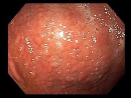
Gastric nodularity

Gastric mucosa with mild nodularity
Images hosted on other servers:

Nodular gastric mucosa
Gross description
- Nodularity of the gastric corpus is the characteristic endoscopic finding but is not seen in all cases (World J Gastrointest Endosc 2015;7:265)
- Depressed mucosa between the nodules shows atrophy and deposition of subepithelial collagen, whereas the nodular lesions themselves show unaffected mucosa (Dig Dis Sci 2007;52:995)
- Other mucosal findings include erythema, erosions and exudate
- Occasionally there are no findings (akin to collagenous colitis)
Microscopic (histologic) description
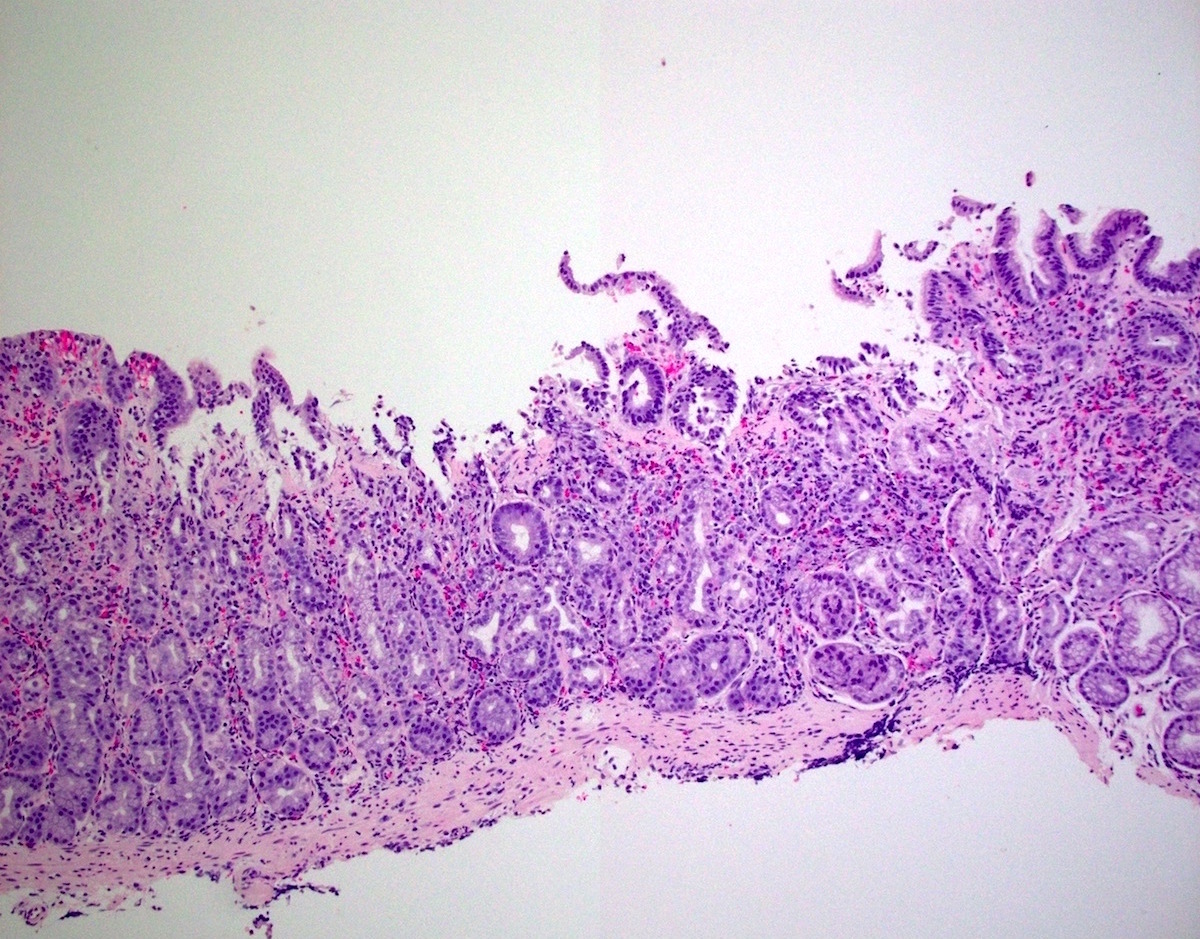
- Patchy increased subepithelial collagen, having a thickness of ≥ 10 µm
- Maximum thickness on average ranges from 15 to 115 μm, mean of 55.1 μm (Mod Pathol 2015;28:533)
- Often associated with the entrapment of red blood cells, inflammatory cells and superficial capillaries (Am J Surg Pathol 2001;25:1174)
- Associated with the denudation or separation of the surface gastric epithelium
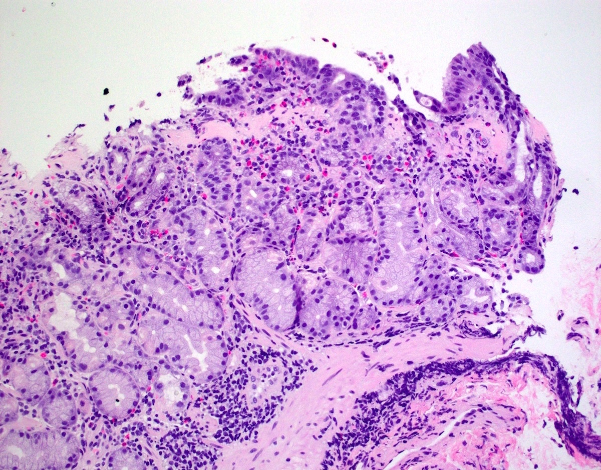
- Increased chronic inflammatory infiltrate of the lamina propria including increased eosinophils and oftentimes neutrophils
- Increased intraepithelial lymphocytes but fewer than in lymphocytic gastritis
- Histologic features may persist for years (Mod Pathol 2015;28:533)
Microscopic (histologic) images
Contributed by Matthew Morrow, M.D.

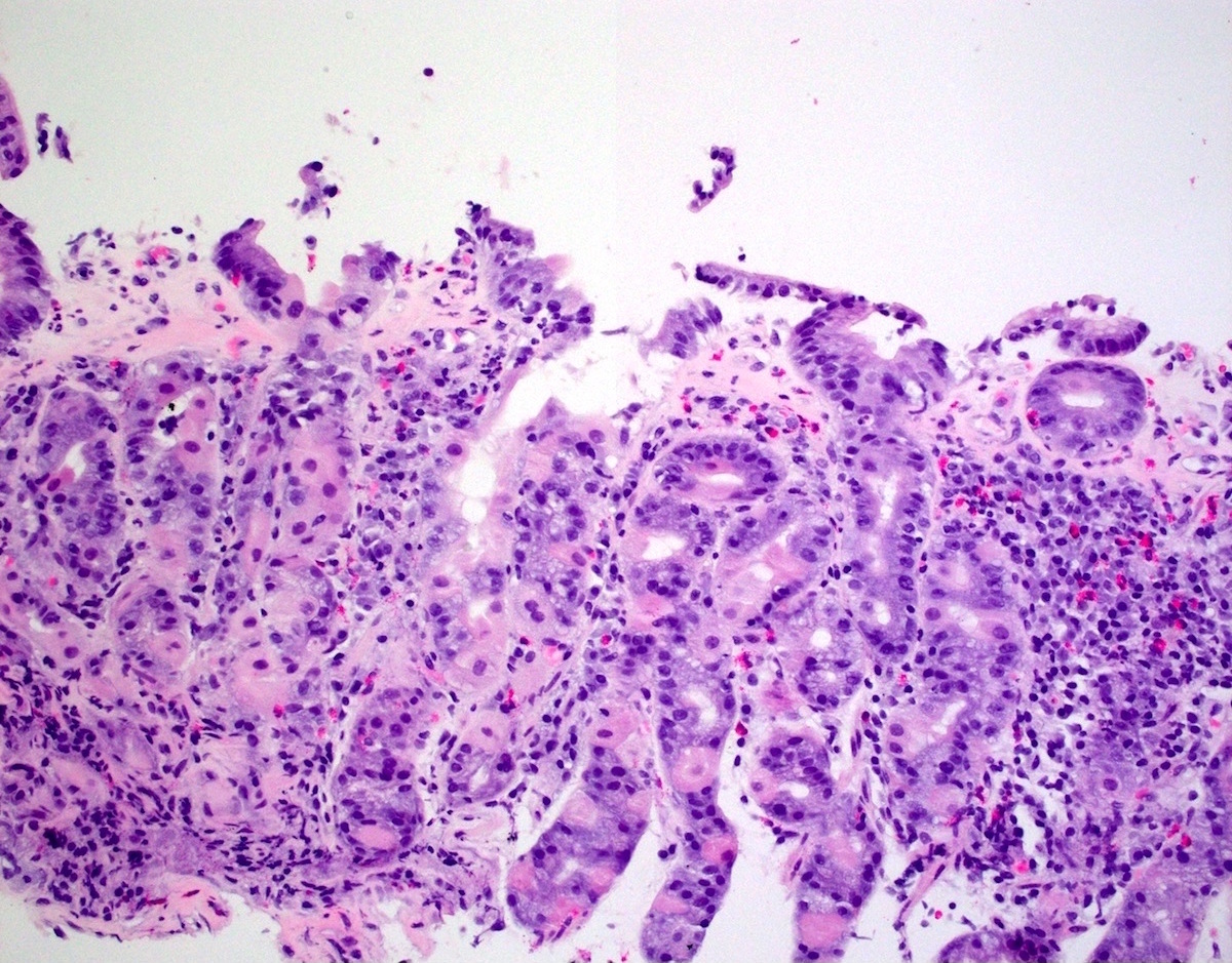
Increased subepithelial collagen with lamina propria chronic inflammation

Denudation of surface gastric epithelium
Abundant eosinophils
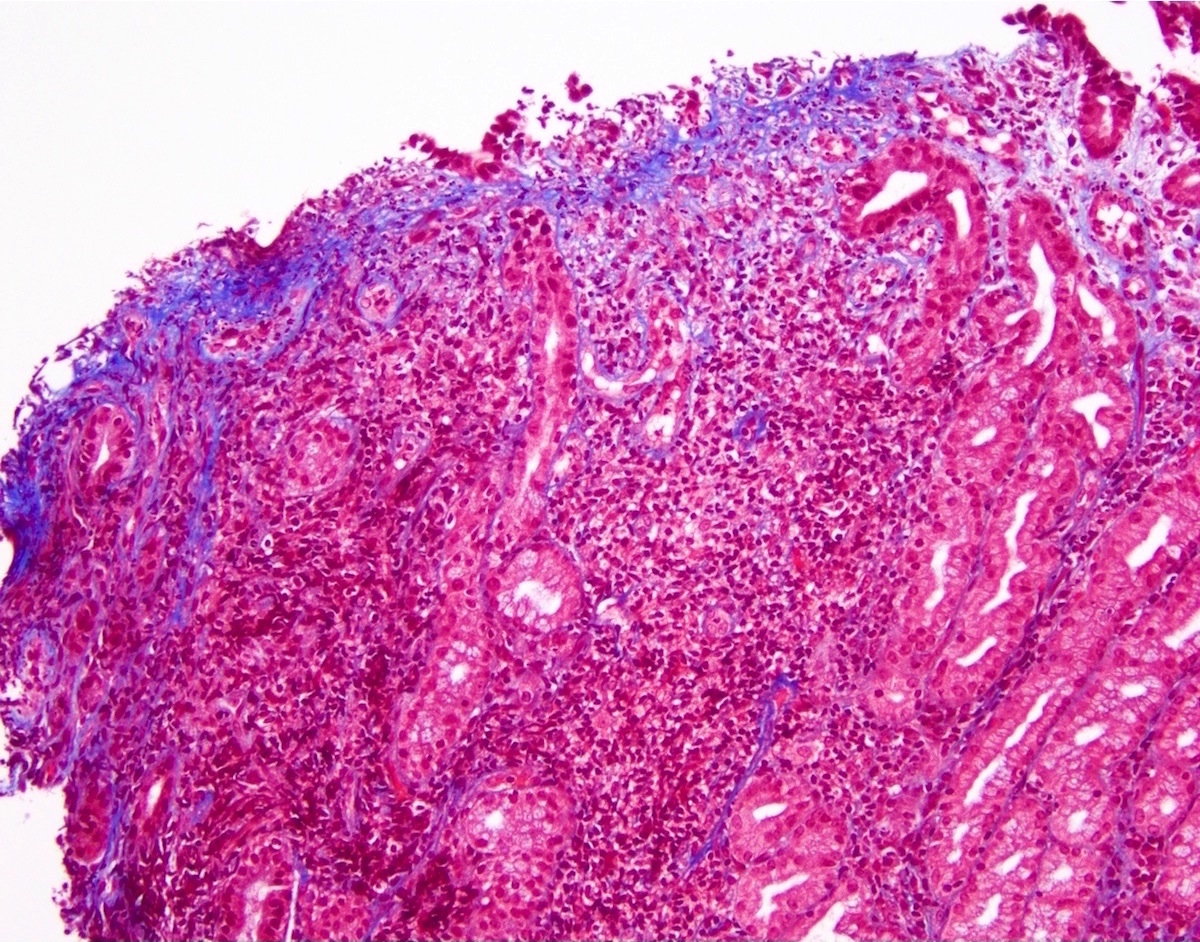
Thickened subepithelial collagen
Positive stains
- Masson trichrome stain highlights the thickened subepithelial layer of collagen
Negative stains
Sample pathology report
- Stomach, antrum, biopsy:
- Collagenous gastritis (see comment)
- Comment: A trichrome special stain highlights a thickened basement membrane.
Differential diagnosis
- Amyloid deposition:
- Positive for Congo red
- Autoimmune gastritis or radiation therapy effect:
- May show diffuse collagen deposition, not specifically located in a subepithelial location
- Scleroderma:
- Fibrosis at all levels of the mucosa and possibly also deep to the mucosa (Ital J Anat Embryol 2010;115:115)
- Tangential sectioning of normal subepithelial collagen:
- Other features (e.g., increased inflammation) not seen
- May need to correlate with clinical presentation
Additional references
Practice question #1
A 10 year old boy presents with chronic abdominal pain and anemia. The image above is a biopsy from the stomach. Which of the following statements is true?
- Absence of both HLA-DQ2 and HLA-DQ8 haplotypes essentially excludes the diagnosis
- Histologic features may persist for years
- Immunosuppression is established standard therapy
- Pediatric type of the disease is classically characterized by accompanying collagenous colitis
Practice answer #1
B. Histologic features may persist for years. The image shows collagenous gastritis. In one study, of the patients who had follow up biopsies, 75% had persistent histologic features, sometimes lasting up to 10 years. Answer A is incorrect because HLA haplotypes are not known to influence collagenous gastritis. Answer C is incorrect because there is currently no standard therapy, though experimental treatments are being investigated. Answer D is incorrect because the adult type of this disease, not the pediatric one, is classically characterized by accompanying collagenous colitis (Mod Pathol 2015;28:533).
Comment Here
Reference: Collagenous gastritis
Comment Here
Reference: Collagenous gastritis
Practice question #2
Which of the following statements about collagenous gastritis is true?
- If nodular gastric mucosa is seen, for diagnosis of this entity it is best to biopsy at the tips of the nodules
- Increase in collagen is seen in the deep lamina propria and bowel wall
- It can be associated with celiac disease
- It is mainly a disease of the elderly
Practice answer #2
C. It can be associated with celiac disease. This can occur in both pediatric and adult patients. Answer A is incorrect because the disease is best appreciated in tissue between the mucosal nodules, not within them. Answer B is incorrect because the increase in collagen is seen in the subepithelial layer. Answer D is incorrect because collagenous gastritis can affect patients of any age range.
Comment Here
Reference: Collagenous gastritis
Comment Here
Reference: Collagenous gastritis
