Testis & paratestis
General
Anatomy & histology
Author: Turki Al-Hussain, M.D.

Last author update: 1 August 2012
Last staff update: 20 February 2024
Copyright: 2002-2025, PathologyOutlines.com, Inc.
PubMed Search: Testis and epididymis - anatomy and histology
Table of Contents
Definition / general | Terminology | Embryology | Diagrams / tables | Gross images | Microscopic (histologic) description | Microscopic (histologic) images | Positive stains | Electron microscopy descriptionCite this page: Al-Hussain T. Anatomy & histology. PathologyOutlines.com website. https://www.pathologyoutlines.com/topic/testisanatomy.html. Accessed October 3rd, 2025.
Definition / general
- Birth to age 4 (static phase): seminiferous tubules filled with small cuboidal cells with no definite lumen present, Leydig cells usually not visible
- Age 4 - 10 (growth phase): tubules, tubular lumina and cells enlarge; tubules become tortuous
- Age 10 to puberty (maturation phase): tubular cells have mitoses; Leydig cells prominent; spermatocyte differentiation visible
- Adult (postpuberty): each testis weights 15 - 19g, measures 5 × 3 cm; takes 70 days for cells to mature from spermatogonium to primary spermatocyte to secondary spermatocyte to spermatid to spermatozoa; maturation is orderly along length of tubule but often not present in biopsy cross section
- Normal spermatogenesis:
- Not every tubule has complete spermatogenesis
- Number of late spermatids correlates best with sperm counts
- Some patients have normal sperm counts but low motility or duct obstruction
- Testes is paired organ suspended in scrotum by spermatic cord
- Each testis is attached to an epididymis, which connects rete testis to vas deferens
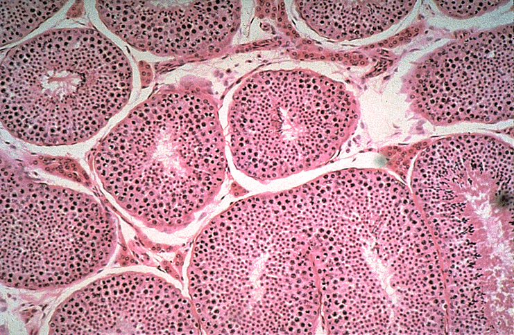
- Testis is composed of convoluted seminiferous tubules in a stroma with Leydig cells
- Three layers: outer serosa (tunica vaginalis, extension of peritoneal cavity) with mesothelial cells; tunica albuginea (tough fibrous septa that extends into testis and separates it into 250 lobules of 1 - 4 seminiferous tubules), inner tunica vasculosa
- Seminiferous tubules converge into rete testis at hilum, anastomose into efferent tubules that penetrate tunica albuginea to form head of epididymis
Terminology
- Efferent ducts: see micro images below
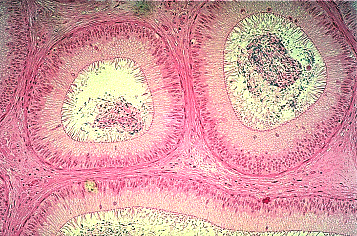
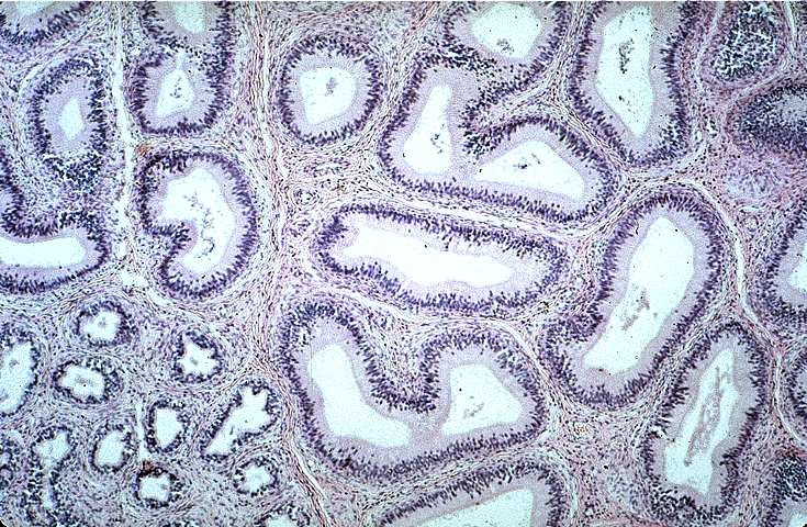
- Epididymis:
- Connects efferent ductules to vas deferens
- Has head, body and tail
- Composed of columnar cells (tall, ciliated with PAS+ nuclear inclusions), clear cells and basal contractile cells (actin positive)
- May have "monster" cells similar to seminal vesicle (no significance, Am J Surg Pathol 1981;5:483)
- Tubules have thick muscular coat
- References: Am J Surg Pathol 2003;27:469
- Nonpathologic morphologic variations:
- Intranuclear eosinophilic inclusions: 72%, usually older patients
- Lipofuscin pigment: 33% usually in efferent ducts and associated with obstructive changes
- Cribriform hyperplasia: 42% usually NOT in normal testis
- Paneth cell-like metaplasia: 8% with hyalin-like globules that are positive for PAS with and without diastase digestion, associated with obstructive changes
- Nuclear atypia: 14% similar to that in seminal vesicles, associated with older age
- Note: rarely present within hernia sacs (Am J Surg Pathol 1999;23:880)
- References: Am J Surg Pathol 1998;22:990
- Interstitium: contains Leydig cells and stromal elements (collagen and myoid cells that surround seminiferous tubules)
- Leydig cells:
- Single (20 microns) or in clusters between seminiferous tubules, produce testosterone in response to luteinizing hormone (LH)
- Often associated with nerve fibers and blood vessels
- Have abundant pink cytoplasm with lipid, lipochrome pigment, Reinke crystalloids (hexagonal prisms by EM), round nuclei with distinct nucleoli
- Fewer Leydig cells in elderly
- Mediastinum: posterior testicular capsule with vasculature, nerves, mediastinum of rete testis (where tubules converge)
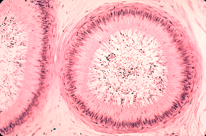
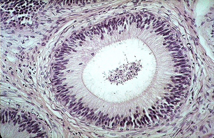
- Rete testis:
- At testicular hilum
- Complex tubular architecture may resemble teratoma
- Connects testicular tubules to 12 - 15 ciliated efferent ducts, which merge into a single duct, the epididymis at its head
- Rete lined by flattened to columnar epithelium with numerous microvilli
- Efferent duct lumina are narrower than epididymis, lined by ciliated columnar cells with microvilli
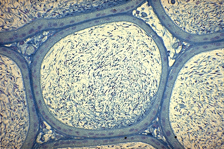
- Seminiferous tubules:
- 150 - 250 microns in diameter, the average total length in each testes is 540 m (range 299 - 981 m)
- Lined by multilayered epithelium with most mature cells towards lumina
- Have basal lamina, outer myoid cells (positive for desmin, muscle specific actin, vimentin) and collagen
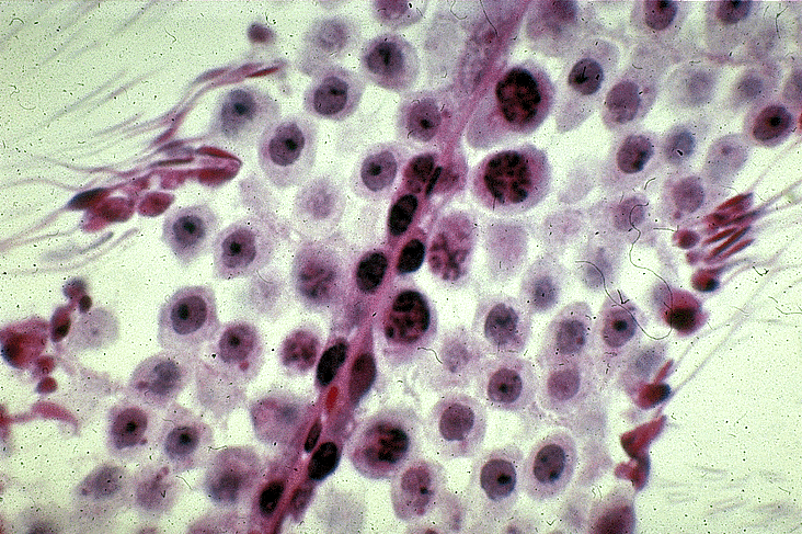
- Contain Sertoli cells, spermatogonia (types A and B), primary spermatocytes, secondary spermatocytes, spermatids and spermatozoa
- All except spermatozoa are held together by a narrow cytoplasmic bridge
- Immature tubules are positive for alpha inhibin
- Sertoli cells:
- Columnar, on basement membrane, surround germ cell elements with cytoplasmic extensions, form blood - testis barrier
- 7% of tubular cells
- May contain Charcot-Bottcher crystalloids (bundles of microfilaments)
- Have irregular, highly folded nuclei with prominent nucleoli
- Produce anti-Müllerian hormone, which causes regression of Müllerian duct structures in utero
- After birth, secrete androgen binding protein and are responsive to FSH
- Also produce inhibin
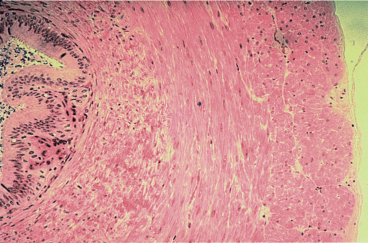
- Vas deferens:
- Also called ductus deferens
- 30 - 40 cm long tubular structure from tail of epididymis to prostatic urethra at level of verumontanum; distal vas deferens joins seminal vesicle to form ejaculatory duct
- Rarely present within hernia sacs (Am J Surg Pathol 1999;23:880)
- Should see complete transection in vasectomy specimens
- References: Am J Surg Pathol 2003;27:469
Vestigial remnants:
- Appendix epididymis: remnant of mesonephric duct
- Appendix testis (hydatid of Morgagni):
- Remnant of Müllerian duct; attached to tunica albuginea at upper testicular pole; present in 90% of males
- May undergo hemorrhagic infarction from twisting on its pedicle
- Gross: round / oval, 1 - 10 mm, pedunculated
- Micro: columnar epithelium with vascular fibrous core with smooth muscle cells; may have glandular-like structures due to surface invaginations
- Paradidymis (organ of Giraldes): remnant of mesonephric tubules
- Vas aberrans (organ of Haller): remnant of mesonephric tubules
Embryology
- Presence of Y chromosome (possibly sex determining region Y) determines formation of testis
- Germ cells migrate to genital ridge, tubules formed by day 45; Wolffian ducts (form epididymis, vas deferens, seminal vesicles and ejaculatory ducts) develop by day 25, Müllerian ducts (form uterus and upper vagina) by day 43
- If no Y, gonad becomes an ovary; if Y present becomes a testis
- If Y present, Sertoli cells develop from genital ridge and secrete anti-Müllerian hormone / Müllerian inhibiting factor (AMH) by day 62, causing ipsilateral regression of Müllerian duct by day 75 - 80; lack of Sertoli cells means no AMH, no regression and presence of uterus and upper vagina
- If Y present, Leydig cells arise by day 64 and produce testosterone, which causes development of Wolffian duct structures (epididymis, vas deferens, seminal vesicles and ejaculatory ducts) if functional androgen receptors are also present
- If no testosterone is produced, Wolffian duct structures degenerate
- Exogenous androgens or their production by maternal tumor or congenital adrenal hyperplasia cause development of Wolffian duct system regardless of presence of Y chromosome
- Development of external genitalia requires testosterone plus androgen receptors plus 5 alpha reductase, which converts testosterone to dihydrotestosterone (DHT), which causes development of external genitalia between days 120 - 140, including elongation of phallus
- If ovaries or no gonads present, internal ducts are female
- If DHT not present, external genitalia is female
Diagrams / tables
Images hosted on other servers:


Scrotum

Transverse section

Right testis

Vertical section

Transverse section

Section of genital cord

Spermatic cord

Subcutaneous inguinal ring

Cremaster

Spermatic veins

Section of epididymis of guinea pig

Fundus of bladder
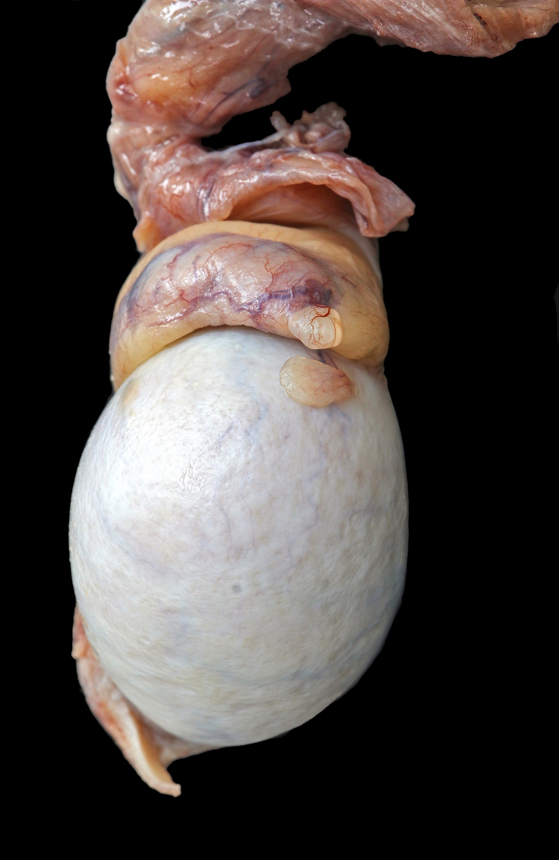
Gross images


Microscopic (histologic) description
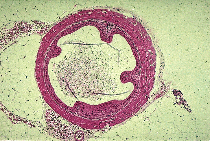
- Spermatic cord:
- Ciliated, pseudostratified epithelium with prominent nuclear inclusions resting on basal cell layer
- Markedly thickened muscle coat of 3 layers (inner longitudinal, outer longitudinal, middle circular layer)
- May contain psammoma bodies
Microscopic (histologic) images
Contributed by Asmaa Gaber Abdou, M.D. and @Andrew_Fltv on Twitter


Testis histology
Images hosted on other servers:

Normal testis

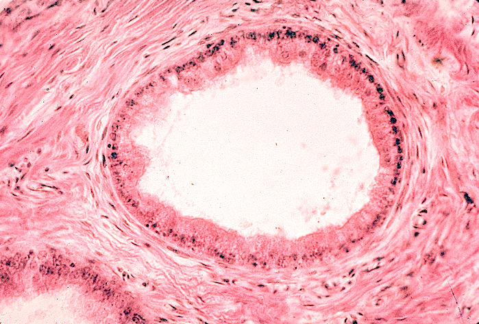
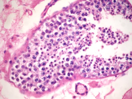
Efferent ducts

Leydig cells
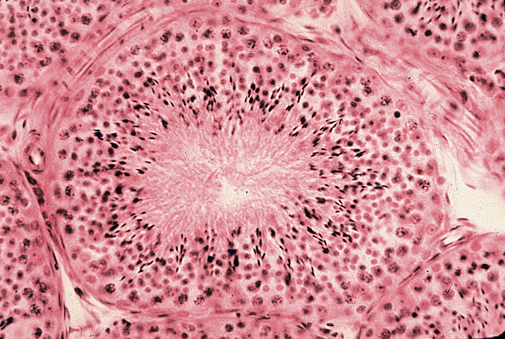

Seminiferous tubules
Various images of epididymis

Various images of spermatic cord
Positive stains
- Epididymis and spermatic cord: CD10
Electron microscopy description
- Spermatogenesis: EM may show round headed sperm or immotile cilia syndrome
