
Colon
Other nonneoplastic
Melanosis coli
Author: Raul S. Gonzalez, M.D.

Last author update: 1 February 2021
Last staff update: 3 May 2022
Copyright: 2003-2024, PathologyOutlines.com, Inc.
PubMed Search: melanosis coli


Table of Contents
Definition / general | Essential features | Sites | Etiology | Clinical features | Diagnosis | Case reports | Gross description | Microscopic (histologic) description | Microscopic (histologic) images | Positive stains | Negative stains | Videos | Sample pathology report | Differential diagnosis | Board review style question #1 | Board review style answer #1Cite this page: Gonzalez RS. Melanosis coli. PathologyOutlines.com website. https://www.pathologyoutlines.com/topic/colonmelanosis.html. Accessed April 26th, 2024.
Definition / general
- Deposition of dark melanin-like pigment in colonic macrophages
Essential features
- Pigment deposition in colon with striking gross and macroscopic features but minimal direct clinical consequences
- Linked to use of anthraquinone laxatives
- Common endoscopic finding
Sites
- Can involve all parts of colon and rectum but typically spares mucosal regions with lymphoid nodules, polyps or carcinomas
- Should biopsy nonpigmented regions in these patients
Etiology
- Linked to use of anthraquinone laxatives but may be secondary to any increase in colonic epithelium apoptosis (Histopathology 1997;30:160)
Clinical features
- Not associated with malignant changes, although it allows small polyps to be more easily identified (Z Gastroenterol 1997;35:313)
Diagnosis
- Colonoscopy
Case reports
- 40 year old woman with colonic lymphoid hyperplasia in melanosis coli (Arch Pathol Lab Med 2001;125:1110)
- 50 year old woman taking laxatives (N Engl J Med 2013;368:2303)
- 83 year old man with melanosis coli involving pericolonic lymph nodes associated with the herbal laxative Swiss Kriss (Arch Pathol Lab Med 2004;128:565)
Gross description
- Diffuse brown-black discoloration of colonic mucosa
Microscopic (histologic) description
- Diffuse deposition of melanized ceroid in macrophages of lamina propria (Gastrointest Endosc 1997;46:131)
- Silver stains may show abnormalities of myenteric plexus
Microscopic (histologic) images
Contributed by Raul S. Gonzalez, M.D. and Yuri Tachibana, M.D.
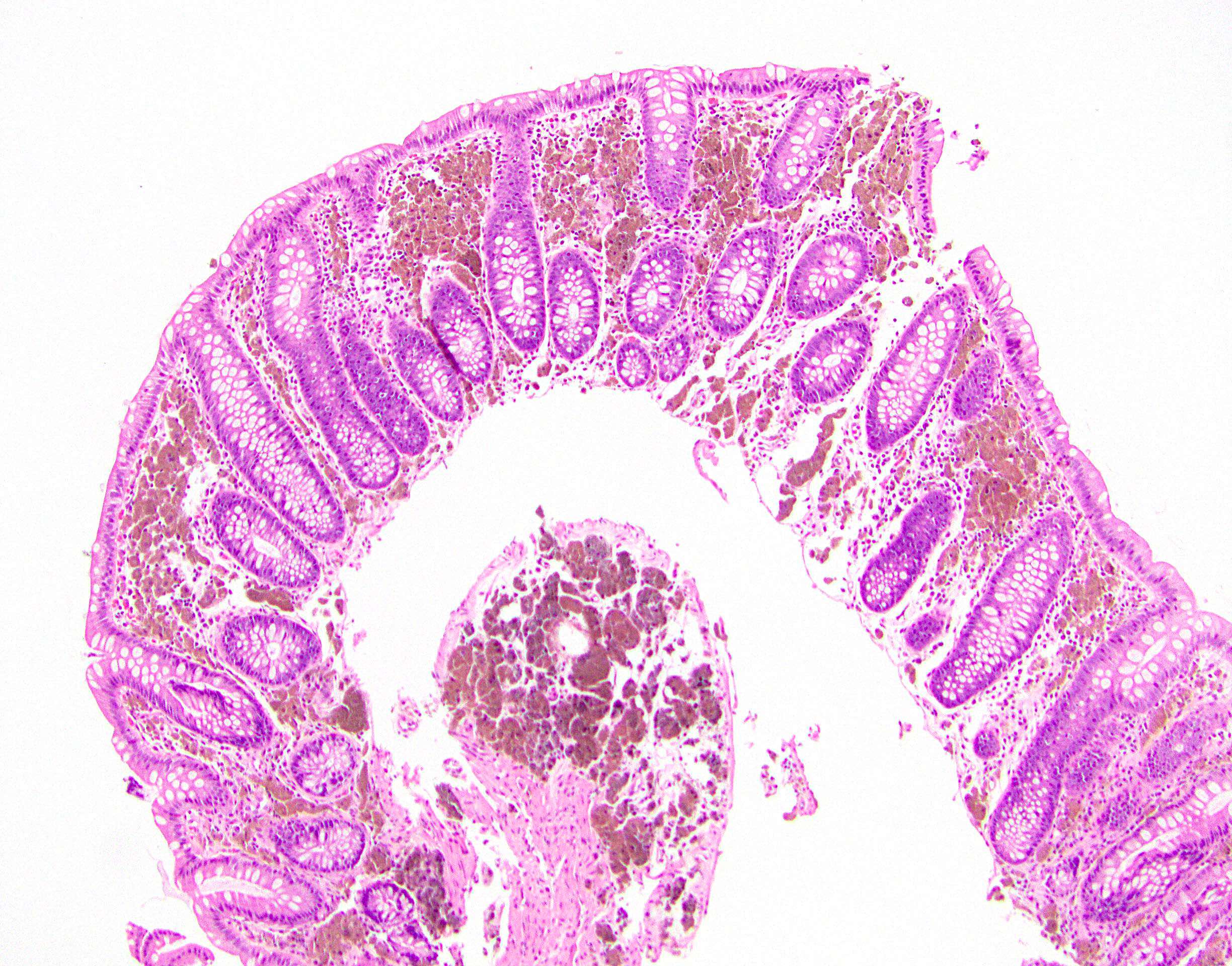
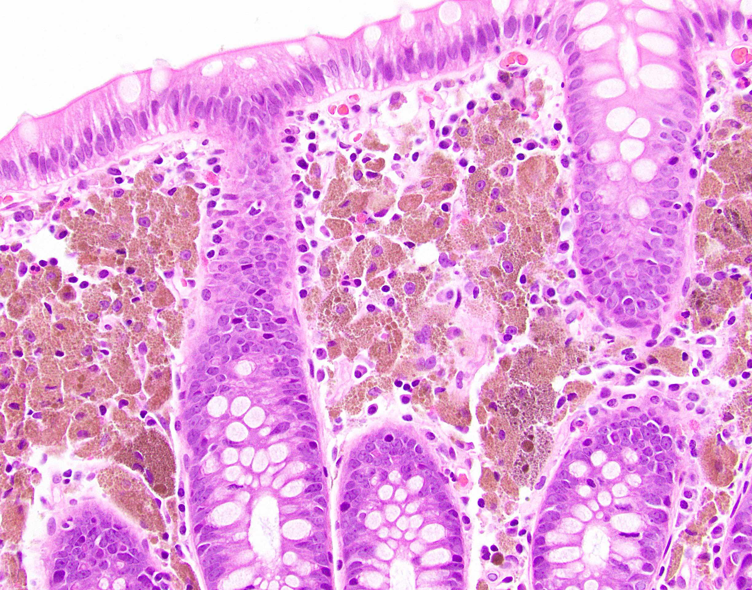
Melanosis coli

Pigment in lamina propria
Positive stains
Negative stains
Videos
Melanosis coli
Sample pathology report
- Random colon, biopsy:
- Colonic mucosa with melanosis coli, otherwise within normal limits
Differential diagnosis
- Blue / green / red bowel:
- Coloration different
- No histologic changes
- Brown bowel / ceroidosis:
- Pigment in smooth muscle cells
- Hemochromatosis:
- Pigment in epithelial cells
- Pseudomelanosis:
- Black pigment in macrophages of duodenum
Board review style question #1
This finding in the colon is typically secondary to
- Chronic granulomatous disease
- Laxative use
- Metastatic melanoma
- Pica
Board review style answer #1
