

Penis & scrotum
Inflammatory
Sclerosing lipogranuloma
Authors: Alcides Chaux, M.D., Antonio L. Cubilla, M.D.

Last author update: 1 May 2010
Last staff update: 3 November 2022
Copyright: 2002-2024, PathologyOutlines.com, Inc.
PubMed Search: Sclerosing lipogranuloma
Table of Contents
Definition / general | Epidemiology | Sites | Etiology | Clinical features | Clinical images | Gross description | Gross images | Microscopic (histologic) description | Microscopic (histologic) images | Positive stains | Differential diagnosisCite this page: Chaux A, Cubilla AL. Sclerosing lipogranuloma. PathologyOutlines.com website. https://www.pathologyoutlines.com/topic/penscrotumsclerosinglipo.html. Accessed April 26th, 2024.
Definition / general
- See also Tancho nodules / paraffinomas
Epidemiology
- Rare; most patients are young adults
Sites
- Usually affects penis, scrotum, spermatic cord and perineum
Etiology
- Usually due to injection or topical application of oil based substances (paraffin, silicone, oil or wax) for cosmetic or therapeutic use (Arch Pathol Lab Med 1977;101:321)
- Foreign body reaction is response to degenerated or damaged fatty tissue or lipids (Med Mol Morphol 2007;40:108)
- May also be due to trauma and cold weather
- Idiopathic cases with peripheral eosinophilia
Clinical features
- Localized painless or slightly tender, indurated plaque / mass
- Up to several centimeters
Clinical images
AFIP images

Marked deformity of penis
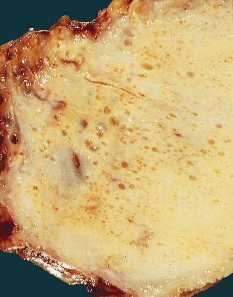
Gross description
- Firm, yellow to grayish white areas
- Solid or solid and cystic
- Often fragmented
Gross images
Contributed by Debra L. Zynger, M.D. and AFIP images
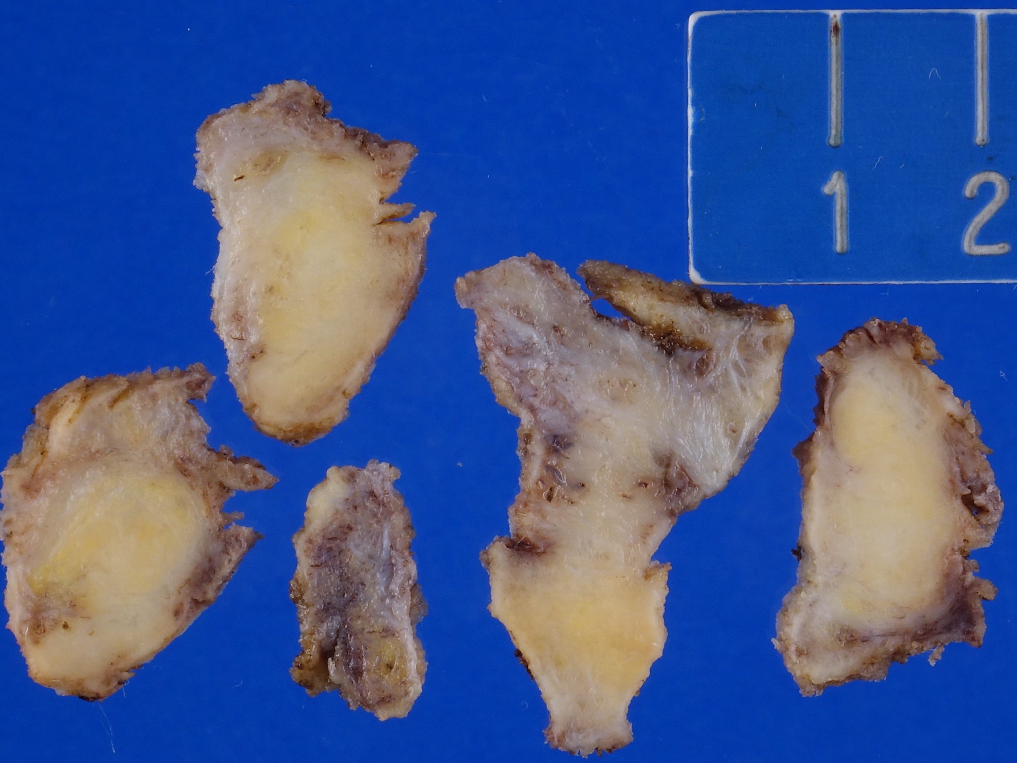
Scrotoplasty

Replacement of entire scrotal wall by solid yellow-white tissue with interspersed cysts
Microscopic (histologic) description
- Fat necrosis, histiocytes, giant cells with extensive fibrosis and hyalinization
- Lipid vacuoles with marked variation in size
- Cysts, if present, lack epithelial lining but may contain giant cells
- Also T lymphocyte infiltrate (Pathol Int 2003;53:121)
Microscopic (histologic) images
AFIP images
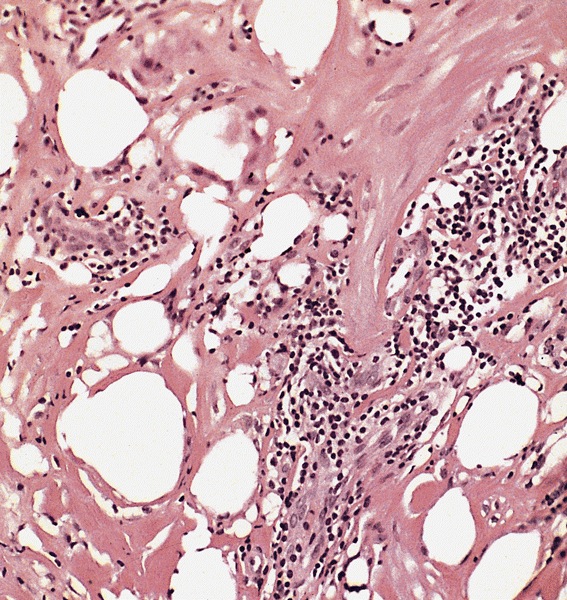
Characteristic vacuoles,
sclerotic stroma and
occasional foreign body
giant cells
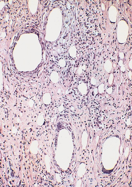
Numerous, variably sized vacuolated spaces

Oil Red O stain
Positive stains
- Lipid stains: Oil Red O for frozen tissue
Differential diagnosis
- Adenomatoid tumor: epitheliod and spindle cells, cystic spaces lined by flat, cuboidal or low columnar cells, no fat necrosis and no giant cells
- Lymphangioma: cystic spaces lined by endothelium; no fat necrosis, no giant cells
- Sclerosing liposarcoma: irregular adipocytes of variable sizes, presence of lipoblasts and usually no giant cells
