
Stains & CD markers
hCG

Copyright: 2002-2024, PathologyOutlines.com, Inc.
PubMed Search: hCG
hCG
Author: Nat Pernick, M.D.
Last author update: 1 August 2012
Last staff update: 1 September 2023
Copyright: 2002-2024, PathologyOutlines.com, Inc.
PubMed Search: hCG

Table of Contents
Definition / general | Uses by pathologists | Microscopic (histologic) images | Positive staining - normal | Positive staining - disease | Negative stainingCite this page: Pernick N. hCG. PathologyOutlines.com website. https://www.pathologyoutlines.com/topic/stainshcg.html. Accessed April 28th, 2024.
Definition / general
- human Chorionic Gonadotrophin
- Also called βhCG or beta hCG
- Glycoprotein with alpha and beta subunits (Wikipedia: Human Chorionic Gonadotropin [Accessed 2 August 2018])
- May cause low TSH levels during early pregnancy because of TSH-like effects of hCG, which has similar alpha subunit as TSH
Uses by pathologists
- Cytoplasmic stain; relatively specific for choriocarcinoma or syncytiotrophoblasts
- Serum levels of beta subunit used to detect pregnancy
- Serum levels also used to stage germ cell tumors and gestational trophoblastic tumors
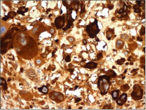
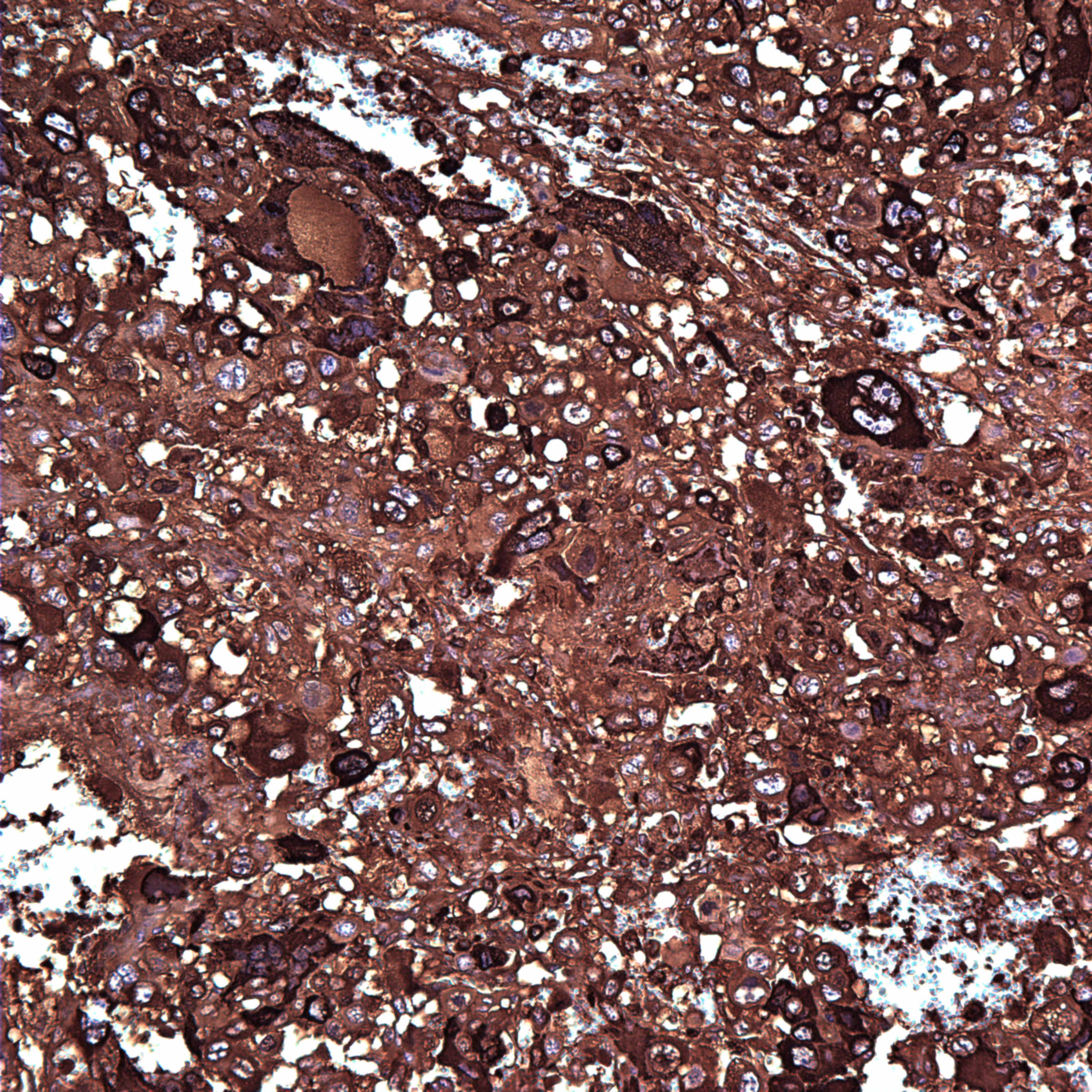
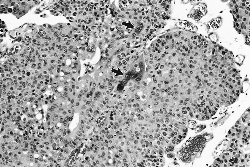
Microscopic (histologic) images

Positive staining - normal
- Placenta (syncytiotrophoblasts)
Positive staining - disease
- Choriocarcinoma → complete mole → partial mole
- Syncytiotrophoblast cells in other tumors, including epithelioid trophoblastic tumors, carcinomas with trophoblastic differentiation, some carcinoids and other tumors
- Serous effusions with reactive mesothelium may be hCG+ (Mod Pathol 2004;17:701)
Negative staining
- Giant cells other than syncytiotrophoblasts; early placenta, cytotrophoblast, intermediate villous trophoblast
- Exaggerated placental site
- Placental site nodule
- Placental site trophoblastic tumor
- Dysgerminoma / seminoma, yolk sac tumor, but may have scattered hCG+ syncytiotrophoblasts

