Placenta
Gestational trophoblastic disease
Nonneoplastic lesions
Exaggerated placental site
Editorial Board Member: Ricardo R. Lastra, M.D.
Deputy Editor-in-Chief: Jennifer A. Bennett, M.D.


Last author update: 20 December 2021
Last staff update: 10 April 2023
Copyright: 2002-2025, PathologyOutlines.com, Inc.
PubMed Search: Exaggerated placental site
Table of Contents
Definition / general | Essential features | ICD coding | Epidemiology | Sites | Pathophysiology | Clinical features | Diagnosis | Radiology description | Prognostic factors | Case reports | Treatment | Microscopic (histologic) description | Microscopic (histologic) images | Virtual slides | Positive stains | Negative stains | Sample pathology report | Differential diagnosis | Practice question #1 | Practice answer #1 | Practice question #2 | Practice answer #2Cite this page: Jabbar A, Lanjewar S, Gupta R. Exaggerated placental site. PathologyOutlines.com website. https://www.pathologyoutlines.com/topic/placentaexagg.html. Accessed August 28th, 2025.
Definition / general
- Exuberant infiltration of the endometrium and myometrium by implantation site intermediate trophoblast
Essential features
- Exaggeration of normal physiologic process, with infiltration of the endometrium and superficial third of myometrium by implantation site intermediate trophoblast
- Can be associated with normal pregnancy or abortions, molar pregnancy and sometimes presents as postpartum bleeding
- Ki67 labeling index is near 0%
- Ki67 labeling index can be 0 - 5 % in exaggerated placental site (EPS) of molar pregnancies (Hum Pathol 1998;29:27)
ICD coding
- ICD-10: O43.899 - other placental disorders, unspecified trimester
Epidemiology
- Incidence is 1.6% of first trimester spontaneous and elective abortions (Int J Gynecol Pathol 2014;33:339, Int J Gynecol Pathol 2001;20:31)
Sites
- Uterus
Pathophysiology
- Exaggeration of normal physiologic process
- Intermediate trophoblasts (IT) invade through endometrium into the superficial third of myometrium (Int J Gynecol Pathol 2001;20:31)
- Normal structure of endometrial glands, myometrium and vessels is usually maintained
- Ki67 indices of near zero indicate increased number of intermediate trophoblasts in EPS is not a result of de novo proliferation of IT in the implantation site (Hum Pathol 1998;29:27)
- Increased Ki67 in molar pregnancy may be a spillover effect IT in the trophoblastic columns of hydatiform moles (Hum Pathol 1998;29:27)
- Although histologically the same, increased Ki67 labelling index in complete molar implantation site represents related but different lesions (Hum Pathol 1998;29:27)
Clinical features
- Can occur in normal pregnancy or following an abortion (J Obstet Gynaecol Res 2008;34:609)
- Causes postpartum bleeding, uterine atony (J Reprod Med 2013;58:448, Tohoku J Exp Med 2014;234:77)
Diagnosis
- Histologic examination
Radiology description
- May present as echogenic lesion in the uterine cavity (J Obstet Gynaecol Res 2008;34:609)
Prognostic factors
- Excellent prognosis following curettage
Case reports
- 26 year old woman presented with complete mole and coexistent exaggerated placental site reaction (Niger Med J 2014;55:180)
- 34 year old woman primigravida with intrauterine fetal demise and suspected placenta previa (Taiwan J Obstet Gynecol 2012;51:440)
- 35 year old woman with intractable massive postpartum hemorrhage after removal of retained placenta (Tohoku J Exp Med 2014;234:77)
- 39 year old woman with an echogenic mass in uterine cavity following elective abortion (J Obstet Gynaecol Res 2008;34:609)
Treatment
- Curettage is curative; no follow up is required (Int J Gynecol Pathol 2001;20:31)
Microscopic (histologic) description
- Extensive infiltration of endometrium and myometrium by implantation site intermediate trophoblasts which are often multinucleate (Int J Gynecol Pathol 2001;20:31)
- Endometrial glands and spiral arterioles can show invasion by trophoblasts
- Smooth muscle cells of myometrium are separated by cords, nests and individual trophoblastic cells
- Despite diffuse infiltration, there is not necrosis or mitoses
- Trophoblastic cells share similar morphologic features of normal implantation site intermediate trophoblasts; these cells contain abundant eosinophilic cytoplasm with hyperchromatic and irregular nuclei (Int J Gynecol Pathol 2001;20:31)
- Chorionic villi are morphologically unremarkable
Microscopic (histologic) images
Contributed by Raavi Gupta, M.D., Absia Jabbar, M.D. and Sonali Lanjewar, M.D., M.B.B.S.
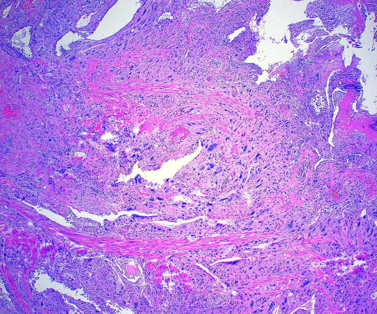
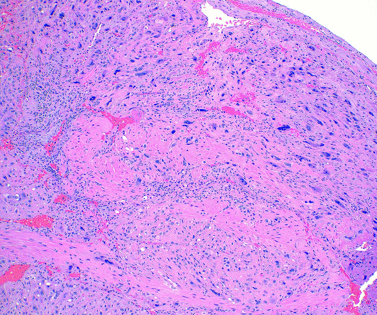
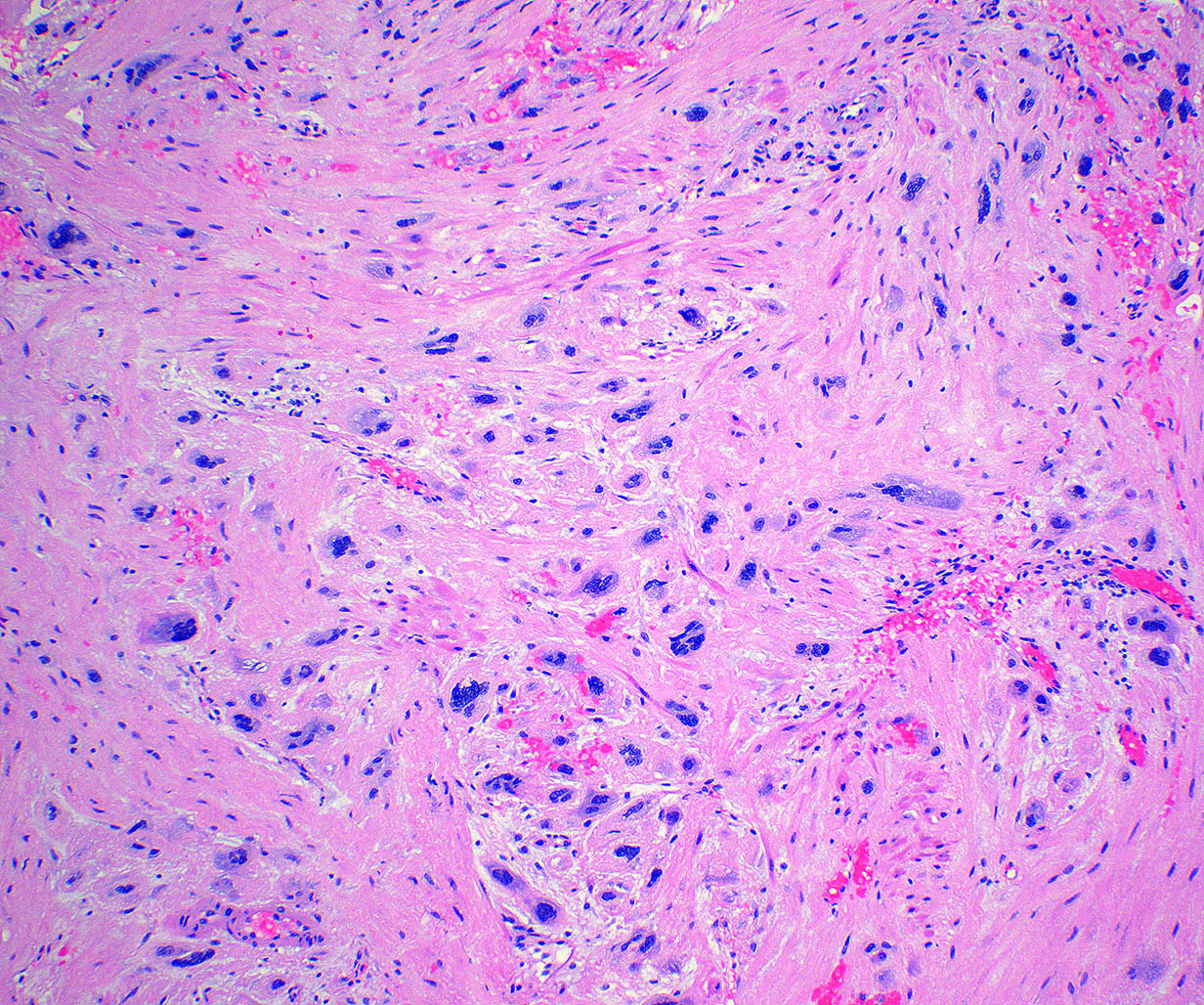
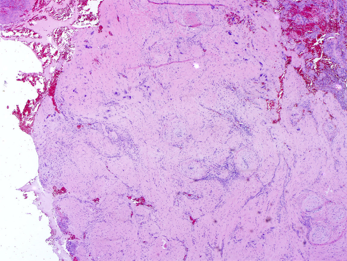
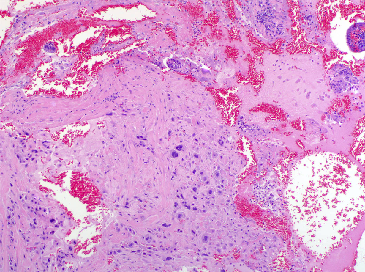
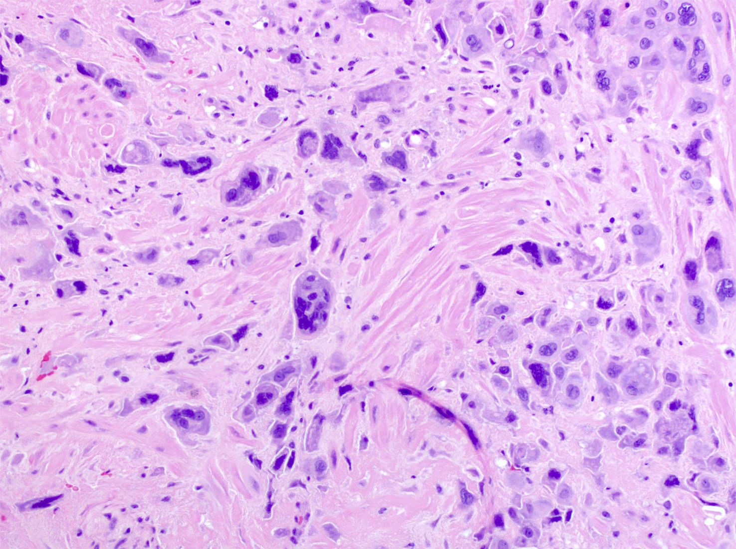
Intermediate trophoblasts invading myometrium
Virtual slides
Images hosted on other servers:

Exaggertated placental site
Positive stains
- HPL (diffuse), MUC4, CK18, HLA-G (Int J Gynecol Pathol 2014;33:339)
- MEL-CAM / CD146, inhibin, PLAP (weak) (Hum Pathol 1998;29:27)
- Double staining of Ki67 and HLA-G is a useful method in the differential diagnosis of exaggerated placental site versus placental site trophoblastic tumor and placental site trophoblastic tumor versus choriocarcinoma (Hum Immunol 2007;68:272)
Sample pathology report
- Uterus, endometrium, dilatation and curettage:
- Exaggerated placental site
Differential diagnosis
- Choriocarcinoma:
- Comprised of syncytiotrophoblasts and cytotrophoblasts
- Extensive hemorrhage and necrosis
- hCG positive
- Epithelioid trophoblastic tumor:
- Well circumscribed nodular expansile masses with necrosis
- Mononucleate trophoblastic cells arranged in nests and cords (Int J Gynecol Pathol 2001;20:31)
- Strongly positive for p63, focally positive for HPL and MEL-CAM / CD146; negative for MUC4
- Placental site trophoblastic tumor (PSTT):
- Confluent sheets of trophoblastic cells
- Increased mitoses (Ki67 is 10 - 15%) (Hum Pathol 1998;29:27)
- PSTT can be confused with molar implantation site; Ki67 labelling index of molar implantation site is 3 - 5% (Hum Pathol 1998;29:27)
- HLA-G immunostaining highlights growth patterns of intermediate trophoblasts and can distinguish placental site trophoblastic tumor and an exaggerated placental site
- In a placental site trophoblastic tumor, the cells form confluent masses, whereas in an exaggerated placental site, the IT cells infiltrate the endomyometrium as single cells and cords of cells (Am J Surg Pathol 2002;26:914)
- Placental site nodule:
- Nodular with well circumscribed, surrounding thin rim of chronic inflammatory cells and occasionally decidual cells
- Ki67: trophoblast nuclear staining < 5% in placental site nodule (Hum Pathol 1998;29:27)
- p63 positive
Practice question #1
Exaggerated placental site (shown in the image above) is characterized by which of the following features?
- Absence of villi
- Presence of necrosis and mitoses
- Proliferation of chorionic type intermediate trophoblasts with invasion of myometrium
- Proliferation of implantation type intermediate trophoblasts with invasion of myometrium
Practice answer #1
D. Proliferation of implantation type intermediate trophoblasts with invasion of myometrium
Comment Here
Reference: Exaggerated placental site
Comment Here
Reference: Exaggerated placental site
Practice question #2
Ki67 labeling index in exaggerated placental site is
- Near 0%
- Near 5%
- Near 15%
- Near 100%
Practice answer #2
