Colon
Other nonneoplastic
Solitary rectal ulcer syndrome
Author: Raul S. Gonzalez, M.D.
Deputy Editor-in-Chief: Catherine E. Hagen, M.D.

Last author update: 28 October 2021
Last staff update: 19 May 2023
Copyright: 2003-2025, PathologyOutlines.com, Inc.
PubMed search: solitary rectal ulcer syndrome

Table of Contents
Definition / general | Essential features | Terminology | Epidemiology | Sites | Etiology | Clinical features | Case reports | Treatment | Gross description | Microscopic (histologic) description | Microscopic (histologic) images | Sample pathology report | Differential diagnosis | Additional references | Practice question #1 | Practice answer #1 | Practice question #2 | Practice answer #2Cite this page: Gonzalez RS. Solitary rectal ulcer syndrome. PathologyOutlines.com website. https://www.pathologyoutlines.com/topic/colonsolitaryrectalyulcer.html. Accessed September 17th, 2025.
Definition / general
- Solitary or multiple ulcerated or polypoid lesions 4 - 10 cm from anal margin
Essential features
- Not always solitary, not always rectal, not always ulcerated, not really a syndrome
- Mucosal prolapse type change resulting in rectal lesions
Terminology
- Also called mucosal prolapse syndrome (may be a better term since not necessarily solitary, ulcerated or rectal)
Epidemiology
- Uncommon (incidence of 1 per 100,000 per year)
- Usually third and fourth decade
- More common in women
- Rarely in children (Pediatrics 2002;110:e79)
Sites
- Usually in rectosigmoid colon
Etiology
- Abnormal function of anal and pelvic floor musculature during defecation, causing rectal mucosal prolapse or intussusception
Clinical features
- Symptoms include constipation, blood and mucus from rectum, change in bowel habits, pain
- Associated with histologic changes of sessile serrated polyps (38%), which often have focal loss of hMLH1 gene expression (Arch Pathol Lab Med 2005;129:1037)
- Related to inflammatory cloacogenic polyp (see Pediatr Pathol 1993;13:409)
Case reports
- 45 year old man complaining of constipation and rectal bleeding is found to have an ulcerated rectal lesion on colonoscopy (Case of the Month #527)
Treatment
- High fiber diet, laxatives, topical steroids
- Possibly resection
Gross description
- Well demarcated irregular ulcer(s) on rectal wall
- Also polypoid, rough, erythematous lesions
- Mucosal thickening
Microscopic (histologic) description
- Superficial mucosal ulceration and villiform change
- Crypt hyperplasia and elongation with focal dilation (some glands diamond shaped)
- Fibromuscular hyperplasia of lamina propria
- Thickened muscularis mucosae with splayed fibers
- Ectatic capillaries
- Minimal inflammation
- May have inflammatory pseudomembranes
- Late changes resemble colitis cystica profunda
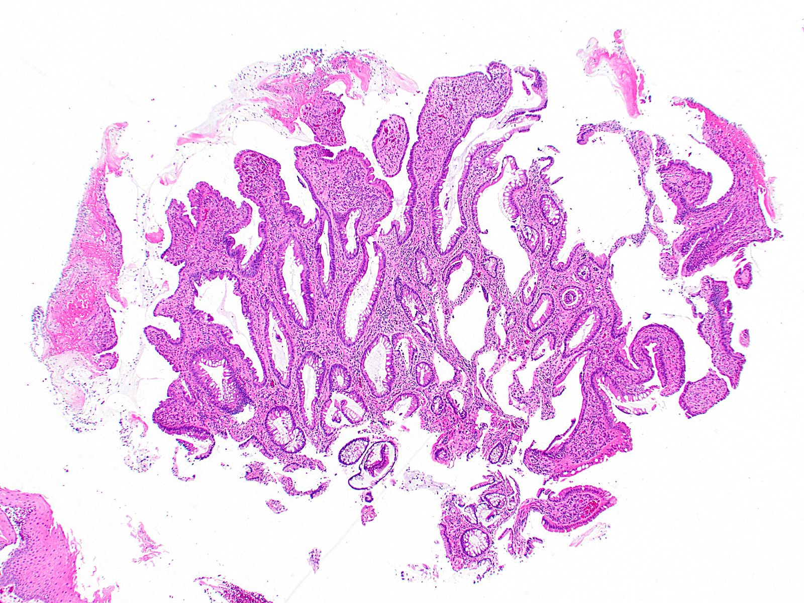
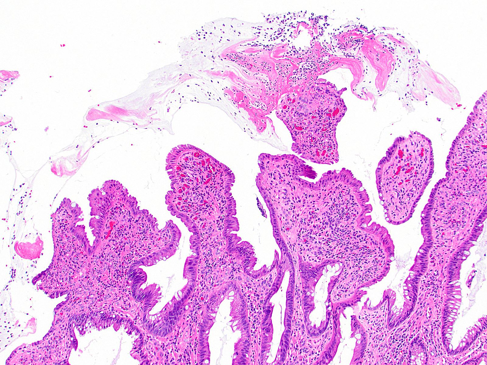
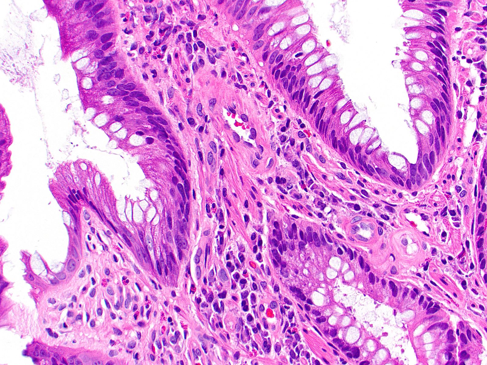
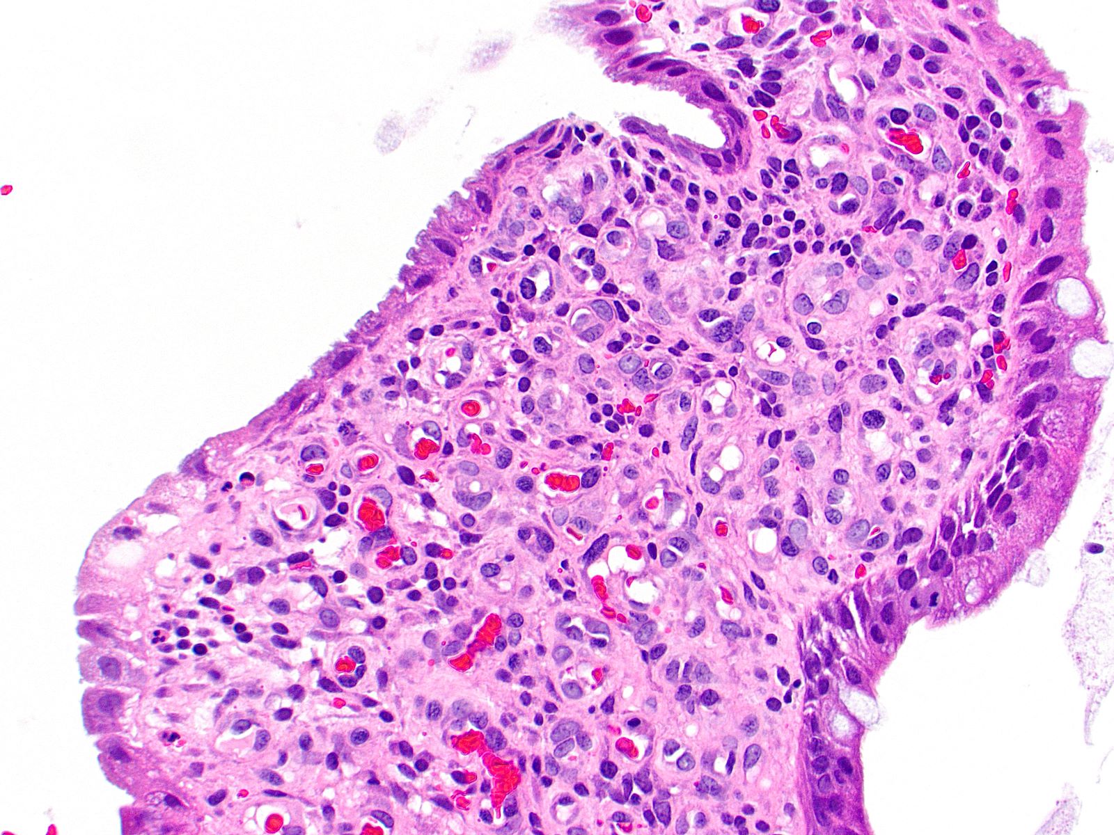
Microscopic (histologic) images
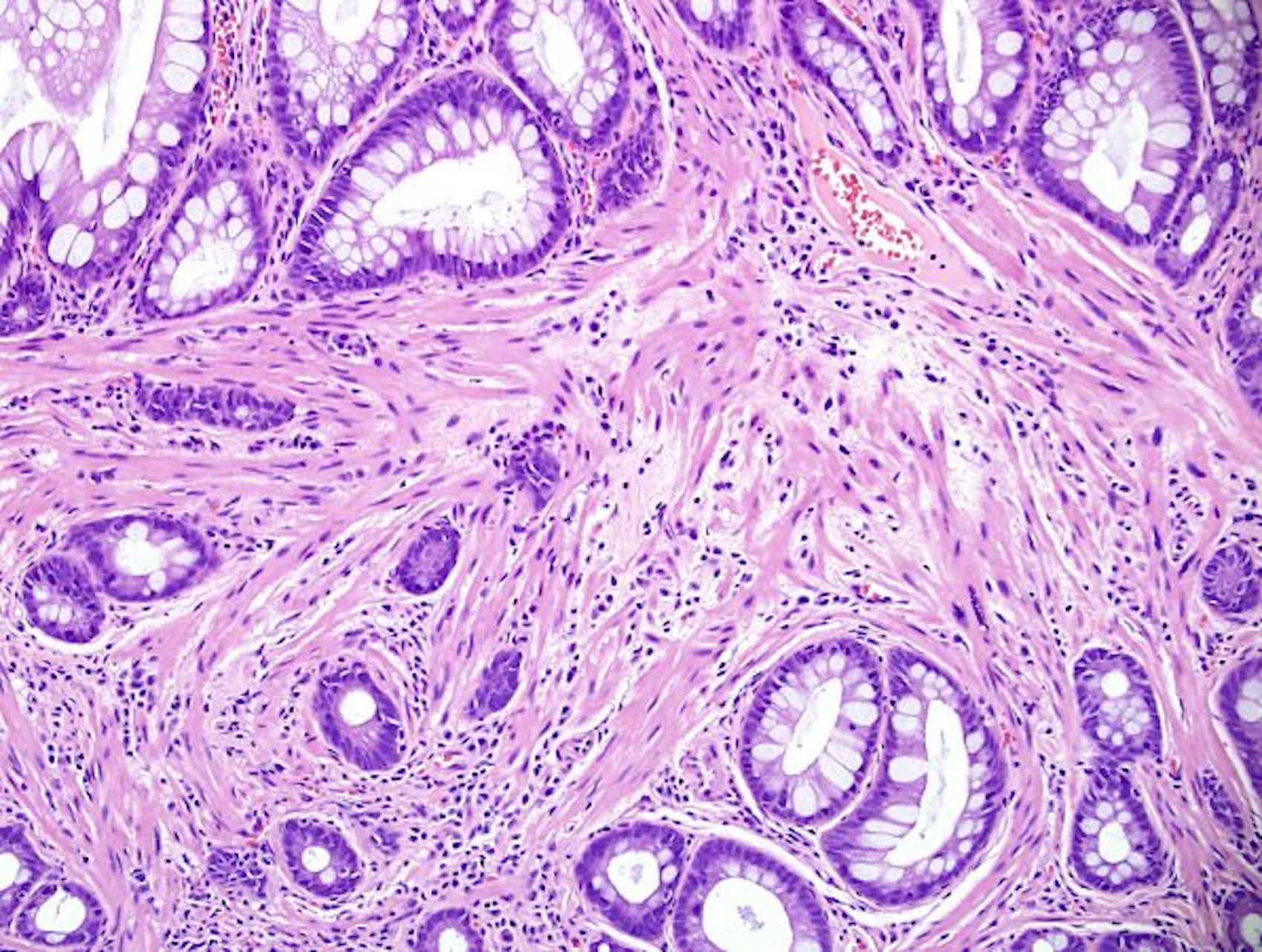
Contributed by Andrey Bychkov, M.D., Ph.D., Jian-Hua Qiao, M.D. and Raul S. Gonzalez, M.D. (Case #527)
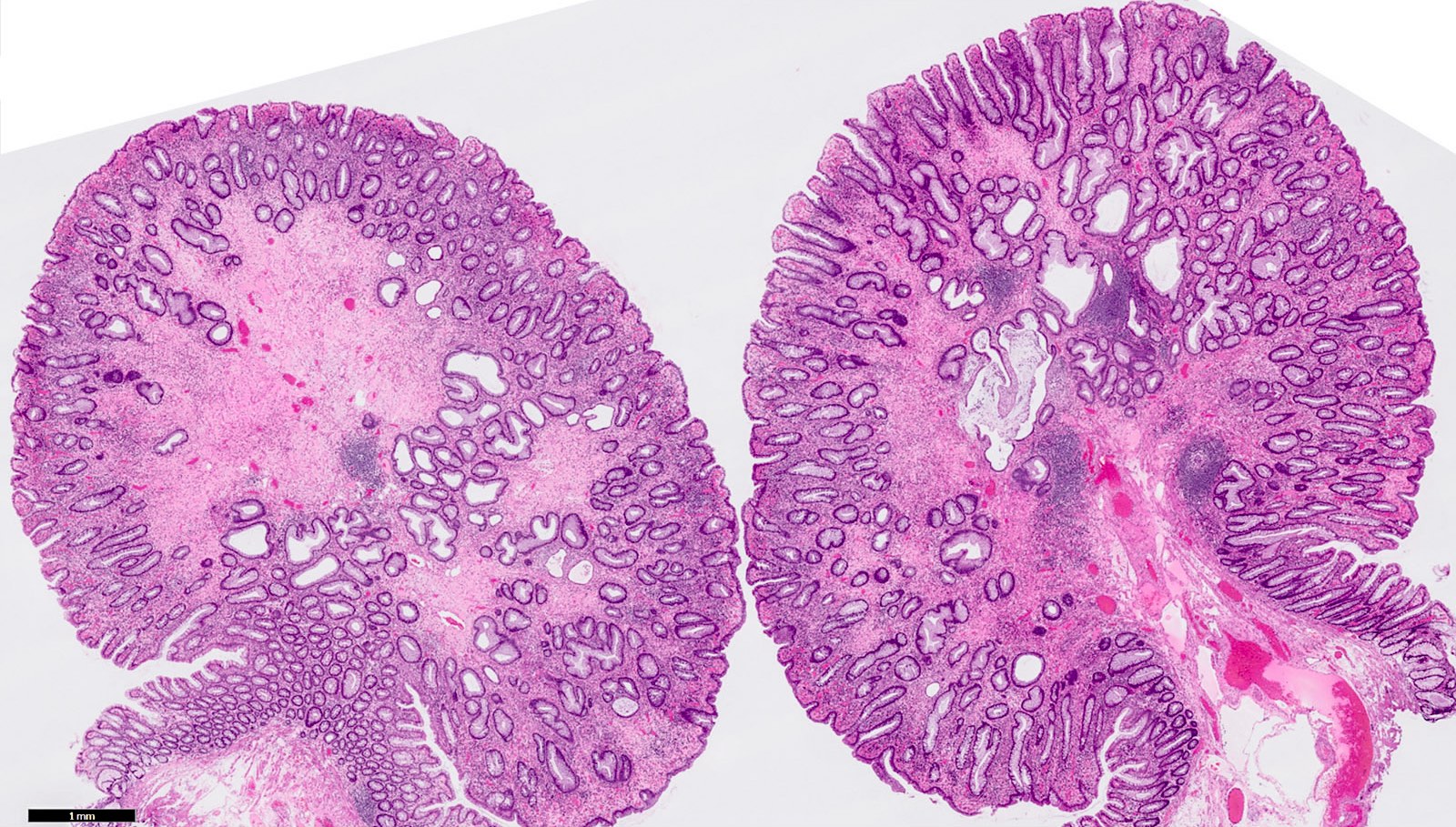
Polypoid shape

Solitary rectal ulcer
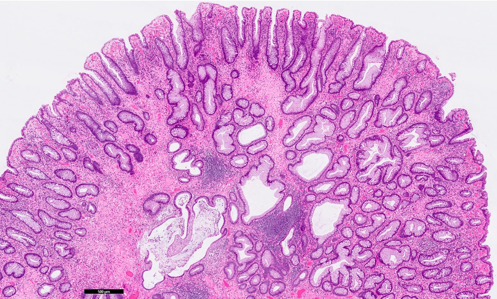
Crypt hyperplasia
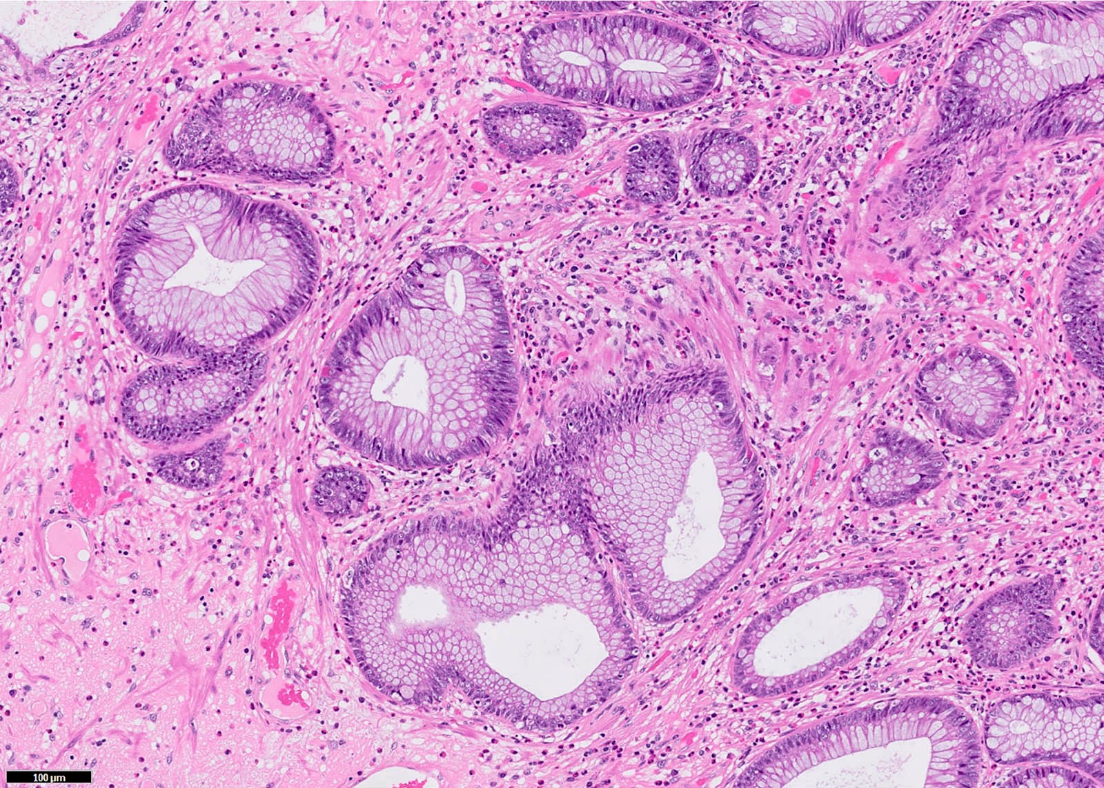
Disarrayed muscularis mucosa

Haphazardly arranged benign colonic crypts
Distorted diamond shaped glands
Ulcerated, reactive mucosa
Smooth muscle wisps
Granulation tissue
Sample pathology report
- Rectum, ulcer, biopsy:
- Colonic mucosa with prolapse type change, mild acute inflammation and focal erosion (see comment)
- Comment: The findings are compatible with so called solitary rectal ulcer syndrome.
Differential diagnosis
- Cowden disease:
- Similar histology, different clinical features
- Crohn's disease
- Mucinous adenocarcinoma:
- Irregular mucin pools, epithelium floating in mucin, complex glandular proliferation, variable atypia, desmoplasia, usually no hemorrhage
- Rectal endometriosis (see Mod Pathol 1995;8:599)
- Ulcerative proctitis
- Ulcers due to ergotamine suppositories
Additional references
Practice question #1
Solitary rectal ulcer syndrome most frequently occurs in what patient population?
- Elderly men
- Female infants
- Middle aged women
- Young adult men
Practice answer #1
Practice question #2
Which of the following is true about solitary rectal ulcer syndrome?
- Patients demonstrate typical associated systemic symptoms
- Prolapse of intestinal mucosa is specific to this disease entity
- Roughly 30% of patients have multiple lesions
- The diagnosis cannot be made without microscopic ulceration
Practice answer #2
C. Roughly 30% of patients have multiple lesions. Despite the name, "solitary rectal ulcer syndrome" is not always a solitary finding. Approximately 30% of patients will demonstrate multiple rectal ulcers on colonoscopy. Answer A is incorrect because solitary rectal ulcer syndrome is not a systemic syndrome, meaning patients will not have associated systemic symptoms. Answer B is incorrect because prolapse of intestinal mucosa can be seen histologically in other diseases, such as diverticulosis. Answer D is incorrect because microscopic ulceration is often but not always encountered, meaning it is not required to establish the diagnosis.
Comment Here
Reference: Solitary rectal ulcer syndrome
Comment Here
Reference: Solitary rectal ulcer syndrome
