Gallbladder & extrahepatic bile ducts
Cholecystitis
Hyalinizing / porcelain gallbladder
Editorial Board Member: Wei Chen, M.D., Ph.D.
Deputy Editor-in-Chief: Aaron R. Huber, D.O.


Last author update: 9 July 2024
Last staff update: 9 July 2024
Copyright: 2003-2025, PathologyOutlines.com, Inc.
PubMed Search: Hyalinizing / porcelain gallbladder
Table of Contents
Definition / general | Essential features | Terminology | ICD coding | Epidemiology | Pathophysiology | Etiology | Clinical features | Diagnosis | Radiology description | Radiology images | Prognostic factors | Case reports | Treatment | Clinical images | Gross description | Gross images | Frozen section description | Frozen section images | Microscopic (histologic) description | Microscopic (histologic) images | Sample pathology report | Differential diagnosis | Practice question #1 | Practice answer #1Cite this page: Chow A, Esnakula AK. Hyalinizing / porcelain gallbladder. PathologyOutlines.com website. https://www.pathologyoutlines.com/topic/gallbladderporcelaingb.html. Accessed September 24th, 2025.
Definition / general
- Porcelain gallbladder is primarily a radiologically defined entity, referring to dystrophic calcification of the gallbladder wall associated with chronic and repeated inflammation; it was originally described as a hard and brittle gallbladder with a bluish hue, hence the term porcelain (Diagnostics (Basel) 2021;11:1073)
- Degree of calcification may vary from partial deposition (hyalinizing cholecystitis / incomplete porcelain gallbladder) to diffuse / complete full thickness involvement of the gallbladder wall (complete porcelain gallbladder) (Am J Surg Pathol 2011;35:1104)
Essential features
- Porcelain gallbladder is a rare disorder characterized by dystrophic calcification of the gallbladder wall and very commonly associated with cholelithiasis
- Calcification can be partial mucosal calcification or diffuse intramural calcification
- Porcelain gallbladder is associated with an increased incidence of gallbladder carcinoma, especially hyalinizing cholecystitis / incomplete porcelain gallbladder
- Recent evidence indicates that the incidence of gallbladder carcinoma associated with porcelain gallbladder is ~6%, much lower than previously reported
- Risk of malignancy with diffuse intramural calcification is minimal and prophylactic cholecystectomy is not routinely recommended due to the high incidence of surgical complications
Terminology
- Calcified gallbladder, calcifying cholecystitis, cholecystopathia chronica calcarea, hyalinizing cholecystitis / incomplete porcelain gallbladder
ICD coding
Epidemiology
- Rare entity with an incidence of < 1% of gallbladder specimens (Diagnostics (Basel) 2021;11:1073)
- More common in older patients (> 60 years old)
- F:M = 5:1
- Strongly associated with the presence of cholelithiasis (90 - 95%)
- ~6% of cases are associated with gallbladder carcinoma, a rate much lower than that previously reported (J Gastrointest Surg 2013;17:1161)
Pathophysiology
- Pathogenesis is not fully understood but is thought to arise due to chronic inflammation and mucosal wall scarring; repair and hyalinization of injured tissue segments promote the deposition of calcium crystals from the bile (Diagnostics (Basel) 2021;11:1073)
- Since porcelain gallbladder is also associated with gallstones, another proposed mechanism is that the buildup and supersaturation of bile leads to the precipitation and buildup of calcium carbonate crystals on the mucosal surface; this leads to calcification of the tissue without prior injury
- Stages of pathogenesis
- Hyalinizing cholecystitis / incomplete porcelain gallbladder (Am J Surg Pathol 2011;35:1104, Am Surg 2001;67:7)
- Hyalinization is patchy or diffuse
- Calcifications are usually focal, involving the mucosa or walls of the gallbladder and are likely related to calcifications of Rokitansky-Aschoff sinuses
- Complete porcelain gallbladder
- Diffuse or complete intramural calcifications involving most of the gallbladder (> 80% of the organ)
- Hyalinizing cholecystitis / incomplete porcelain gallbladder (Am J Surg Pathol 2011;35:1104, Am Surg 2001;67:7)
Etiology
- Cholelithiasis and chronic cholecystitis
- Bile stasis
- Stasis contributes to calcium carbonate crystal precipitation and deposition onto mucosal surface
Clinical features
- Most patients are asymptomatic (Dis Mon 2021;67:101130)
- Symptoms related to cholelithiasis and chronic cholecystitis
- Right upper quadrant pain
- Common bile duct obstruction related symptoms such as jaundice, fever, pancreatitis
- Rarely, it can present as a nontender, firm, right upper quadrant mass
Diagnosis
- Diagnosed incidentally on abdominal imaging (Xray, ultrasound and computed tomography) and confirmed on the resected cholecystectomy specimen (Dis Mon 2021;67:101130)
Radiology description
- Xray and computed tomography (Abdom Radiol (NY) 2017;42:322, Surg J (N Y) 2017;3:e145)
- Calcification / mineralization outlining gallbladder wall
- Ultrasound (3 types) (Radiology 1984;152:137)
- Type I: hyperechoic semilunar structure with posterior acoustic shadowing (diffuse gallbladder calcification)
- Type II: biconvex curvilinear echogenic structure with acoustic shadowing (focal mucosal wall calcification)
- Type III: irregular clumps of echoes with posterior acoustic shadowing (focal mucosal wall calcification)
Radiology images
Images hosted on other servers:

Xray and CT diffuse intramural calcification

Xray with thin calcifications

CT with calcifications

CT with calcifications and stones

Xray and CT
Prognostic factors
- Porcelain gallbladder is associated with an increased incidence of gallbladder carcinoma, especially the hyalinizing cholecystitis / incomplete porcelain gallbladder (Diagnostics (Basel) 2021;11:1073, Am Surg 2001;67:7, Am J Surg Pathol 2011;35:1104)
- Recent evidence indicates the incidence of gallbladder carcinoma in association with porcelain gallbladder is ~6%, which is much lower than previously reported (J Gastrointest Surg 2013;17:1161)
- Risk of malignancy with diffuse intramural calcification (complete porcelain gallbladder) is minimal and prophylactic cholecystectomy is not routinely recommended due to the high incidence of surgical complications (J Am Coll Surg 2018;226:1064)
Case reports
- 53 year old woman with epigastric and right hypochondrial pain for 6 months (BMJ Case Rep 2021;14:e240319)
- 55 year old woman with epigastric and right hypochondrial pain for 1 month (Ann Med Surg (Lond) 2022;84:104947)
- 57 year old woman with right upper quadrant abdominal pain and nausea for 1 week (J Surg Case Rep 2023;2023:rjac600)
- 86 year old woman with a 3 month history of intermittent epigastric pain (Clin Gastroenterol Hepatol 2021;19:A30)
Treatment
- Prophylactic cholecystectomy in younger patients with minimal comorbidities and in symptomatic patients (Dis Mon 2021;67:101130, Diagnostics (Basel) 2021;11:1073)
- Conservative management with follow up is recommended in asymptomatic older patients, patients with comorbidities and patients with complete porcelain gallbladder showing diffuse mural calcifications
Clinical images
Images hosted on other servers:

Intraoperative
Gross description
- Variable gallbladder wall thickening and brittleness depending on the extent of hyalinization / calcifications
- Variable blue-gray to pearly white tissue coloration depending on the extent of hyalinization / calcifications
- Frequently associated with cholelithiasis
- Occasionally associated with gallbladder carcinoma presenting as a mass lesion or diffusely thickened wall
- Additional sampling (minimum of 10 blocks) is recommended in cases of hyalinizing cholecystitis / incomplete porcelain findings (Histopathology 2021;79:2)
Gross images
Contributed by Ashwini Kumar Esnakula, M.D., M.S.
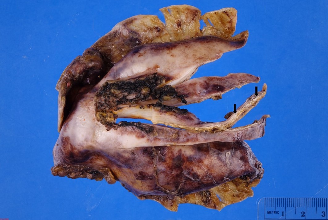
Incomplete porcelain gallbladder
Images hosted on other servers:

Gallbladder with diffuse transmural calcifications

Rigid wall

Diffusely and heavily calcified wall
Frozen section description
- Intraoperative consultation may be requested to exclude the possibility of gallbladder carcinoma
- Variable thick gallbladder wall with effacement of wall architecture, hyaline sclerosis and foci of dystrophic calcifications
Frozen section images
Contributed by Ashwini Kumar Esnakula, M.D., M.S.
Incomplete porcelain gallbladder
Gallbladder adenocarcinoma
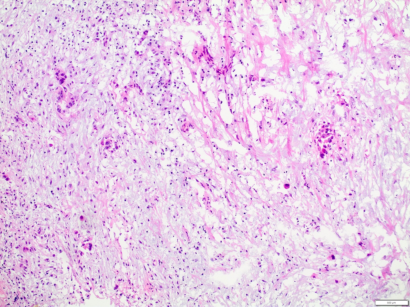
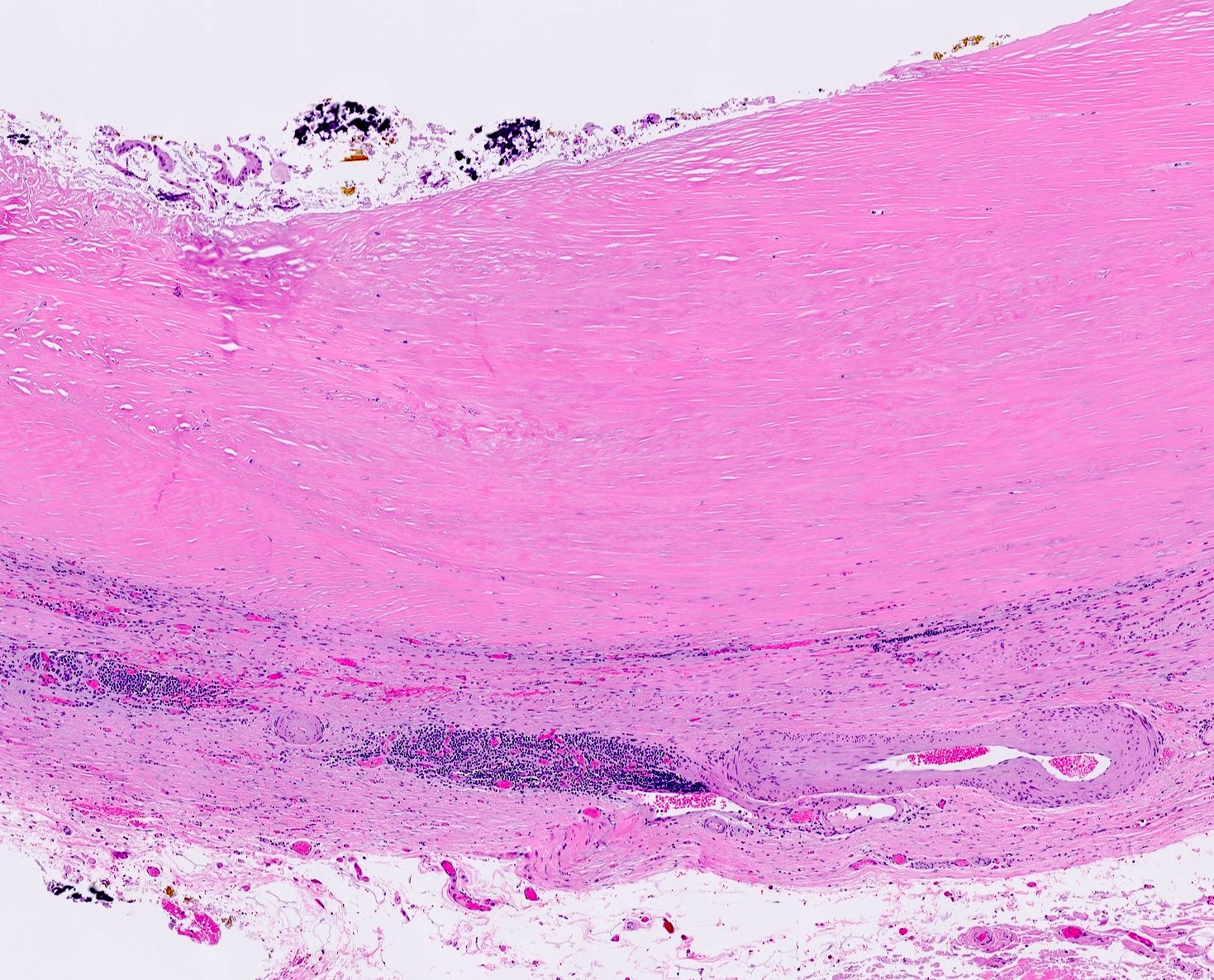
Microscopic (histologic) description
- Hyalinizing cholecystitis / incomplete porcelain gallbladder (Am J Surg Pathol 2011;35:1104)
- Partial or complete effacement of normal gallbladder wall architecture
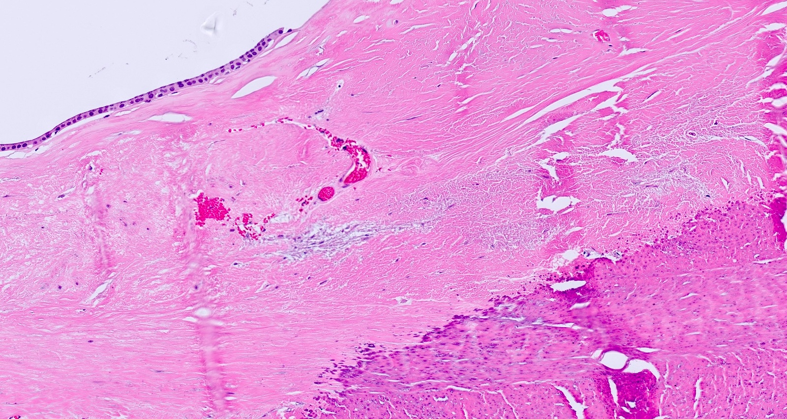
- Partial or complete denudation of the epithelium
- Residual atrophic epithelium with reactive alterations
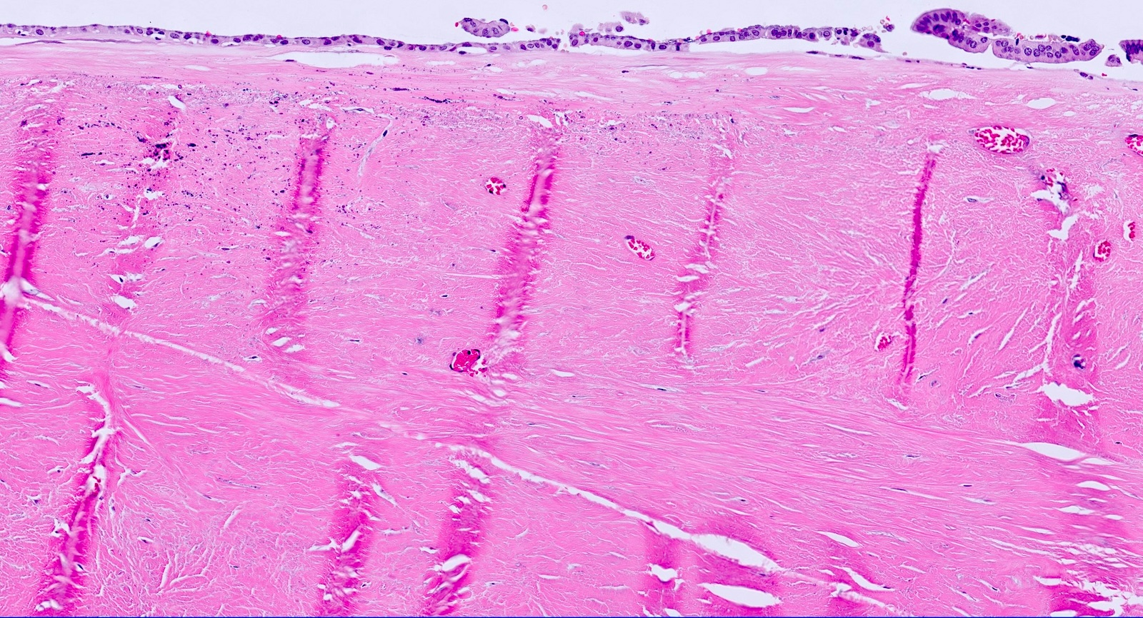
- Dense paucicellular hyaline sclerosis with basketweave-like clefting
- Scattered foci of lymphoid aggregates
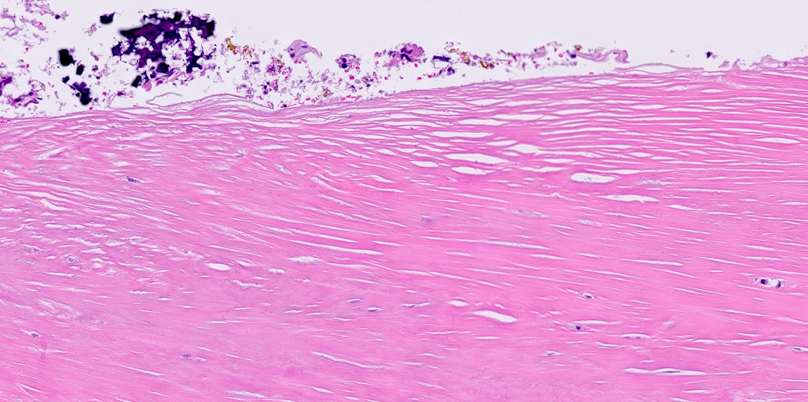
- Foci of mucosal or intramural calcifications
- Complete porcelain gallbladder
- Intramural thick bands of dystrophic calcifications, usually involving > 80% of the gallbladder wall
- Epithelial elements are rare or completely absent
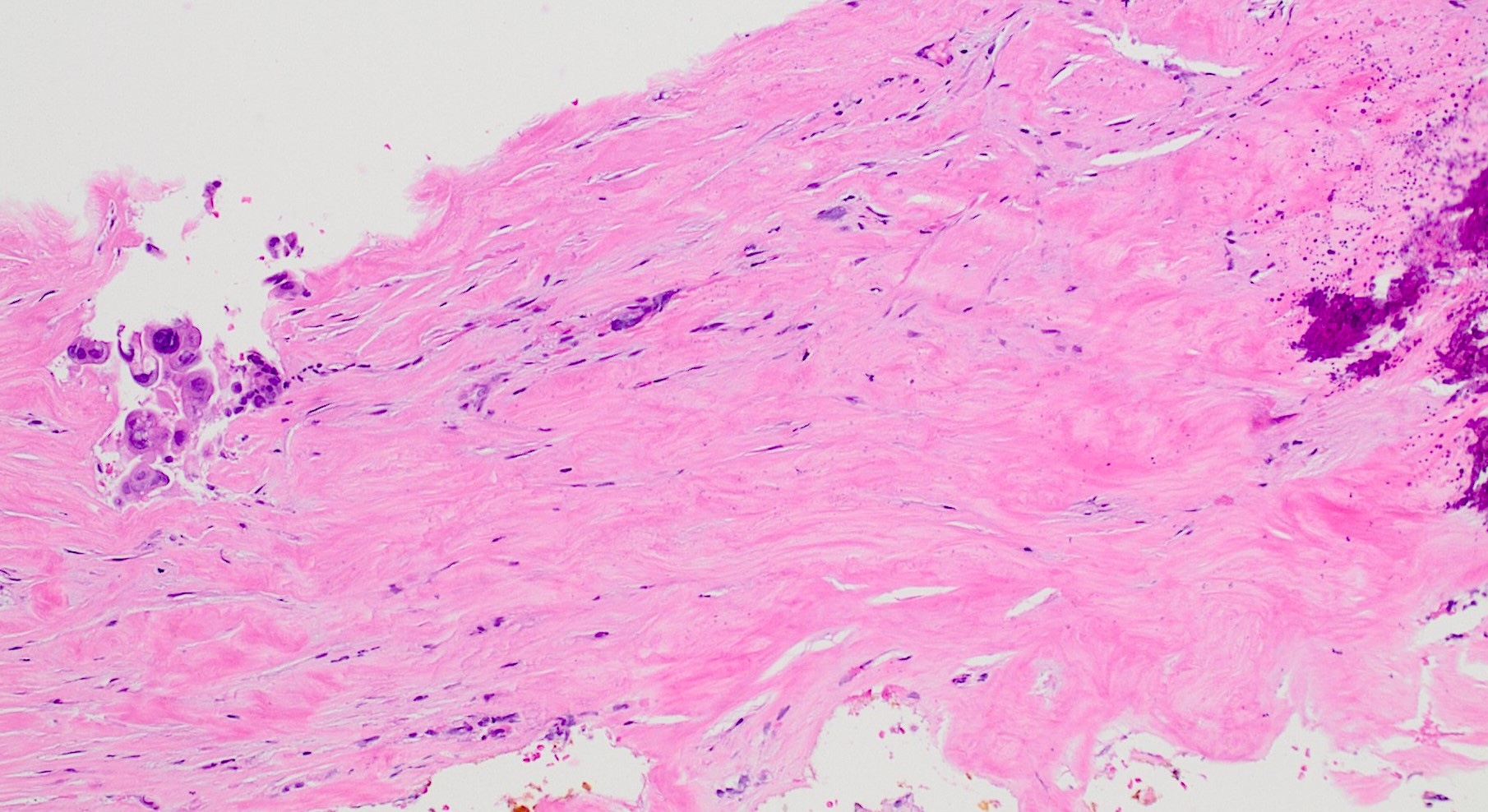
Microscopic (histologic) images
Contributed by Ashwini Kumar Esnakula, M.D., M.S.
Hyalinizing cholecystitis
Mucosal calcifications
Mural calcifications
Dense hyaline sclerosis
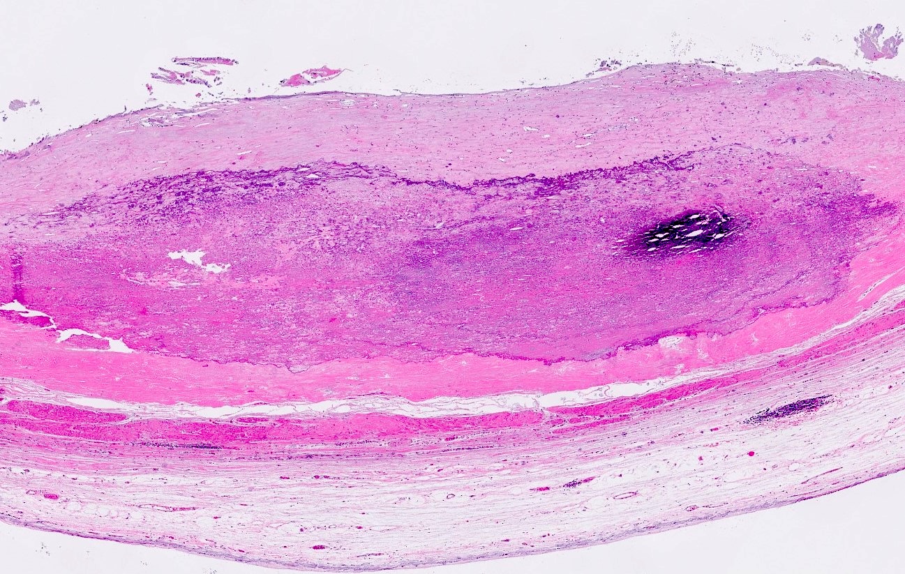
Mural thick calcifications
Gallbladder carcinoma in hyalinizing cholecystitis
Sample pathology report
- Gallbladder, cholecystectomy:
- Cholelithiasis with chronic cholecystitis, diffuse hyalinization and foci of mucosal and mural calcifications, consistent with hyalinizing cholecystitis / incomplete porcelain gallbladder
- Negative for malignancy
Differential diagnosis
- Clinical by imaging:
- Cholelithiasis:
- Calcifications outline individual gallstones
- Emphysematous cholecystitis:
- Echogenic foci in the gallbladder wall
- Absence of gallstones
- Cholelithiasis:
- Pathology:
- Gallbladder adenocarcinoma:
- Commonly infiltrating irregular / angulated glands lined by polygonal cells with prominent cytologic atypia and associated with dense desmoplastic response
- Gallbladder adenocarcinoma:
Practice question #1
What finding in a porcelain gallbladder is associated with a lower risk of malignant progression?
- Diffuse mural calcification in gallbladder wall
- Focal calcifications with diffuse hyalinization
- History of chronic cholecystitis
- History or presence of gallstones
Practice answer #1
A. Diffuse mural calcification in gallbladder wall. Based on historical and more recent studies, the presence of full thickness calcification (complete porcelain gallbladder) in the gallbladder wall has not been associated with any progression to malignancy. Answer B is incorrect because studies demonstrate that hyalinizing cholecystitis / incomplete porcelain gallbladder is associated with the presence of malignancy. Answer D is incorrect because the presence or history of gallstones increases the risk for malignant transformation. Answer C is incorrect because chronic cholecystitis is a risk factor for developing gallbladder malignancies.
Comment Here
Reference: Hyalinizing / porcelain gallbladder
Comment Here
Reference: Hyalinizing / porcelain gallbladder
