

Heart & vascular pathology
Benign tumors / other nonneoplastic
Rhabdomyoma
Author: Nat Pernick, M.D.

Last author update: 1 November 2013
Last staff update: 25 March 2021
Copyright: 2007-2024, PathologyOutlines.com, Inc.
PubMed Search: Rhabdomyoma [title] heart pathology
Table of Contents
Definition / general | Case reports | Treatment | Gross description | Gross images | Microscopic (histologic) description | Microscopic (histologic) images | Positive stains | Negative stains | Electron microscopy description | Electron microscopy images | Differential diagnosis | Additional referencesCite this page: Pernick N. Rhabdomyoma. PathologyOutlines.com website. https://www.pathologyoutlines.com/topic/hearttumorrhabdomyoma.html. Accessed April 26th, 2024.
Definition / general
- 50 - 90% of primary heart tumors in children
- Usually discovered in patients less than 1 year of age, rarely in fetuses (Ital Heart J 2002;3:48)
- May obstruct valvular orifice or cardiac chamber; may present with sudden cardiac death (Pediatr Dev Pathol 2007;10:129)
- Not a true neoplasm, but a hamartoma or malformation due to mutation in TSC1 and TSC2 genes
- 50% of patients have tuberous sclerosis; sporadic cases are occasionally associated with congenital heart disease
Case reports
- 2 year old boy with tuberous sclerosis (Arch Pathol Lab Med 2002;126:1559)
- 13 year old boy with diffuse rhabdomyomatosis (Arch Pathol Lab Med 1977;101:78)
Treatment
- Many tumors regress spontaneously
- Excision if left ventricular outflow tract obstruction or refractory arrhythmia
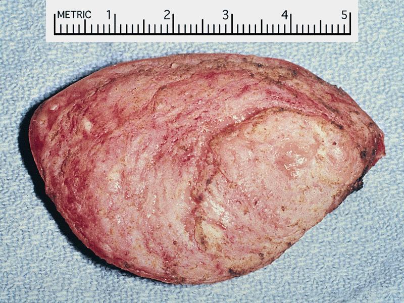
Gross description
- Small, firm, gray-white, well circumscribed myocardial masses (often multiple) that protrude into ventricles
- Average size is 3 - 4 cm, up to 10 cm, particularly in sporadic cases
- Rhabdomyomatosis: numerous miliary nodules less than 1 mm
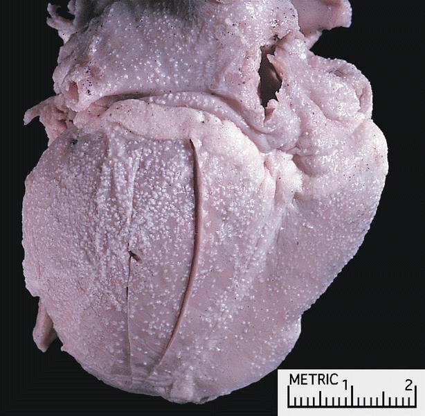
Gross images
AFIP images
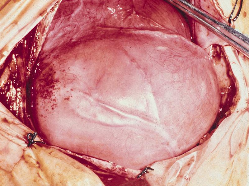
Right ventricular mass
with tuberous sclerosis
and multiple minute tumors
studding epicardium
Images hosted on other servers:


Left ventricular tumor causing sudden death in children ages 1-2
Microscopic (histologic) description
- Clear cells and large, rounded, polygonal cells ("spider cells") with glycogen vacuoles separated by strands of cytoplasm extending between cell membrane and nucleus
- No mitotic activity
- Adult tumors: more cellular with smaller cells, few spider cells and more cellular proliferation (Hum Pathol 2002;33:1092)
Microscopic (histologic) images
AFIP images
and intracellular
myofilaments
(oil emersion)
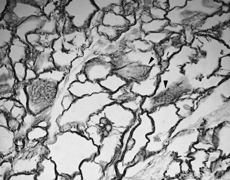
highlights spider
cells and cross
striations (arrowheads)
Images hosted on other servers:

Drawings from 1938 report
Positive stains
Negative stains
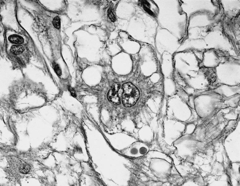
Electron microscopy description
- Altered myocytes with abundant glycogen, small and sparse mitochondria
- Cellular junctions resembling intercalated disks are extensive and randomly distributed, not just at poles of cell as in normal myocytes (Hum Pathol 1977;8:700)
Electron microscopy images
AFIP images
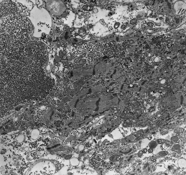
sparse mitochondria, fragmented
irregular myofilaments with Z bands
Differential diagnosis
- Glycogen storage disease: no well formed nodules, cells have intercalated disks at poles by EM
- Granular cell tumor: epicardial, no vacuoles, no myofibers, S100+, desmin-, myoglobin-
- Histiocytoid myopathy: small tumor nodules, finely granular cells, no large vacuoles, no spider cells; a variant of rhabdomyomatosis
- Lipoma: usually epicardial, no myofibers, no glycogen
Additional references
Back to top
