Salivary glands
Other tumors
Lipoma / sialolipoma
Author: Adriana Handra-Luca, M.D., Ph.D.

Last author update: 1 September 2012
Last staff update: 2 November 2020
Copyright: 2003-2025, PathologyOutlines.com, Inc.
PubMed Search: Lipoma[TI] OR sialolipoma[TI] salivary
Table of Contents
Definition / general | Clinical features | Case reports | Treatment | Gross description | Gross images | Microscopic (histologic) description | Microscopic (histologic) images | Cytology description | Immunohistochemistry & special stains | Molecular / cytogenetics description | Differential diagnosisCite this page: Handra-Luca A. Lipoma / sialolipoma. PathologyOutlines.com website. https://www.pathologyoutlines.com/topic/salivaryglandslipoma.html. Accessed August 29th, 2025.
Definition / general
- Lipoma:
- Benign tumor; rare in salivary glands but usually involves parotid gland (Laryngol Rhinol Otol 1986;65:485)
- More common than lipomatosis
- Clinical findings are nonspecific (J Oral Maxillofac Surg 2006;64:1583)
- Sialolipoma:
- Uncommon lipoma variant composed of mature adipose tissue mixed with acinar, ductal, basal and myoepithelial cells of normal salivary gland
- First described in 2001 by Nagao (Histopathology 2001;38:30)
- Lipoadenoma:
- Slow growing tumor with glandular structures with sertoliform features and adipose tissue; oncocytic and sebaceous differentiation
- Initially described by Yau in 1997 (Mod Pathol 1997;10:242)
Clinical features
- Lipoma:
- 3% of parotid tumors - #2 most common benign mesenchymal neoplasms of major salivary glands (#1 is schwannoma)
- Often incidental (Rev Stomatol Chir Maxillofac 1988;89:117)
- Ages 40+, usually men, occasionally children
- Sialolipoma:
- Mean age 61 years but wide age range at presentation
- Female gender predominance for minor salivary gland location
- Parotid and submandibular glands, hard and soft palate
- May be due to entrapment of salivary gland elements by lipoma
- Benign behavior, no recurrences reported
Case reports
- Lipoma:
- Infant girl with parotid angiolipoma (Laryngoscope 1988;98:818)
- 36 year old woman with 3.5 cm parotid lipomatous pleomorphic adenoma (Pathol Res Pract 1999;195:247)
- 44 year old man with slow growing, asymptomatic parotid mass (Diagn Cytopathol 2013;41:171)
- 47 year old man with spindle cell lipoma of parotid gland diagnosed by fine needle aspiration (Arch Pathol Lab Med 2001;125:820)
- 55 year old woman with oncocytic lipoadenoma (Int J Clin Exp Pathol 2012;5:1000)
- 56 year old man (Br J Oral Maxillofac Surg 2010;48:203)
- 64 year old man with oncocytic lipoadenoma (Pathol Res Pract 2010;206:66)
- 66 year old woman with 11 cm oncocytic tumor (Hum Pathol 1998;29:410)
- Sialolipoma:
- Newborn girl with slight facial asymmetry (Head Neck Pathol 2008;2:36)
- Infant girl with congenital sialolipoma (Int J Pediatr Otorhinolaryngol 2005;69:429)
- 3 year old girl with sialolipoma with diffuse sebaceous differentiation (Pediatr Dev Pathol 2007;10:138)
- 3 year old boy with submandibular gland tumor (J Pediatr Surg 2011;46:408)
- 18 year old man with submandibular sialoangiolipoma (Natl J Maxillofac Surg 2012;3:98)
- 68 year old woman with sensation of "large foreign body" in throat (Case #49)
- 72 year old woman with painless swelling of hard palate (Head Neck Pathol 2010;4:249)
- 73 year old man with submandibular mass (Indian J Pathol Microbiol 2009;52:379)
- 77 year old woman with submandibular mass (Br J Oral Maxillofac Surg 2008;46:599)
Treatment
- Simple excision, usually do not recur (J Korean Med Sci 1996;11:522, Arch Otolaryngol 1976;102:230, Ann Diagn Pathol 2011;15:6)
Gross description
- Well circumscribed, resembles lipoma at other sites
- Median 2 cm (range, 1 - 4 cm)
Gross images
Case #49

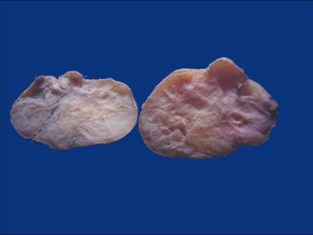
Well circumscribed tumor
Images hosted on other servers:

Parotid sialoangiolipoma
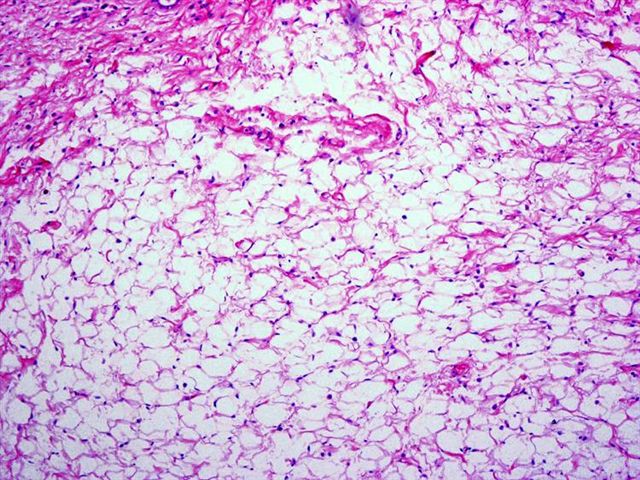
Microscopic (histologic) description
- Lipoma: bland appearing adipose tissue; osteolipoma or angiolipoma variants exist
- Lipoadenoma: mature adipose cells (> 90% mass) and proliferated glandular tissue (sharply demarcated, duct - acinar units or proliferated glands, may resemble sertoliform tubules), oncocytic change, sebaceous differentiation, squamous metaplasia
- Sialolipoma:
- Mature adipose tissue mixed with acinar, ductal, basal and myoepithelial cells of normal salivary gland
- Also duct ectasia with fibrosis, prominent lymphoid infiltrates with nodular aggregates in stroma, oncocytic changes, sebaceous differentiation
- Vascular variant is sialoangiolipoma
Microscopic (histologic) images
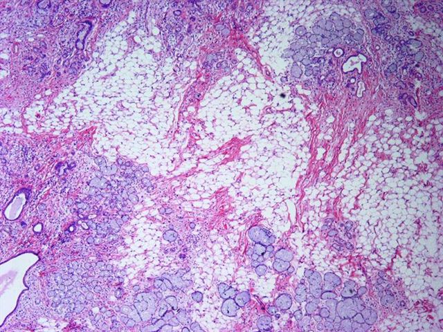
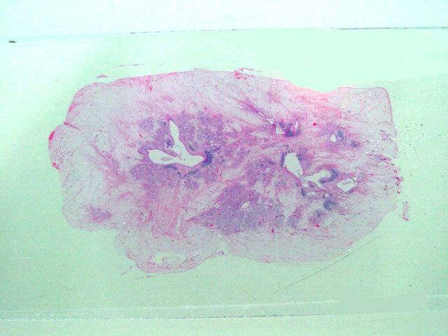
Case #49


Acini and dilated ducts
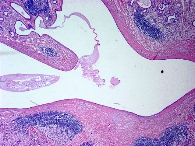

Ducts with fibrosis of wall infiltrated by lymphocytes
Peripheral
lipomatous tissue
with central salivary
gland elements
Lipoma-like areas
Images hosted on other servers:

Lower lip tumor

Submandibular sialoangiolipoma


Parotid sialoangiolipoma

Pleomorphic lipoma with classic floret-like cells

Chondroid lipoma
of lip has chondroid
and lipomatous
features

Oncocytic lipoadenoma of parotid

Spindle cell lipoma of parotid gland
Cytology description
- Nonspecific findings (Eur Arch Otorhinolaryngol 2008;265:S47); spindle cell lipomas have bland appearing spindle cells in myxoid background, CD34+ mature fat cells; S100 may be negative
Immunohistochemistry & special stains
Molecular / cytogenetics description
- t(12,14) / HMGA rearrangements
Differential diagnosis
- Pleomorphic adenoma: may have extensive lipometaplasia / lipomatosis (Am J Surg Pathol 2005;29:1389)
