Skin nontumor
Panniculitis
Lupus panniculitis
Author: Robert E. LeBlanc, M.D.

Last author update: 1 August 2017
Last staff update: 14 July 2023
Copyright: 2002-2025, PathologyOutlines.com, Inc.
PubMed Search: (Lupus erythematosus panniculitis) LEP
Table of Contents
Definition / general | Essential features | Terminology | ICD coding | Epidemiology | Sites | Clinical features | Laboratory | Case reports | Treatment | Microscopic (histologic) description | Microscopic (histologic) images | Positive stains | Negative stains | Differential diagnosis | Additional references | Practice question #1 | Practice answer #1 | Practice question #2 | Practice answer #2Cite this page: LeBlanc RE. Lupus panniculitis. PathologyOutlines.com website. https://www.pathologyoutlines.com/topic/skinnontumorlupuspanniculitis.html. Accessed August 27th, 2025.
Definition / general
- Autoimmune lobular lymphocytic panniculitis which shares features with subcutaneous panniculitis-like T cell lymphoma (SPTCL)
Essential features
- Tender subcutaneous plaques and nodules
- Autoimmune lobular lymphocytic panniculitis
- Patients often do not have or develop systemic lupus
- Must be distinguished from subcutaneous panniculitis-like T cell lymphoma
- Presence of high Ki67 or hotspots favors a diagnosis of subcutaneous panniculitis-like T cell lymphoma (SPTCL)
- Patients with overlapping lupus erythematosus panniculitis and SPTCL features may develop hemophagocytic lymphohistiocytosis (HLH)
Terminology
- Lupus panniculitis
- Lupus erythematosus profundus (when there is concomitant discoid lupus erythematosus)
ICD coding
- L93.2
Epidemiology
- Women more often than men
- Usually young to middle aged adults
- Fewer reported cases in childhood and in patients of advanced age
- Rare manifestation of autoimmune connective tissue disease, representing less than 3% of patients with cutaneous or systemic lupus erythematosus
Sites
- Trunk, breast, proximal extremities (upper more often than lower), face and scalp
- Distal extremities are less commonly involved
Clinical features
- Tender, indurated and erythematous plaques and nodules
- Overlying skin occasionally shows features of discoid lupus (scaling, plugged follicles, scarring and atrophy)
- Lesions may spontaneously regress and recur
- Occasional severe lipoatrophy with resolution of lesions
- Patients often do not have a history of cutaneous or systemic lupus
- Some patients develop mild systemic symptoms but do not meet full criteria for a diagnosis of systemic lupus
Laboratory
- Antinuclear antibodies are identified in a subset of patients with LEP
- The absence of antinuclear antibodies does not exclude LEP
Case reports
- 9 year old girl with linear sclerodermoid lupus erythematosus profundus (Am J Dermatopathol 2016;38:904)
- 40 year old woman with LEP diagnosis who developed HLH (Postepy Dermatol Alergol 2017;34:281)
- 49 year old woman with adalimumab induced LEP (Lupus 2014;23:1443)
Treatment
- Antimalarial agents
- Topical corticosteroids for concomitant discoid lupus
- Systemic corticosteroids and other immunosuppressants are sometimes used temporarily at disease onset
Microscopic (histologic) description
- Lobular or mixed lobular and septal panniculitis
- Hyaline lipomembranous changes punctuated by foci of active lobular panniculitis
- Infiltrate consists of lymphocytes, histiocytes and plasma cells
- Germinal center lymphoid follicles present throughout the lesion
- Plasma cells may be prominent and form large clusters
- Lymphocytes are small, bland and splay apart adipocyte lobules
- Lymphocytic vasculitis, granulomas and dermal mucin are variable
- Features of discoid lupus are present in a subset of patients (this is termed lupus erythematosus profundus)
Microscopic (histologic) images
Contributed by Robert E. LeBlanc M.D.
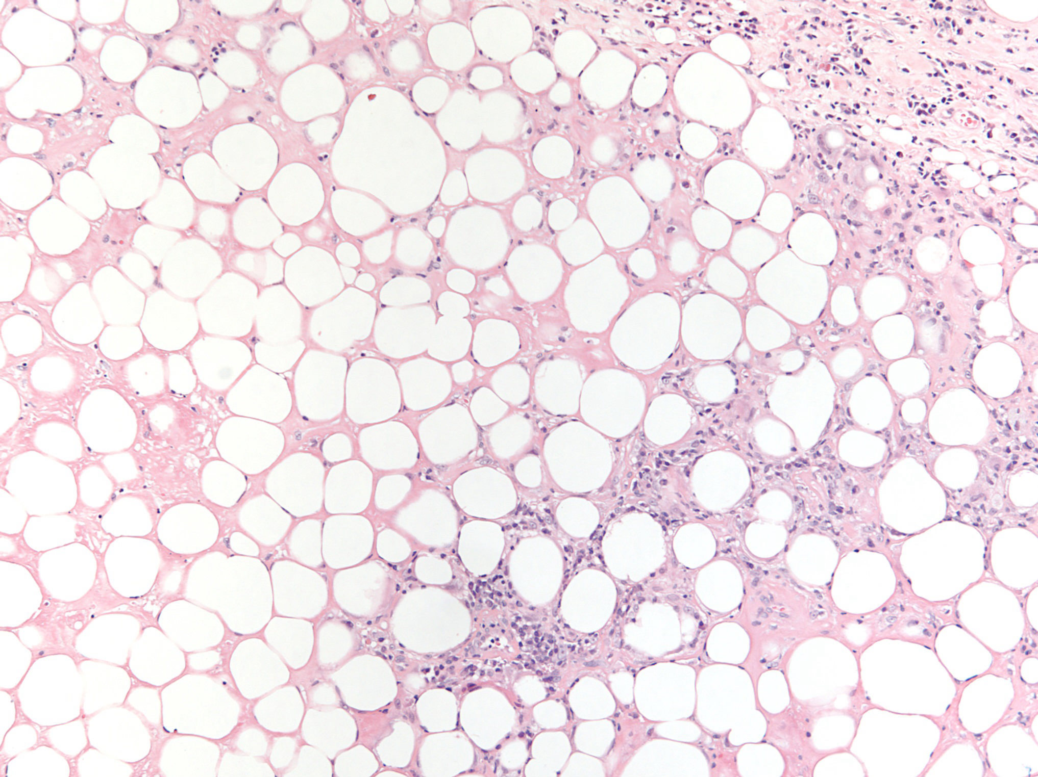
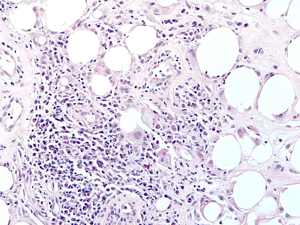
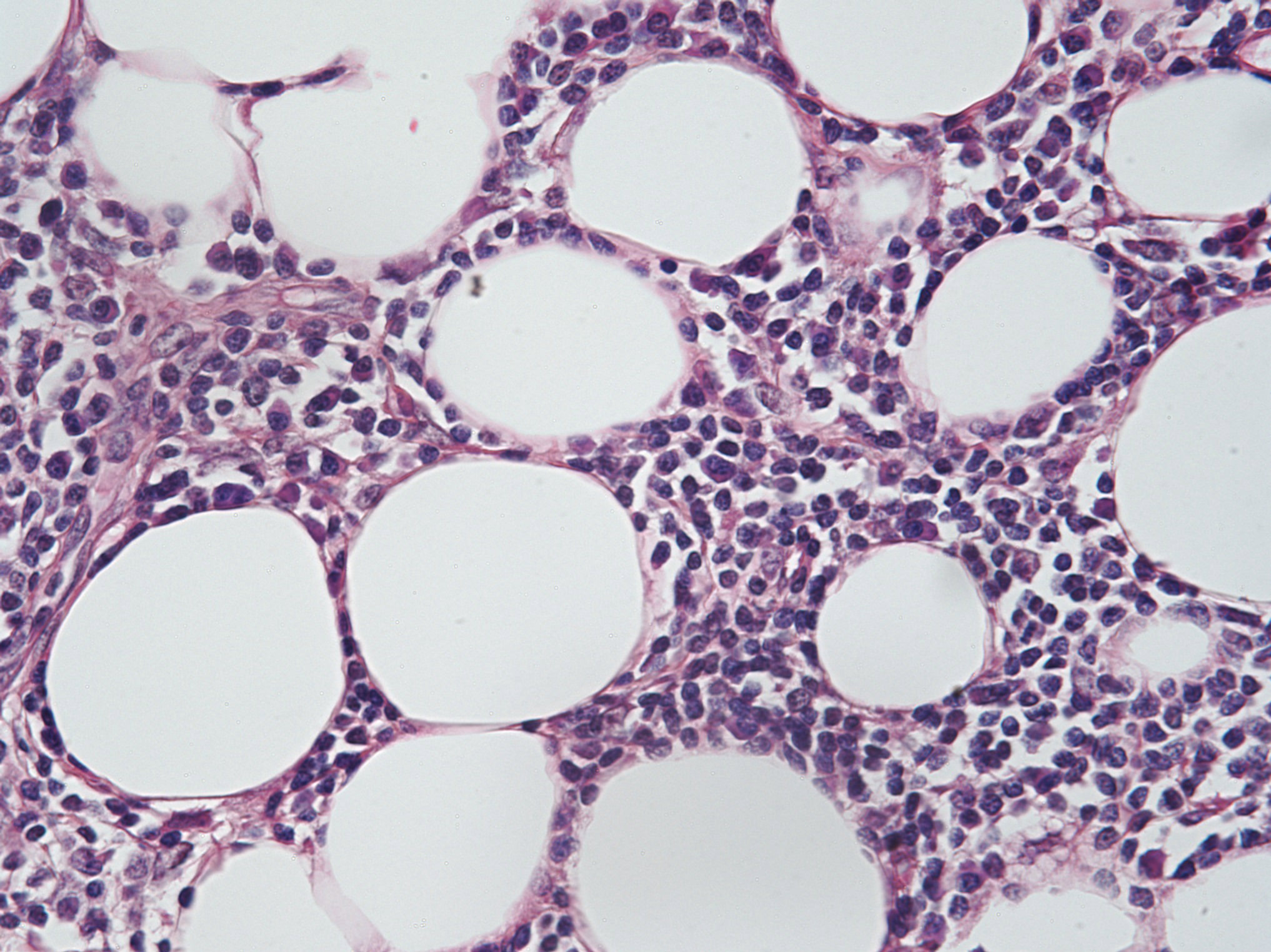
H&E (10x, 20x, 40x)
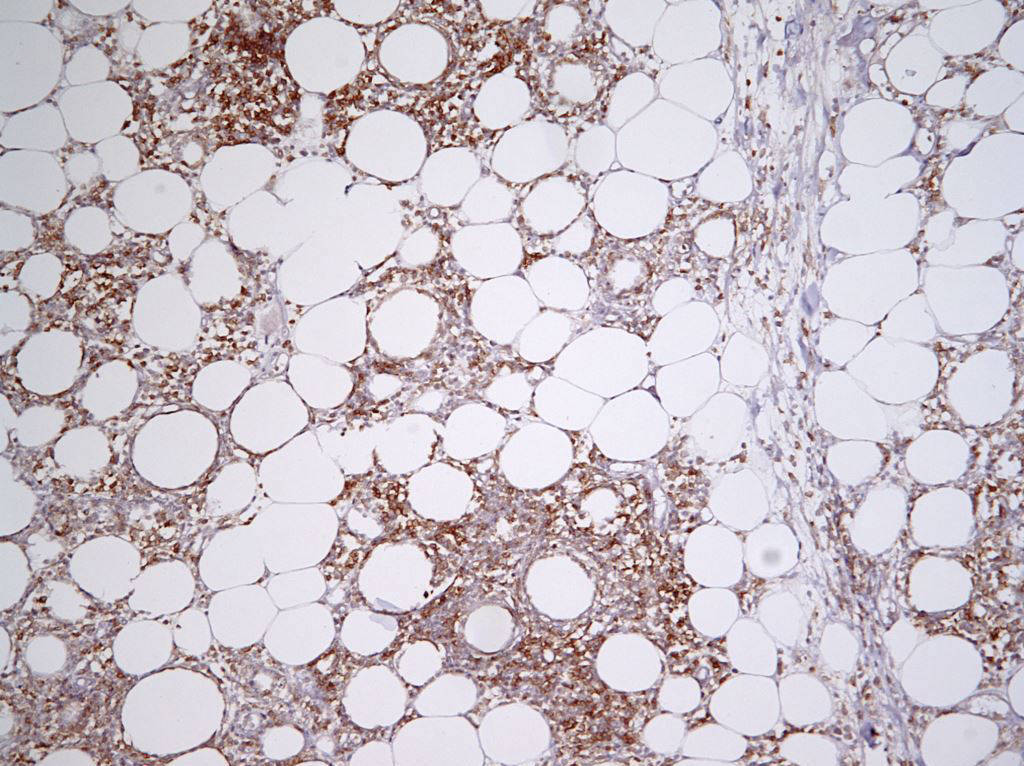

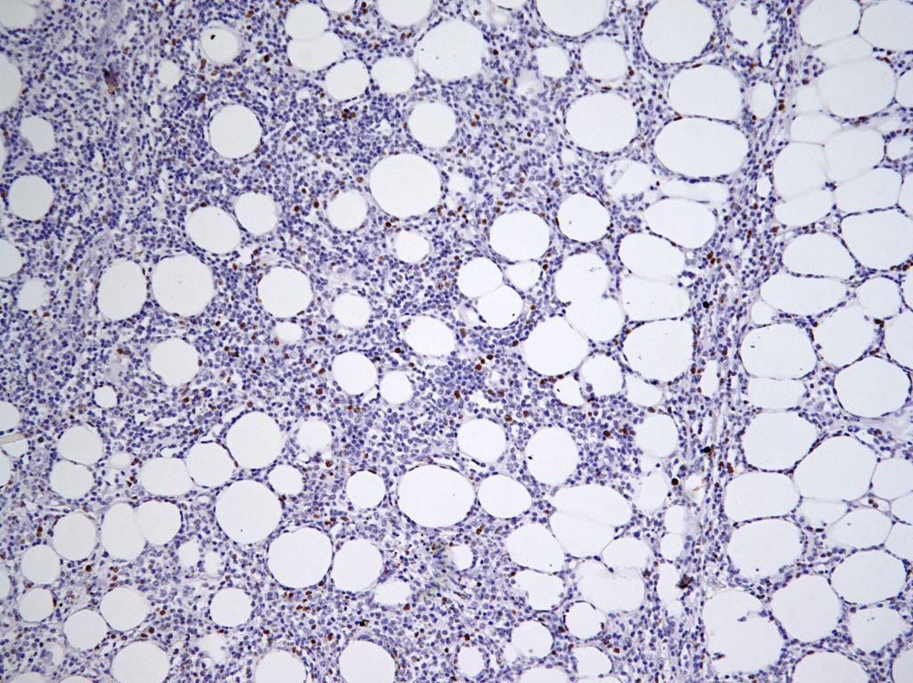
CD4, CD8, Ki67
Positive stains
Negative stains
Differential diagnosis
- Injection site reaction or drug reaction: this distinction relies heavily on clinical correlation
- Other connective tissue disease associated panniculitidies: this distinction relies heavily on clinical correlation
- Subcutaneous panniculitis-like T cell lymphoma:
- Ki67 is either expressed diffusely throughout the lesion or in hotspots enriched with adipotropic cytotoxic CD8+ T cells
- The lymphoma cells are slightly enlarged and hyperchromatic with irregular nuclear contours
- They surround individual fat lobules but also appear within the disrupted cell membranes of involved adipocytes
- Paired T cell gene rearrangement studies may be helpful to assess clonality
Additional references
Practice question #1
Subcutaneous panniculitis-like T cell lymphoma is characterized by which of the following immunophenotypes?
- CD4+ T cells, high Ki67 expression
- CD4+ T cells, low Ki67 expression
- CD8+ T cells, high Ki67 expression
- CD8+ T cells, low Ki67 expression
Practice answer #1
C. SPTCL has a cytotoxic CD8 possitive immunophenotype and high expression of Ki67 that can often permit its distinction from lupus erythematosus panniculitis.
Comment Here
Reference: Lupus panniculitis
Comment Here
Reference: Lupus panniculitis
Practice question #2
Which of the following microscopic findings would favor the diagnosis of lupus erythematosus profundus over lupus erythematosus panniculitis?
- Epidermal vacuolar interface changes
- Hyaline lipomembranous changes
- Lobular lymphohistiocytic panniculitis
- Lymphocytic vasculitis
Practice answer #2
A. Lupus erythematosus profundus is the preferred terminology for lesions of lupus erythematosus panniculitis that have overlying changes of discoid lupus, namely vacuolar interface dermatitis, basement membrane thickening, scale, follicular plugging and both perifollicular and periadnexal inflammation. The other answer options can be present in both lupus erythematosus profundus and lupus erythematosus panniculitis.
Comment Here
Reference: Lupus panniculitis
Comment Here
Reference: Lupus panniculitis
