Skin nontumor
Infectious disorders
Bacillary angiomatosis
Authors: Joel Tjarks, M.D., Sara C. Shalin, M.D., Ph.D.


Last author update: 1 January 2017
Last staff update: 18 July 2025
Copyright: 2001-2025, PathologyOutlines.com, Inc.
PubMed search: bacillary angiomatosis [title] skin
Table of Contents
Definition / general | Essential features | Epidemiology | Sites | Etiology | Clinical features | Laboratory | Case reports | Treatment | Clinical images | Microscopic (histologic) description | Microscopic (histologic) images | Positive stains | Differential diagnosis | Additional referencesCite this page: Tjarks J, Shalin S. Bacillary angiomatosis. PathologyOutlines.com website. https://www.pathologyoutlines.com/topic/skintumornonmelanocyticbacillaryangiomatosis.html. Accessed September 2nd, 2025.
Definition / general
- Reactive vascular proliferation due to infection by Bartonella henselae or Bartonella quintana
- Most commonly affects immunocompromised individuals (especially those with HIV)
Essential features
- Lobular proliferation of capillaries with ectatic vessels lined by prominent endothelial cells
- Neutrophils, lymphocytes and histiocytes are frequently present
- Purplish grey bacterial colonies may be seen
Epidemiology
- May affect adults and children
- Most commonly affects immunocompromised individuals
Sites
- Can occur in any cutaneous site
- Rarely occurs in mucosa or internal organs
Etiology
- B. Henselae is acquired via cat scratch or bite in 2/3 of cases
Clinical features
- Red, smooth papules and nodules which are widely distributed
- May mimic Kaposi sarcoma and pyogenic granuloma clinically
Laboratory
- PCR and IHC may be helpful in identifying organisms
Case reports
- 36 year old man with bacillary angiomatosis presenting with facial tumor and multiple abcesses (Medicine (Baltimore) 2016;95:e4155)
- 43 year old man with bacillary angiomatosis presenting with polymorphic skin lesions (IDCases 2016;6:77)
Treatment
- Antibiotics - typically doxycycline
Clinical images
Images hosted on other servers:

Papules on face, chest, abdomen and back; mass lesion on face

Exophytic lesion on neck

Small, cherry angiomata-like lesions and nodules
Microscopic (histologic) description
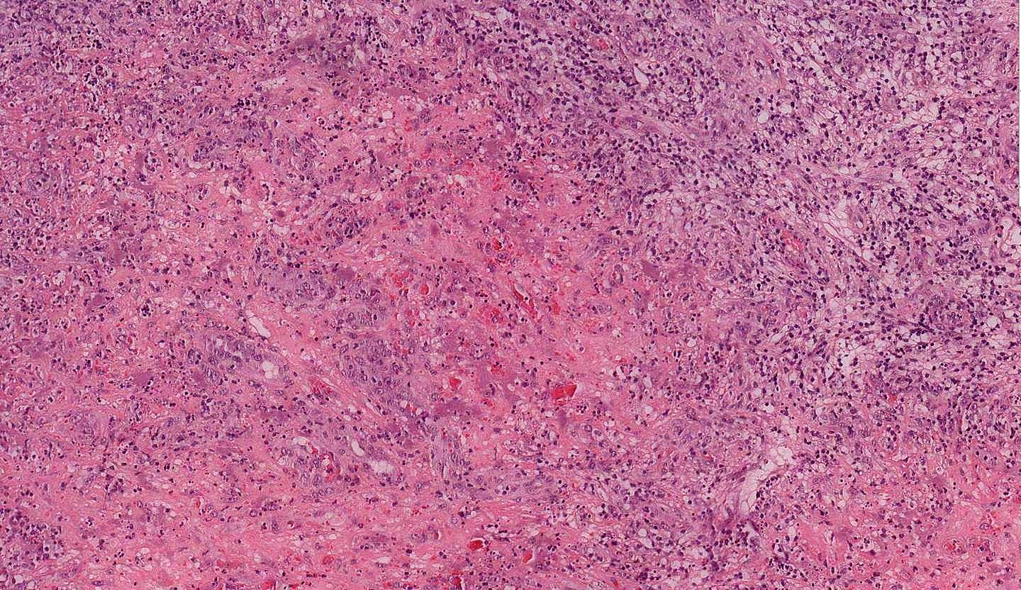
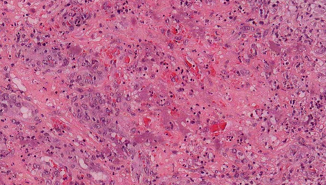
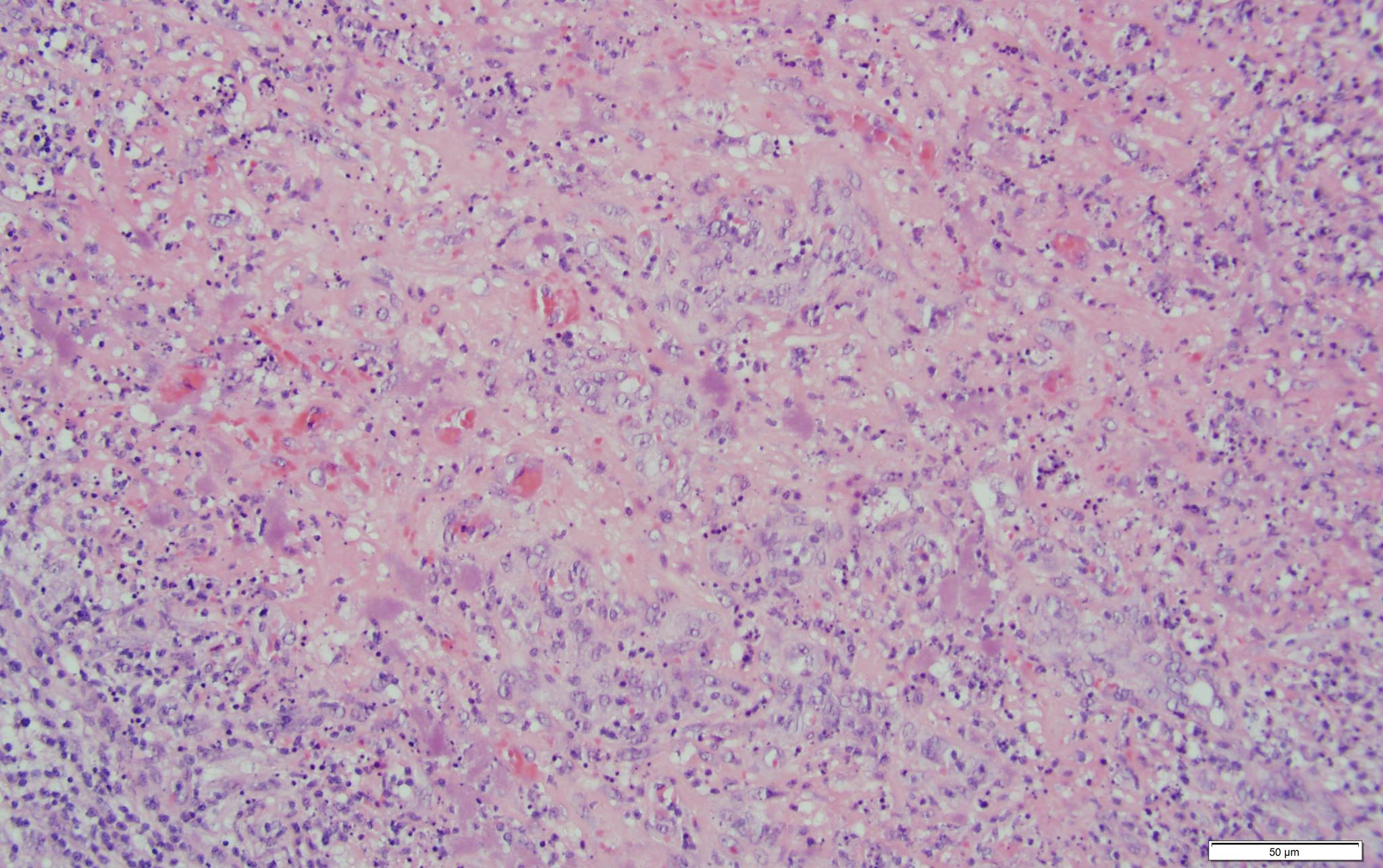
- Lobular proliferation of capillaries with ectatic vessels lined by prominent endothelial cells in edematous stroma
- Neutrophils, lymphocytes and histiocytes are frequently present
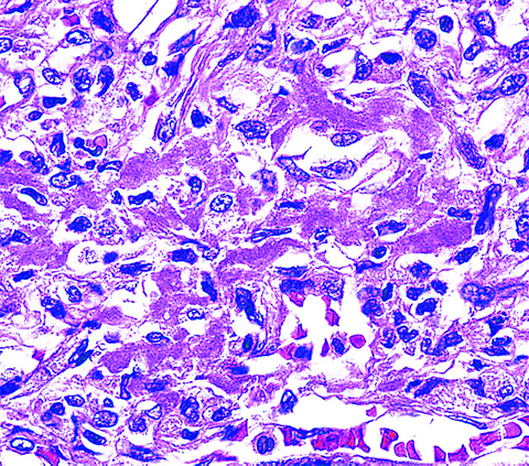
- Purplish grey bacterial colonies may be seen, especially near neutrophils
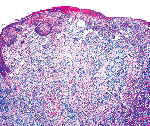
- Peripheral collarette may be seen (low power histologic image mimics pyogenic granuloma)
- Warthin-Starry special stain will highlight Bartonella organisms
Microscopic (histologic) images
Contributed by Tammie Ferringer, M.D., Nathan Lee, M.D. and Mark R. Wick, M.D.
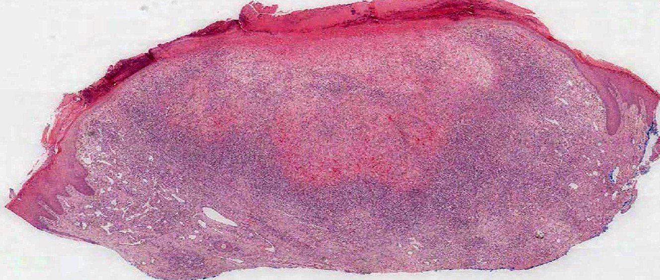
Bacillary angiomatosis profile
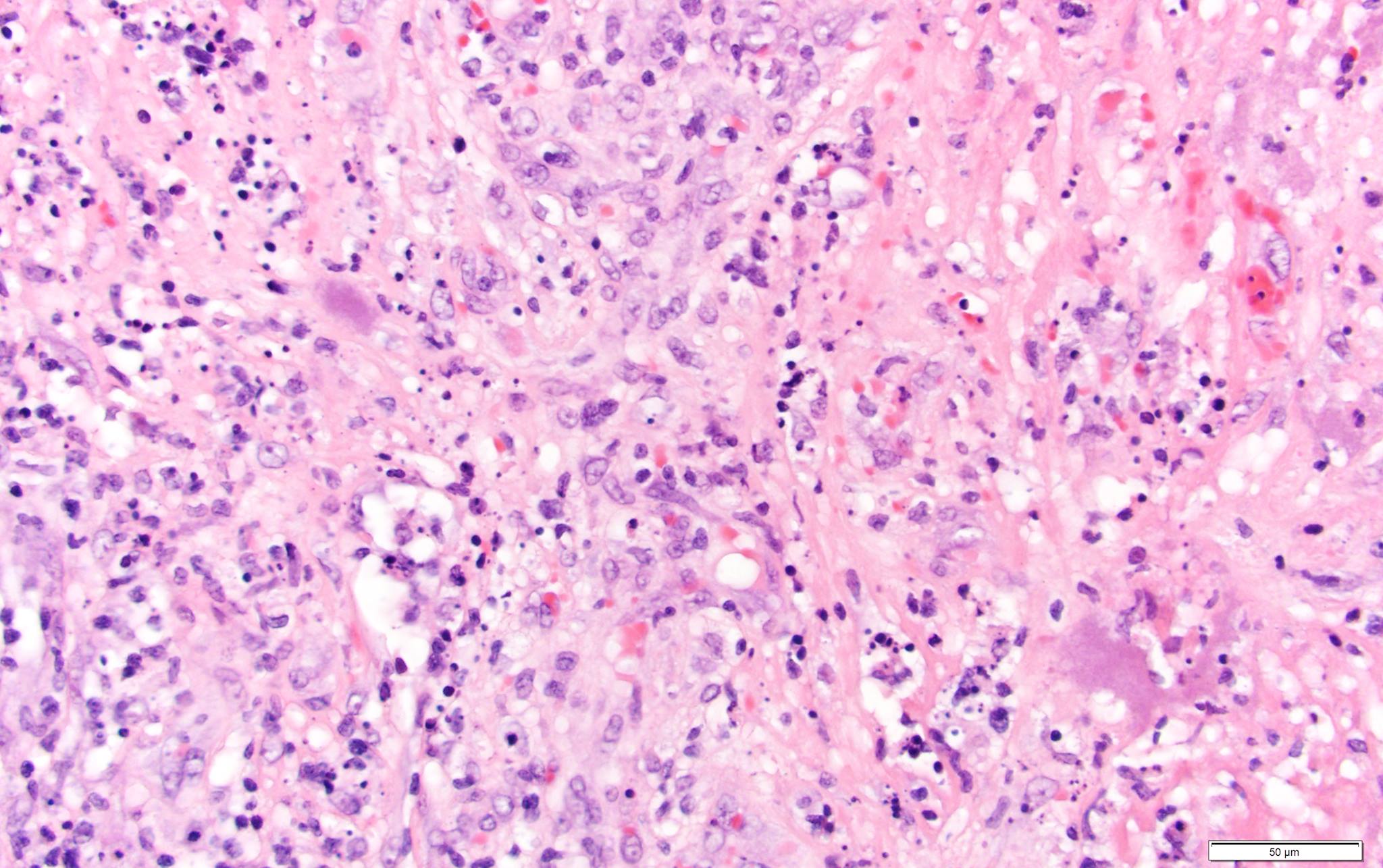
Bacillary angiomatosis

Breast skin

Steiner stain, breast
Positive stains
Differential diagnosis
- Kaposi sarcoma: spindled vascular proliferation with slit-like vascular spaces, plasma cells, HHV8 positive, lacks purplish grey bacterial colonies
- Pyogenic granuloma: lobular proliferation of capillaries, but typically somewhat less inflamed (unless ulcerated) and no purplish grey bacterial colonies
Additional references
