Skin nonmelanocytic tumor
Vascular tumors
Hemangioma variants
Lobular capillary hemangioma
Authors: Jordan T. Hyde, M.D., Jason B. Lee, M.D.
Board of reviewers: Caroline I. Mullins, M.D.


Last author update: 30 October 2024
Last staff update: 30 October 2024
Copyright: 2003-2025, PathologyOutlines.com, Inc.
PubMed Search: Lobular capillary hemangioma skin
Table of Contents
Definition / general | Essential features | Terminology | ICD coding | Epidemiology | Sites | Pathophysiology | Etiology | Clinical features | Diagnosis | Prognostic factors | Case reports | Treatment | Clinical images | Microscopic (histologic) description | Microscopic (histologic) images | Positive stains | Negative stains | Videos | Sample pathology report | Differential diagnosis | Additional references | Practice question #1 | Practice answer #1 | Practice question #2 | Practice answer #2Cite this page: Hyde JT, Lee JB. Lobular capillary hemangioma. PathologyOutlines.com website. https://www.pathologyoutlines.com/topic/skintumornonmelanocyticpyogenicgranuloma.html. Accessed August 25th, 2025.
Definition / general
- Benign vascular lesion that is characterized by the proliferation of capillary sized blood vessels and typically presents as a solitary, rapidly growing, bright red papule that frequently bleeds following trauma
Essential features
- Rapidly growing, benign vascular lesion that frequently ulcerates and bleeds and is often located on sites of frequent trauma like the face, lips, mucosa and fingers
- Histopathologic features include lobules containing numerous capillary sized vessels in the dermis within an edematous stroma in early lesions or fibrotic stroma with fibrous septa in older lesions; secondary changes, such as a mixed cell inflammatory infiltrate, ulceration and granulation tissue formation, are common
- Treatment usually involves shave removal with electrodesiccation or surgical excision; may recur following treatment
Terminology
- Pyogenic granuloma is the term most often used by clinicians; however, it is a misnomer as it is neither infectious nor granulomatous
- Tumor of pregnancy or granuloma gravidarum are names sometimes given when developed during pregnancy
ICD coding
- ICD-10: L98.0 - pyogenic granuloma
Epidemiology
- Demographics
- Affects individuals of all age groups, with the highest prevalence in young adults
- No gender predilection is observed
- Mucosal lesions may be more common in women (J Am Acad Dermatol 2000;42:1012)
- Risk factors
- Majority of cases with no apparent cause
- Recent history of trauma
- Pregnancy
- Oral contraceptives
- Systemic retinoids, BRAF inhibitors and EGFR inhibitors have been associated with lobular capillary hemangiomas, particularly in the periungual regions; however, some believe these lesions represent solely excessive granulation tissue (Open Access Maced J Med Sci 2017;5:423)
Sites
- Face, lips, gingiva, hands and fingers are the most common but may occur anywhere (Open Access Maced J Med Sci 2017;5:423)
Pathophysiology
- Unclear
- There is controversy about whether the proliferation of capillaries is a reactive or neoplastic process (Plast Reconstr Surg Glob Open 2024;12:e6160)
Etiology
- Most cases occur without an apparent cause
- Some cases are associated with recent trauma
- Higher incidence is noted during pregnancy and with the use of oral contraceptives, suggesting a possible hormonal influence
- Reference: Plast Reconstr Surg Glob Open 2024;12:e6160
Clinical features
- Typically appears as a rapidly growing, bright red papule that is often pedunculated or polypoid on the skin or mucosa
- Lesion frequently ulcerates and bleeds
- Rare presentations include
- Multiple lesions with satellite formations
- May occur within a pre-existing capillary malformation, such as a port wine stain (Dermatol Reports 2021;13:9115)
- As a skin colored nodule if present in the deep dermis, subcutaneous tissue or intravascularly
Diagnosis
- Diagnosis is usually made clinically but may be confirmed with a biopsy
Prognostic factors
- Benign but may recur after treatment
Case reports
- 15 year old boy with exophytic papule on the lower lip (Dent Traumatol 2020;36:446)
- 33 year old pregnant woman with bleeding intranasal lesion (Maedica (Bucur) 2024;19:160)
- 36 year old woman with bright red papule on the thumb (Dermatol Ther 2022;35:e15194)
- 40 year old man with exophytic mass on the thumb (J Surg Case Rep 2023;2023:rjad157)
- 45 year old woman with friable pedunculated papule on the upper lip (N Engl J Med 2022;387:1979)
Treatment
- Shave removal followed by electrodesiccation or electrocautery is often sufficient, especially for smaller lesions
- Complete excision
- Other less common treatments include silver nitrate and laser therapy
- Reference: Plast Reconstr Surg Glob Open 2024;12:e6160
Clinical images
Contributed by Jason B. Lee, M.D.
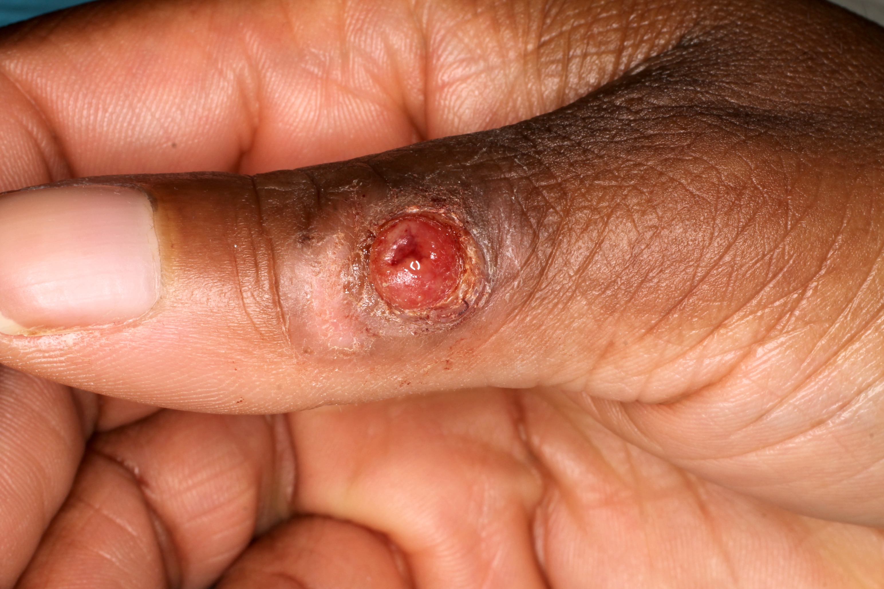
Ulcerated exophytic papule
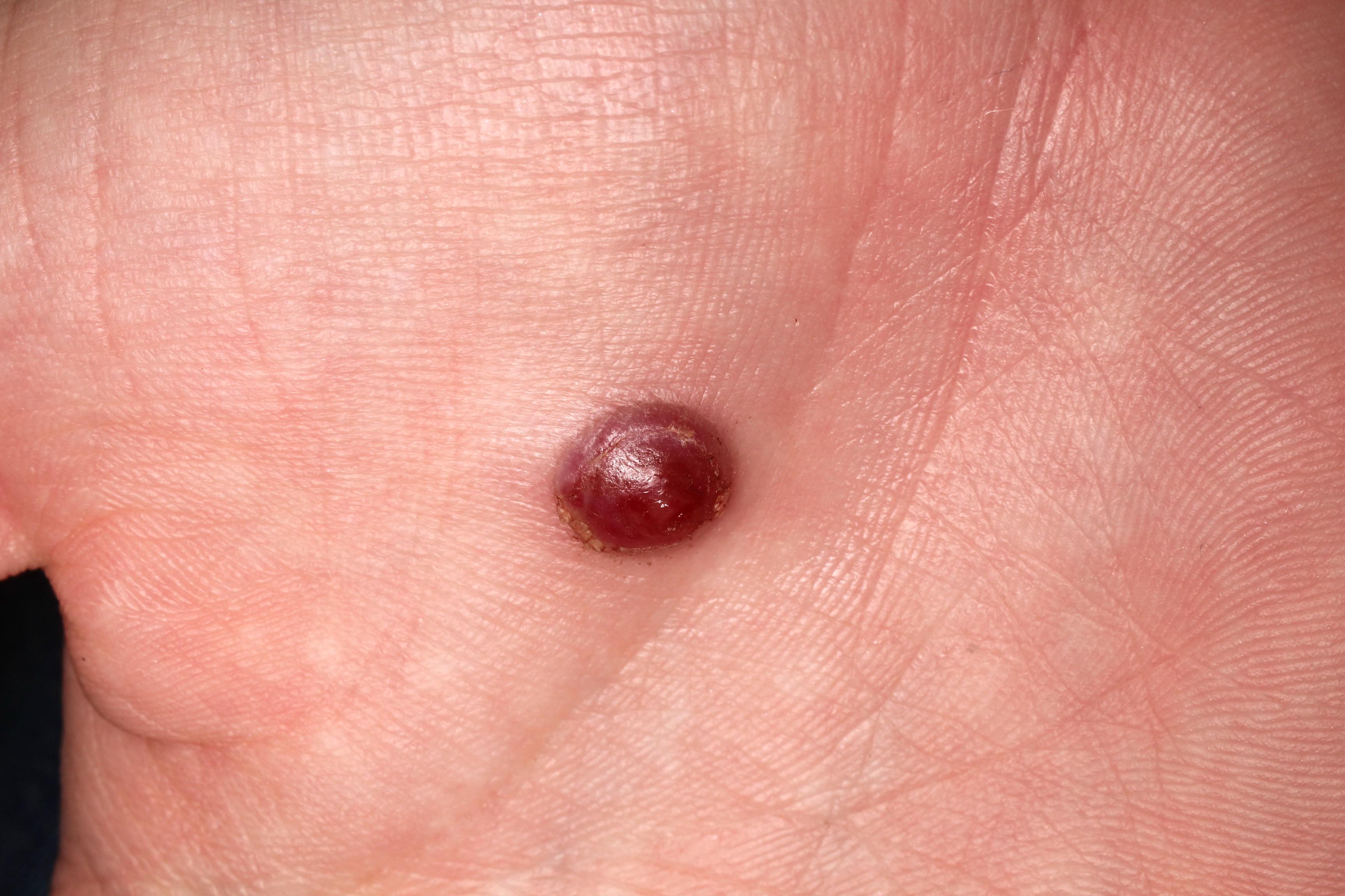
Red papule
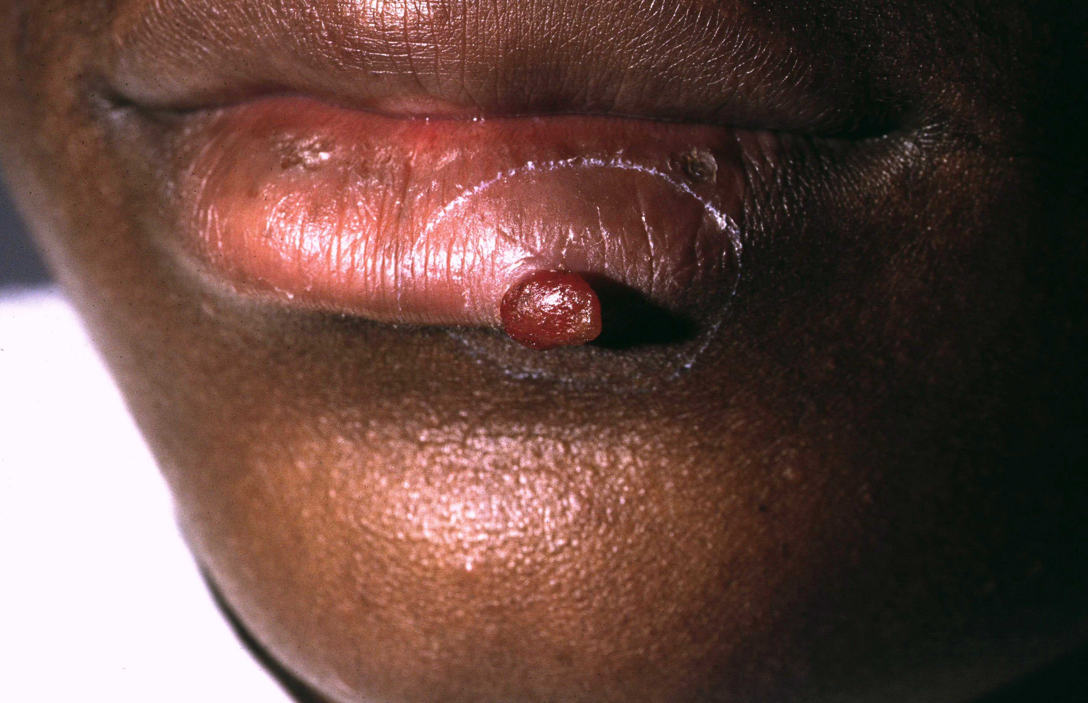
Exophytic pedunculated papule
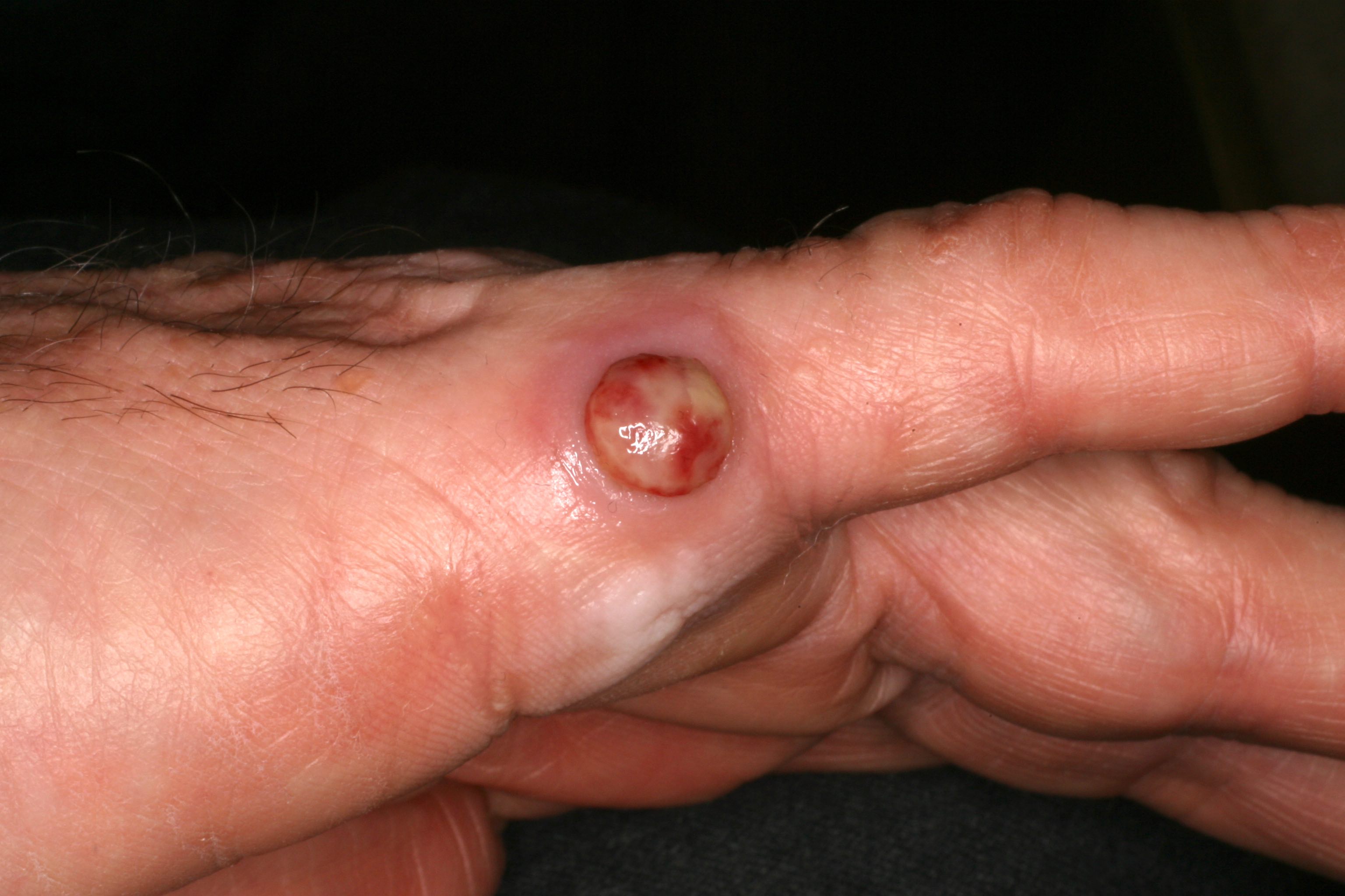
Ulcerated papule
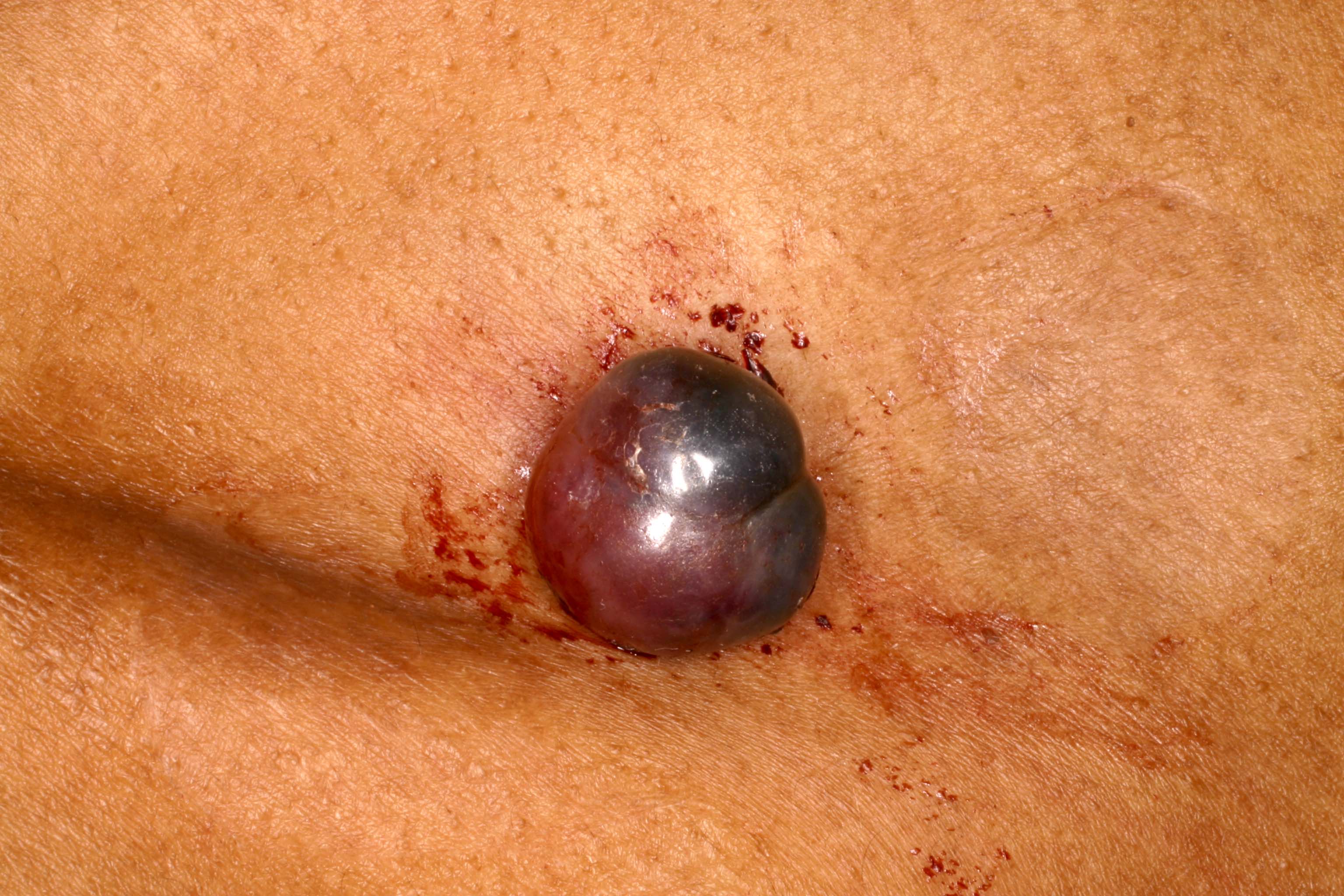
Blue red exophytic papule
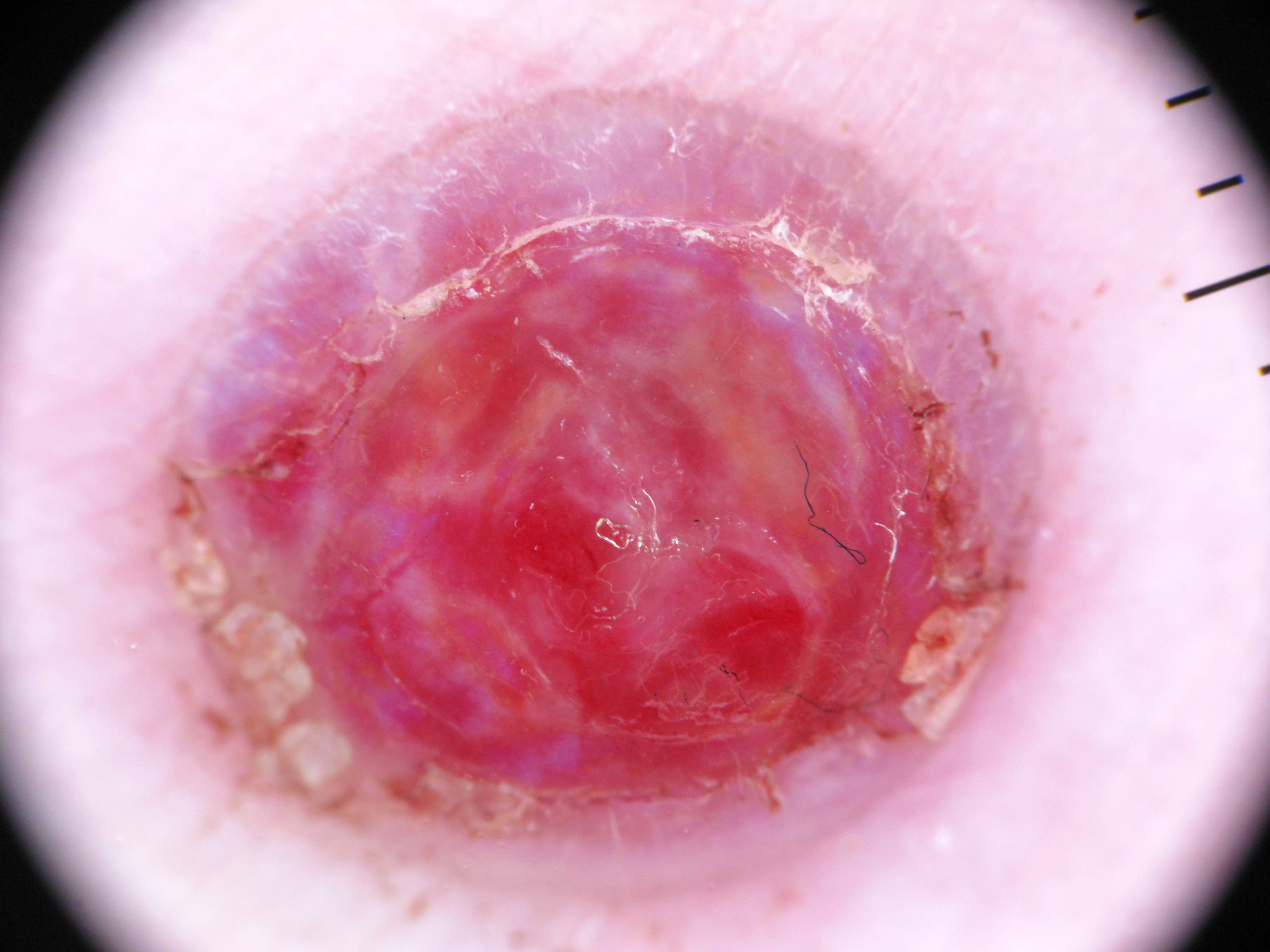
Bright red ulcerated papule
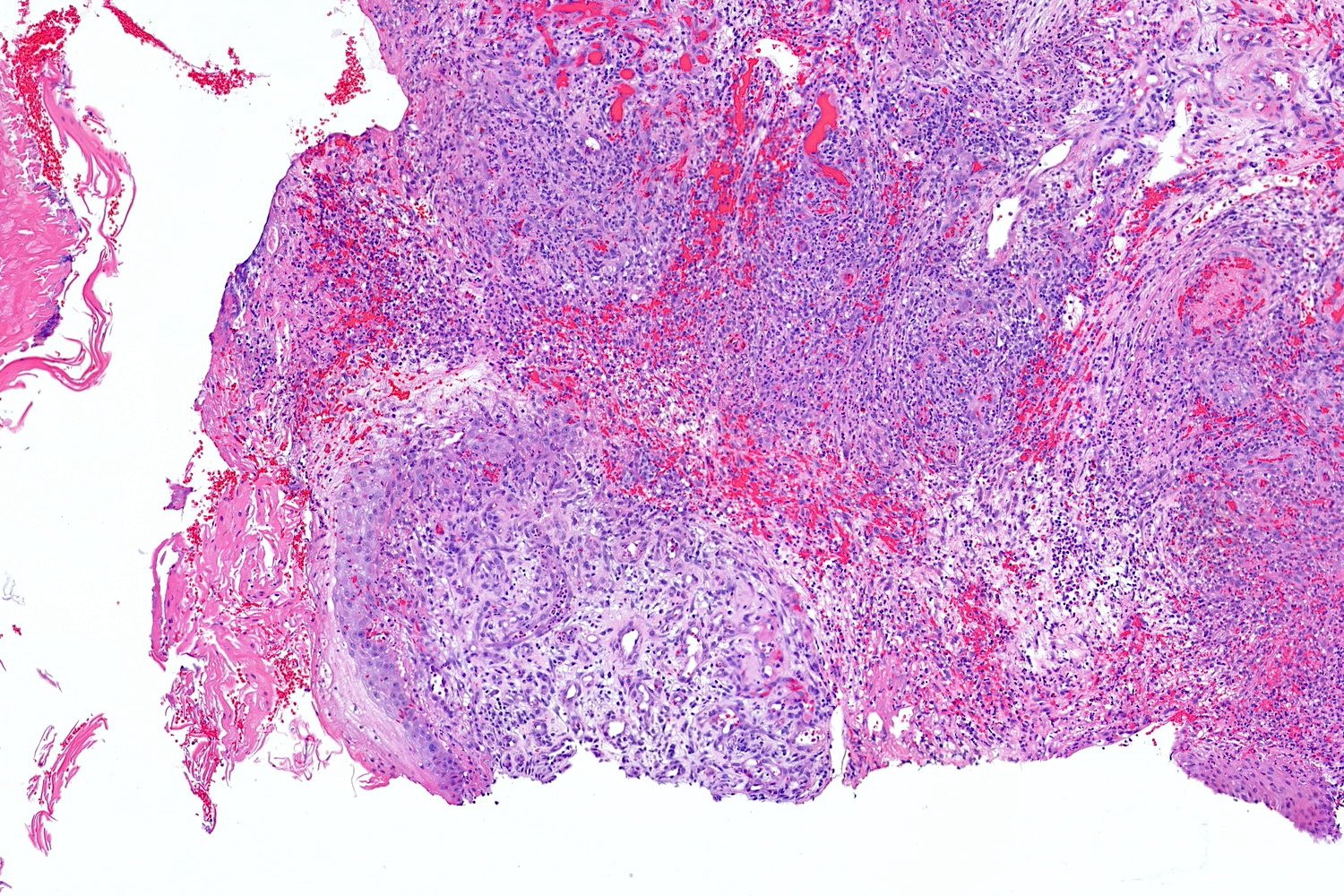
Microscopic (histologic) description
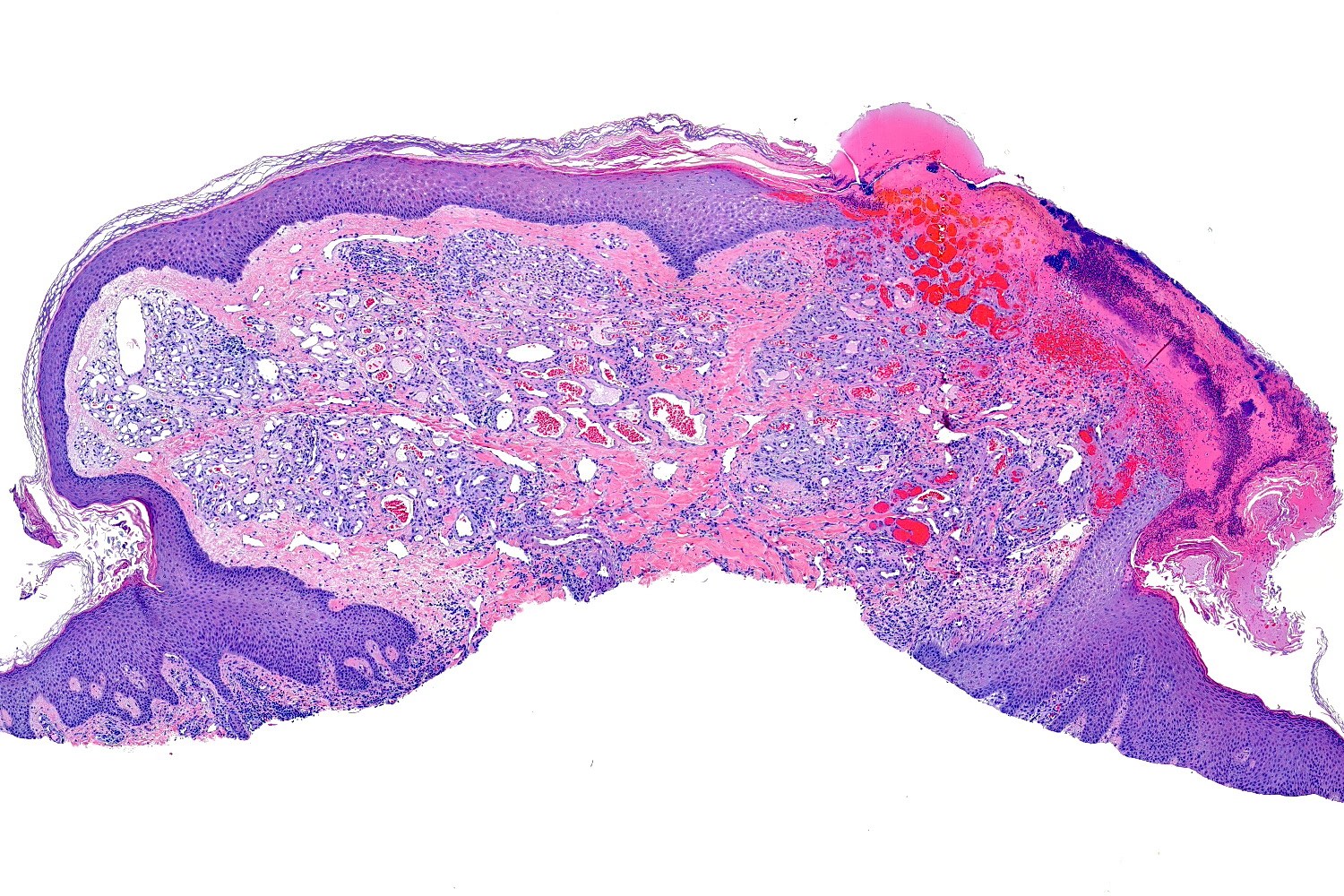
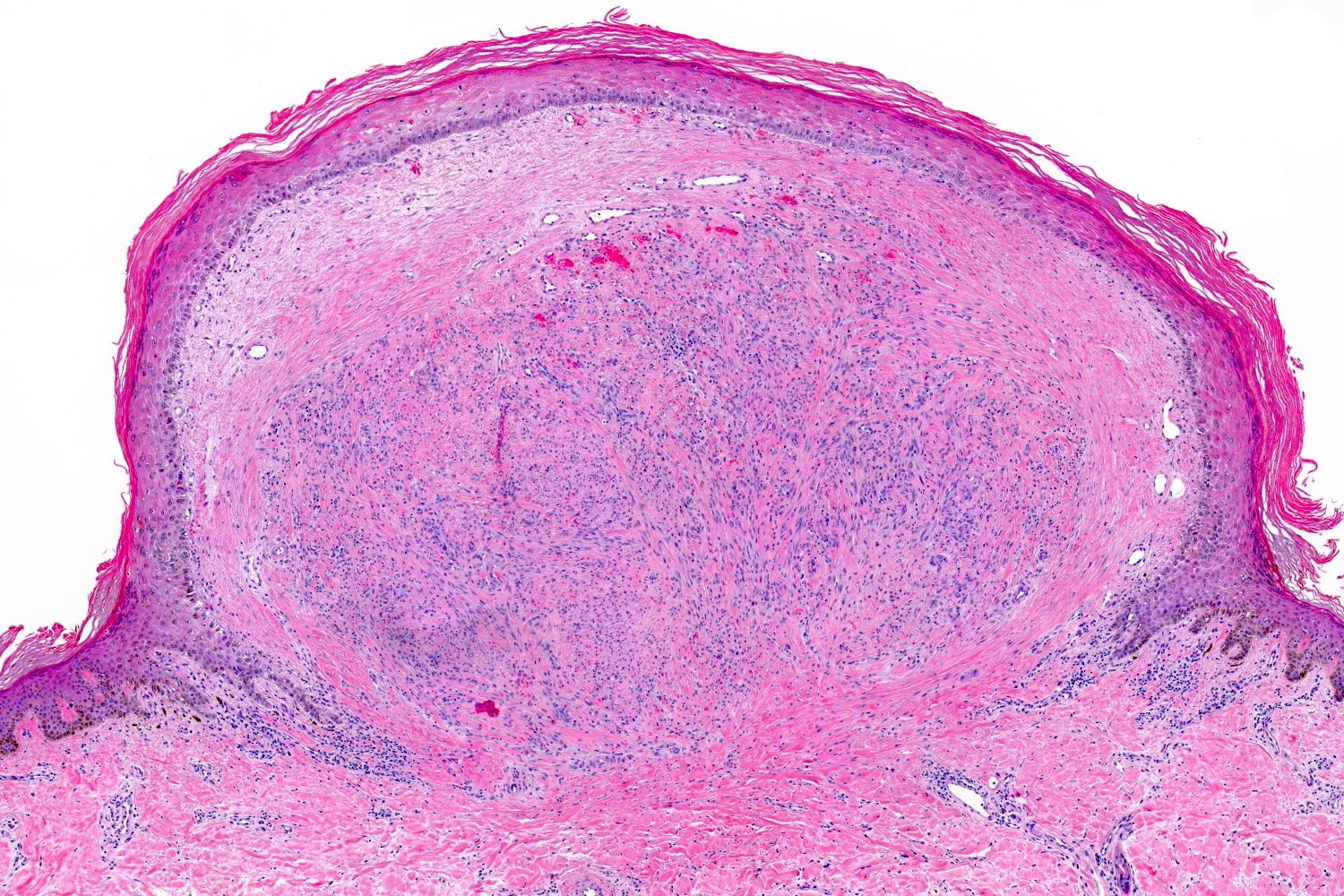
- Frequently exophytic or polypoid, though may also be sessile
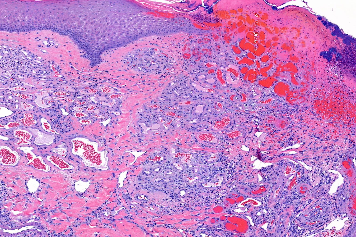
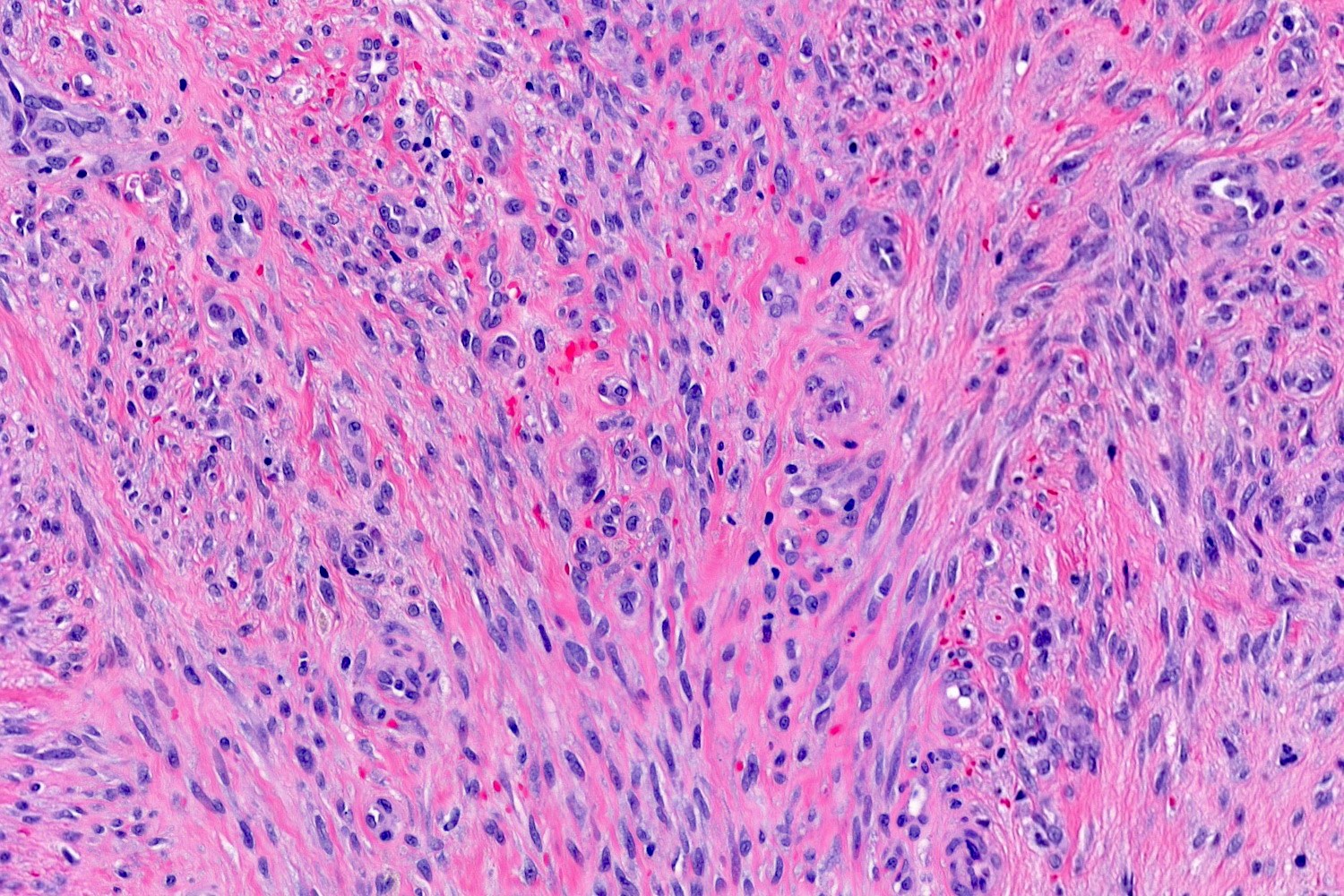
- Characterized by lobules in the dermis that contain numerous capillary sized vessels
- Lobules are separated by fibrous septa or trabeculae, especially in older lesions
- Stroma is often edematous and more mucinous in early lesions, becoming more fibrotic with time
- Epidermal collarette is commonly observed
- Cytologic features include bland endothelial cells that may be plump; mitotic figures may be frequent
- Fibroblasts and pericytes are also present
- Secondary changes may obscure the underlying architecture and morphology, which may only be present at the base of the lesion
- Ulceration at the surface of the lesion is common
- Granulation tissue may be present at the surface of ulcerated lesions
- Secondary mixed cell infiltrate with lymphocytes and neutrophils is common (Am J Surg Pathol 1980;4:470)
- May rarely be present intravascularly, affect the deep dermis or subcutaneous tissue or internal organs (Cureus 2023;15:e45142, Am J Dermatopathol 2007;29:408, Case Rep Pathol 2022;2022:5641608)
Microscopic (histologic) images
Contributed by Jason B. Lee, M.D.
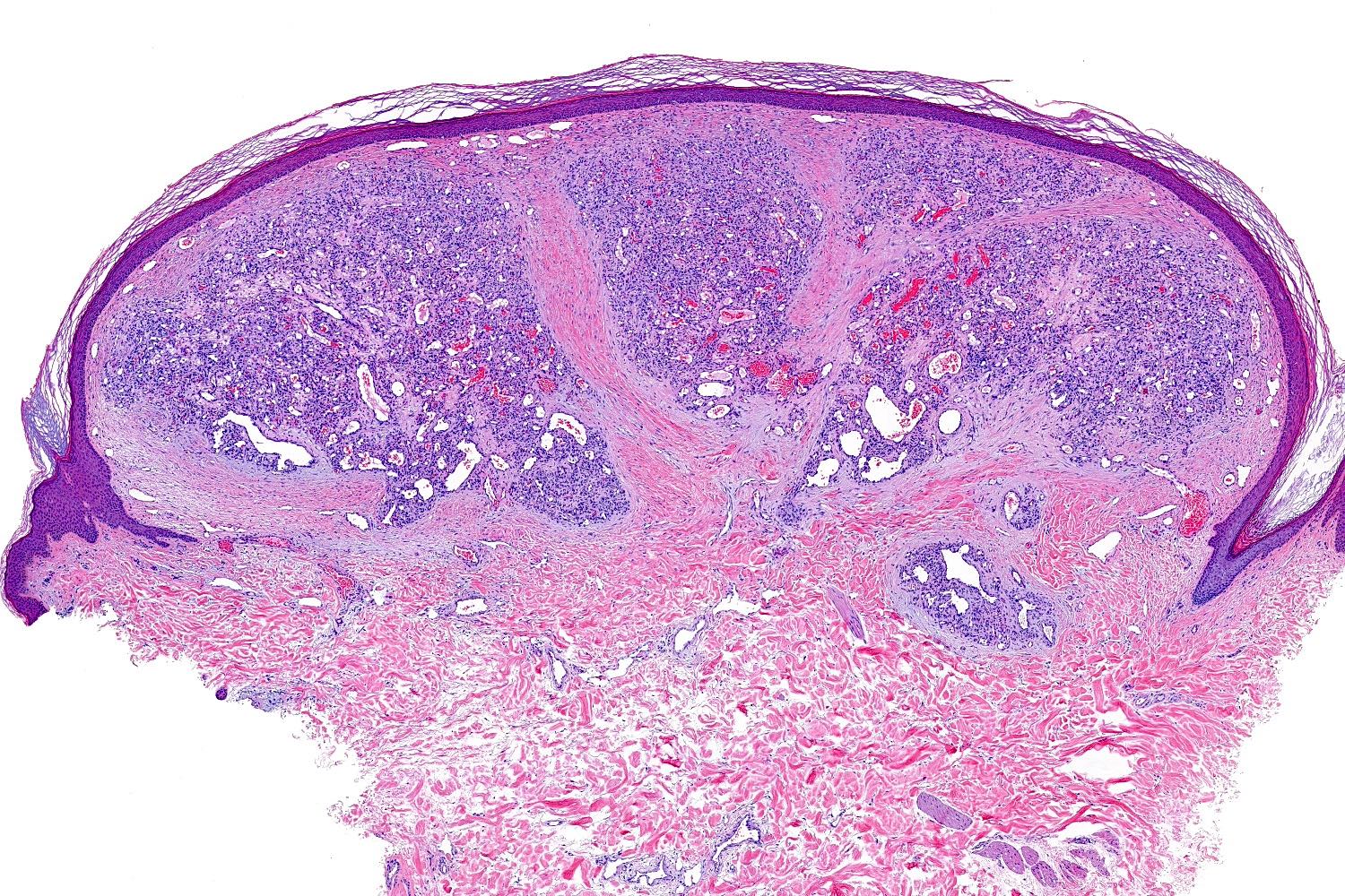
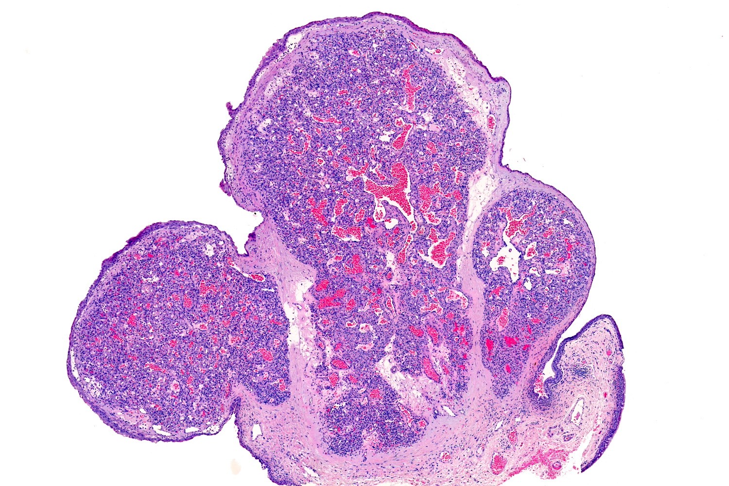
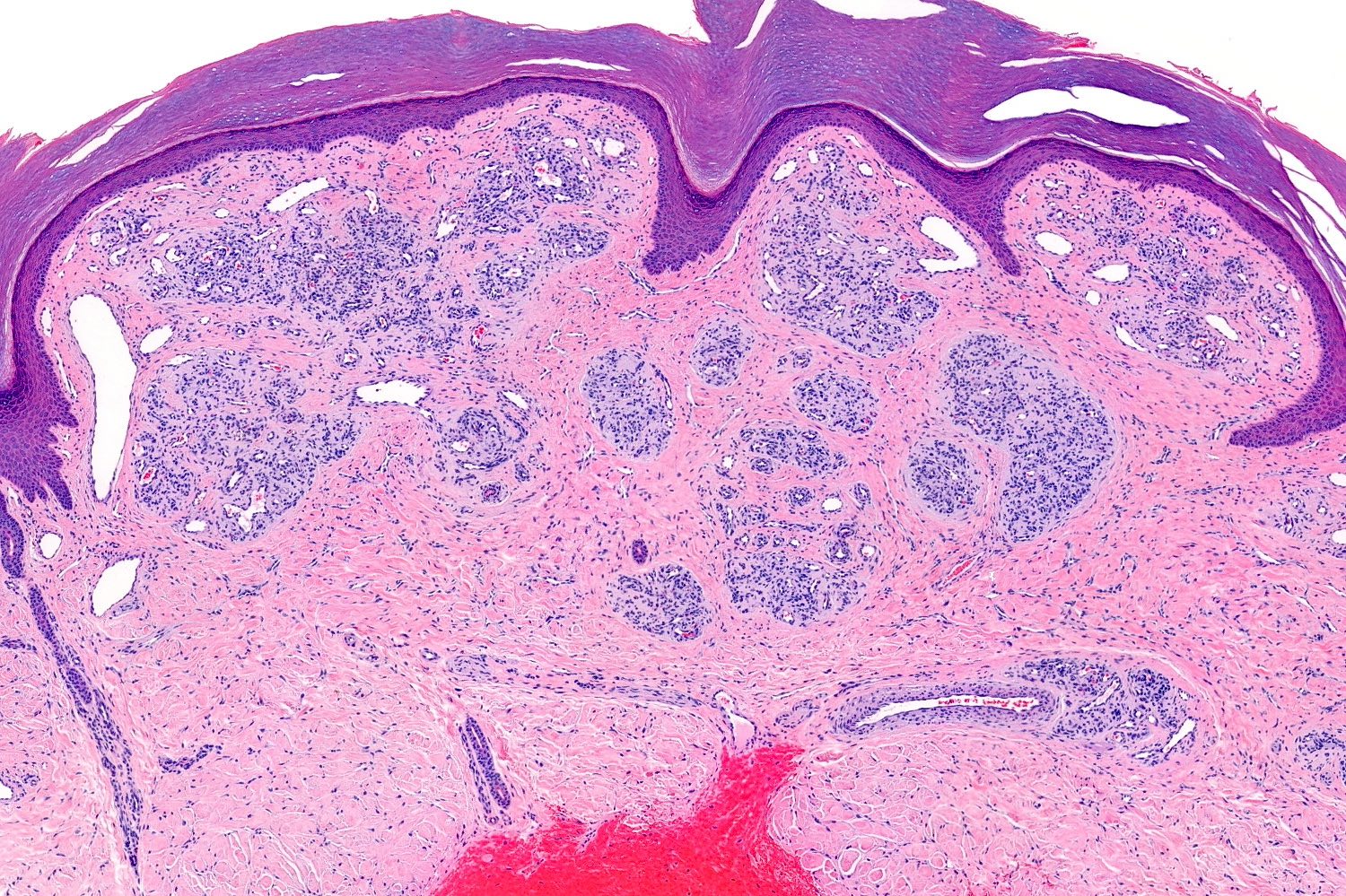
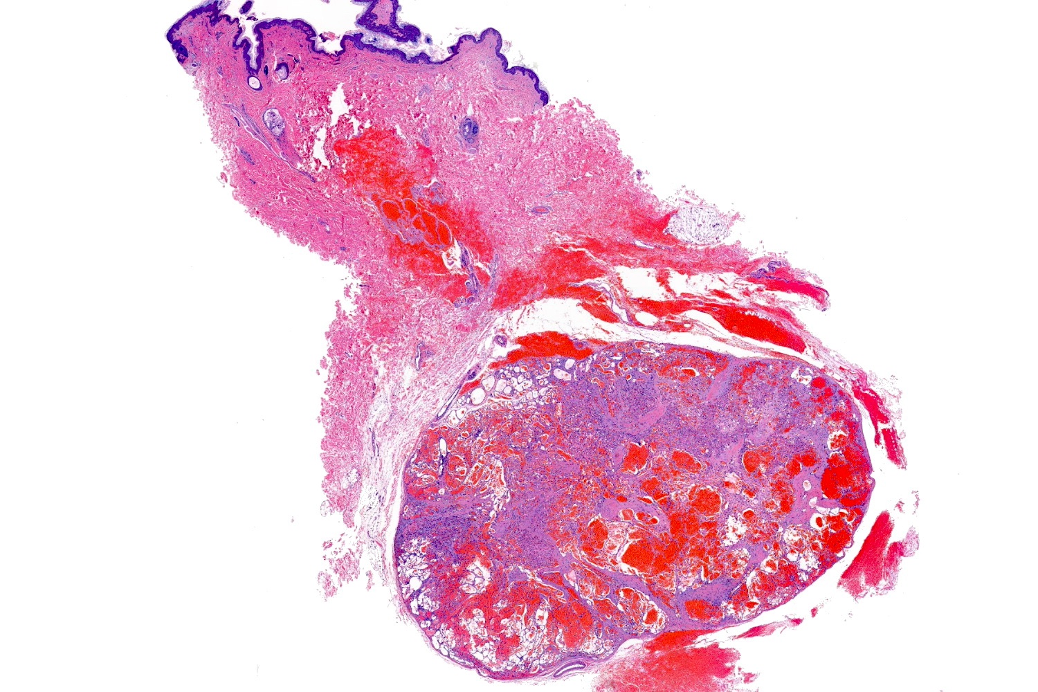
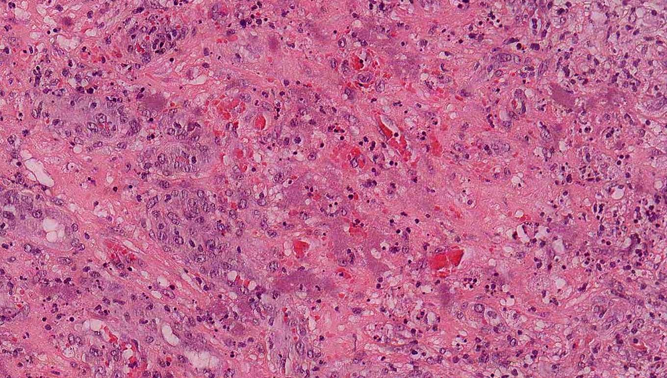
Fully developed lesion
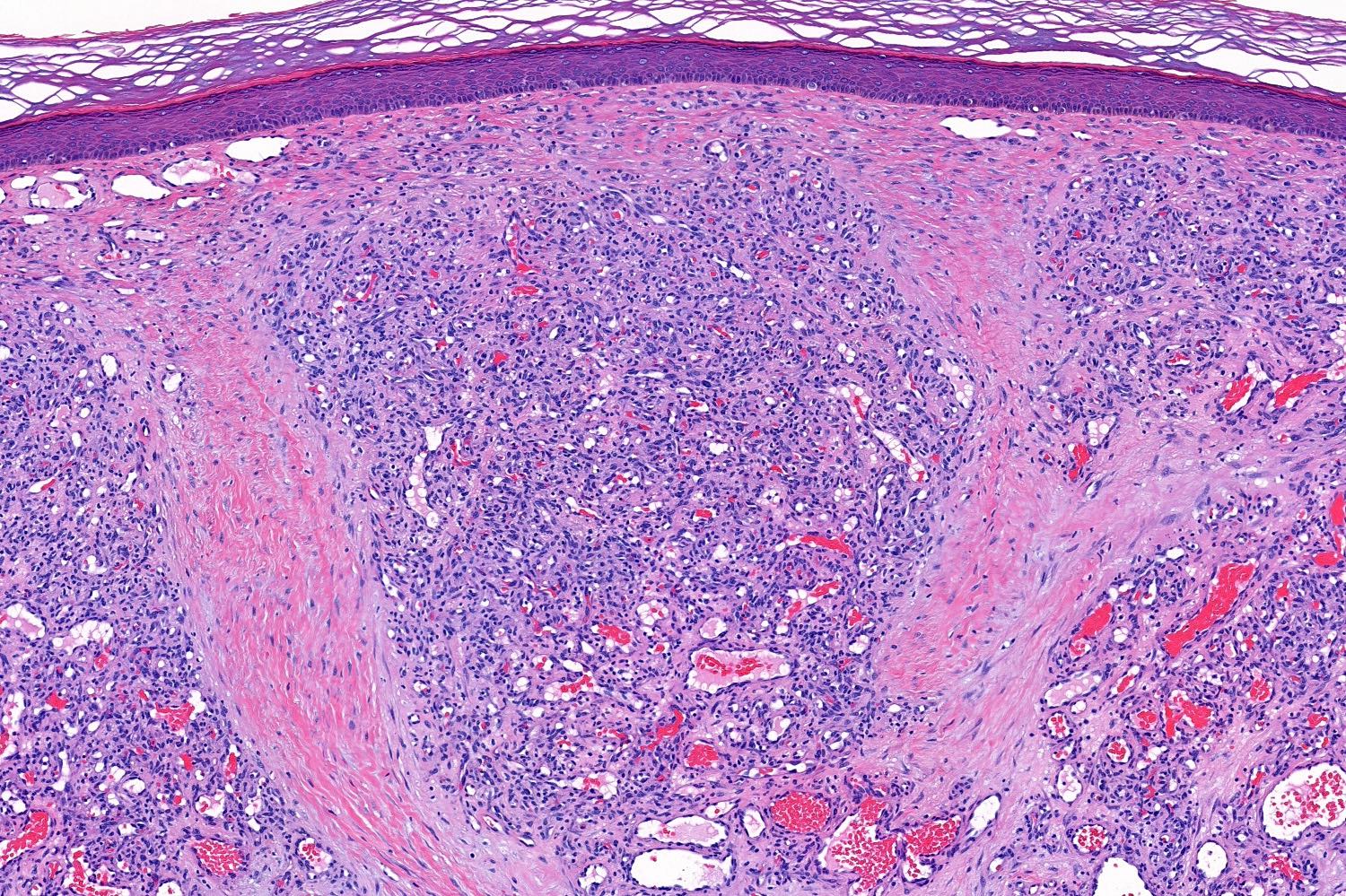
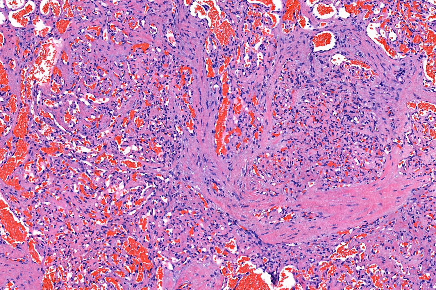
Lobule of capillaries
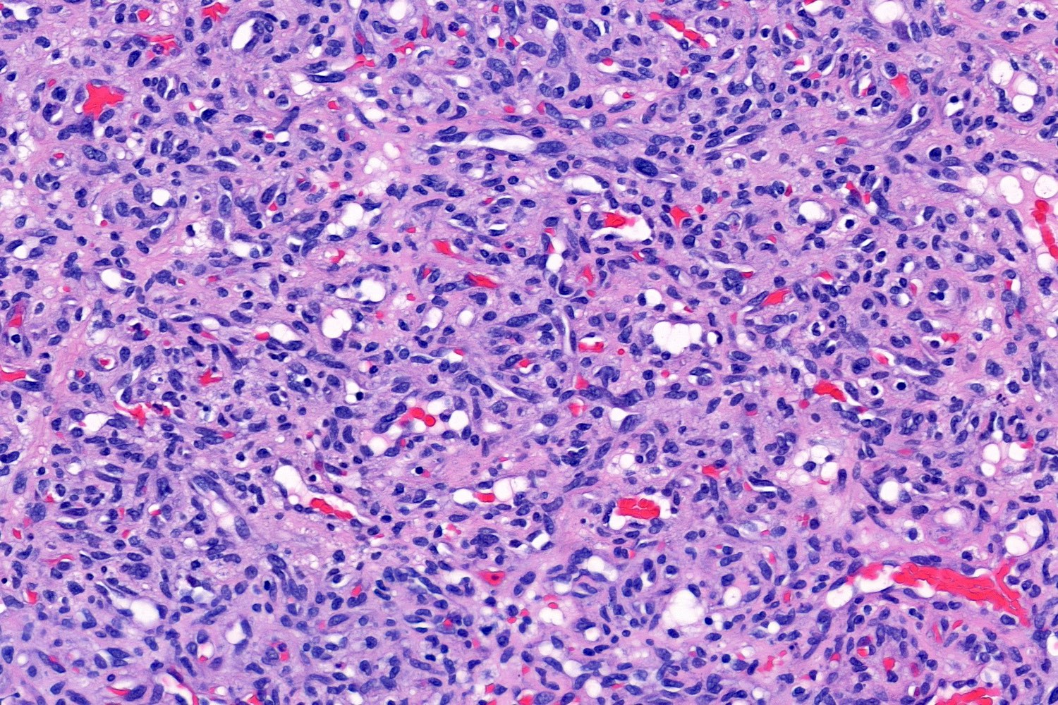
Capillary sized
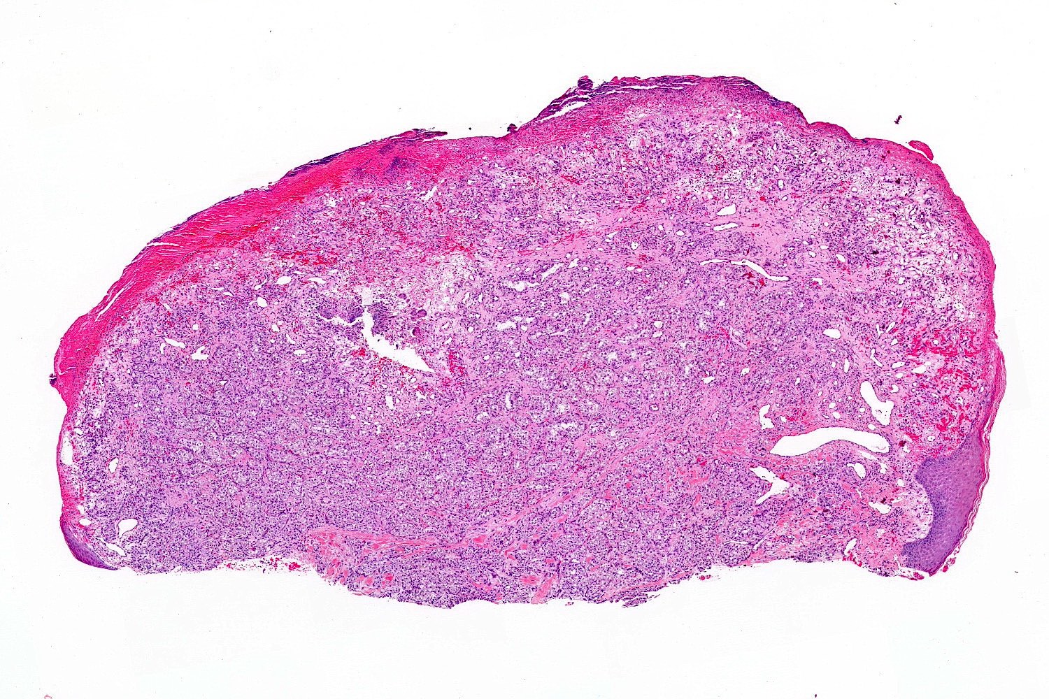
Oral mucosa, polypoid
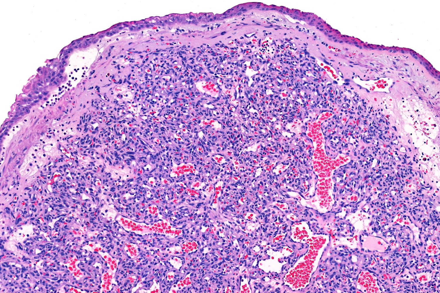
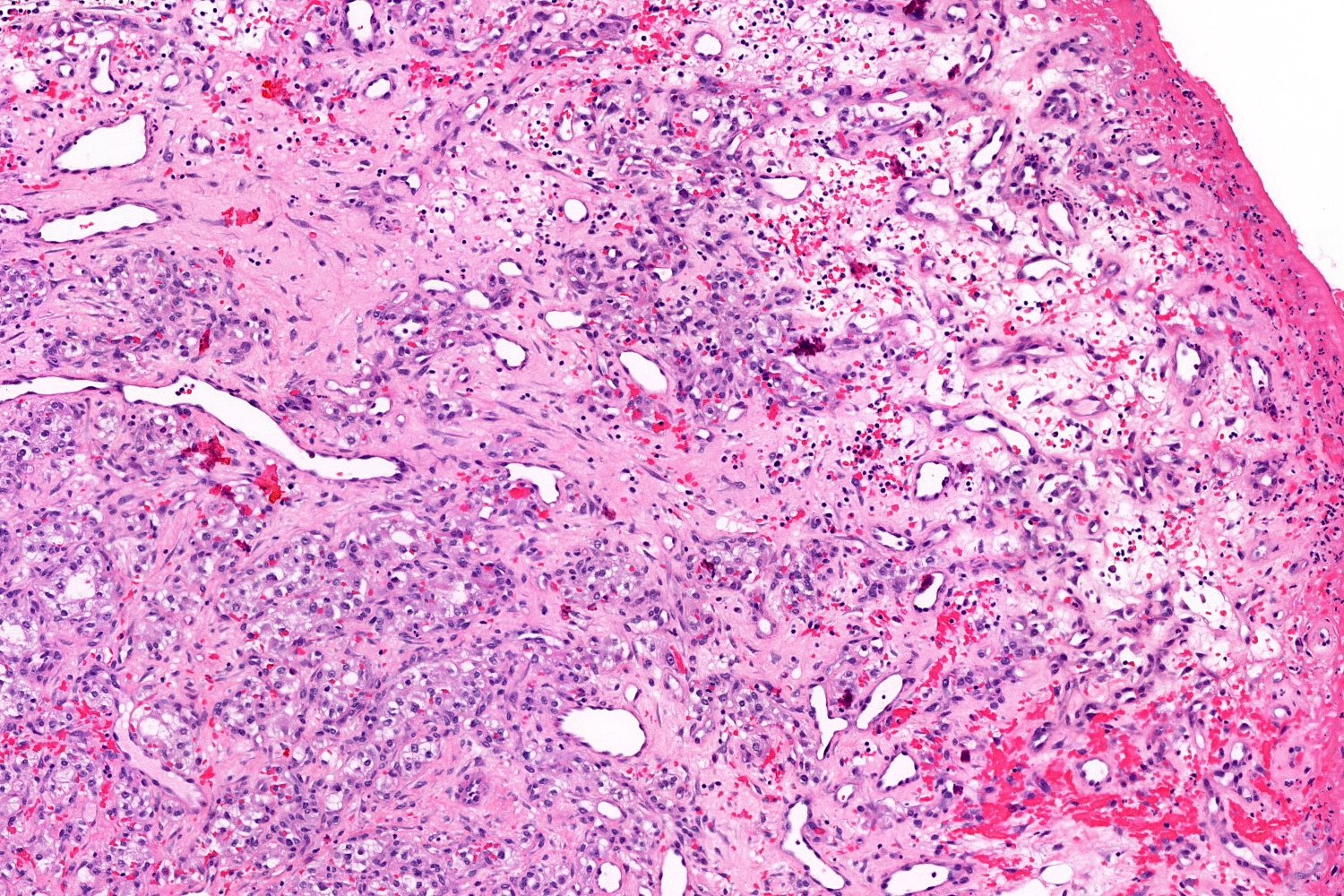
Capillary sized vessels
Prominent fibrosis
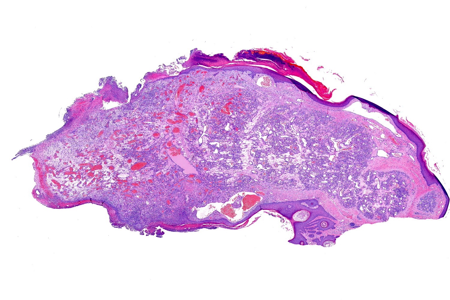
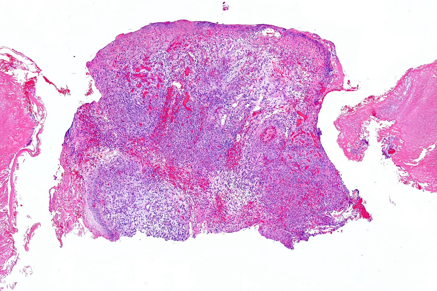
Large and ulcerated
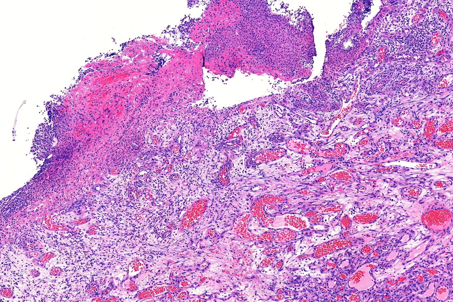
Ulcerated with granulation tissue
Intravascular example
Positive stains
- ERG
- CD31
- CD34
- FLI1
- Factor VIII
- SMA (stains pericytes) (Pathol Int 2003;53:1)
Negative stains
Videos
Pyogenic granuloma under the microscope
(lobular capillary hemangioma)
Sample pathology report
- Skin, shave biopsy:
- Lobular capillary hemangioma (pyogenic granuloma) (see comment)
- Comment: Microscopic examination reveals an exophytic lesion composed of lobules of numerous capillary sized blood vessels separated by fibrous septa consistent with a lobular capillary hemangioma.
Differential diagnosis
- Hemangioma (cherry angioma):
- Traumatized or ulcerated hemangiomas may resemble lobular capillary hemangioma
- Vessels are fewer in number and larger in caliber
- Granulation tissue:
- Irregularly distributed, larger dilated vessels within a loose, edematous stroma
- Secondary granulation may be observed superficially overlying a lobular capillary hemangioma
- Kaposi sarcoma (nodular stage):
- Pyogenic granuloma-like Kaposi sarcoma has been described (Dermatol Online J 2012;18:4)
- Displays areas with erythrocytes and hyaline globules between spindle cells
- Positive for D2-40 (podoplanin) and HHV8 (LNA)
- Bacillary angiomatosis:
- Characterized by basophilic granular aggregations representing the bacteria
- Presence of scattered neutrophils
- Endothelial cells appear more pale pink in color
- Metastatic renal cell carcinoma (cutaneous):
- Commonly located on the scalp
- Demonstrates clear cell differentiation
- Shows marked cytologic atypia
Additional references
Practice question #1
A 30 year old woman presents with a bleeding papule on the right hand for 2 weeks duration. A shave biopsy of the lesion is performed. Based on the histopathologic images above, what is the diagnosis?
- Bacillary angiomatosis
- Granulation tissue
- Lobular capillary hemangioma
- Pyogenic granuloma-like Kaposi sarcoma
- Ulcerated cherry angioma
Practice answer #1
C. Lobular capillary hemangioma. The base of the lesion shows lobules of capillary sized vessels of a lobular capillary hemangioma. Answer B is incorrect because the surface of the lobular capillary hemangioma is ulcerated and granulation tissue is present below the ulcer; however, toward the base of the specimen are capillary sized vessels of a lobular capillary hemangioma. Answer A is incorrect because basophilic aggregates of bacteria are not present. Answer E is incorrect because the capillary sized caliber vessels toward the base of the lesion are too small for a cherry hemangioma. Answer D is incorrect because spindled endothelial cells with intervening erythrocytes are not present.
Comment Here
Reference: Lobular capillary hemangioma
Comment Here
Reference: Lobular capillary hemangioma
Practice question #2
Which of the following is an important distinguishing feature of a lobular capillary hemangioma (pyogenic granuloma)?
- Capillary sized vessels
- Extravasated erythrocytes
- Hyaline globules
- Large caliber vessels
- Presence of basophilic granular material
Practice answer #2
A. Capillary sized vessels. The vessels of a lobular capillary hemangioma are small, numerous and typically in multiple lobules. Answer B is incorrect because many vascular proliferations have extravasated erythrocytes and therefore this is not a feature used to distinguish between various vascular entities. Answer C is incorrect because hyaline globules may be seen in other vascular lesions (such as Kaposi sarcoma) but are not characteristic of lobular capillary hemangiomas. Answer E is incorrect because basophilic granular material is seen in bacillary angiomatosis, which can mimic lobular capillary hemangioma both clinically and histopathologically. .
Comment Here
Reference: Lobular capillary hemangioma
Comment Here
Reference: Lobular capillary hemangioma
