Stomach
General
Anatomy & histology
Authors: Kelsey E. McHugh, M.D., Thomas P. Plesec, M.D.
Editorial Board Member: Raul S. Gonzalez, M.D.


Last author update: 2 February 2024
Last staff update: 6 February 2024
Copyright: 2003-2025, PathologyOutlines.com, Inc.
PubMed Search: Histology stomach
Table of Contents
Definition / general | Essential features | Anatomy | Physiology | Diagrams / tables | Clinical images | Gross description | Gross images | Microscopic (histologic) description | Microscopic (histologic) images | Virtual slides | Cytology description | Cytology images | Positive stains | Sample pathology report | Additional references | Practice question #1 | Practice answer #1 | Practice question #2 | Practice answer #2 | Practice question #3 | Practice answer #3Cite this page: McHugh KE, Plesec TP. Anatomy & histology. PathologyOutlines.com website. https://www.pathologyoutlines.com/topic/stomachnormalhistology.html. Accessed September 1st, 2025.
Definition / general
- A review of the normal constituents of the gastric wall, with a focus on the gastric mucosa, its compartments, its cell types and their cellular products
Essential features
- Anatomic regions: cardia, fundus, body, antrum / pylorus
- Layers: mucosa, submucosa, muscularis propria, subserosa, serosa
- Cell types: mucous cells, parietal cells, chief cells, endocrine cells
- Cardia and antrum: mucinous glands
- Body and fundus: oxyntic glands
Anatomy
- Normal volume is 1.5 liters, capacity is 3 liters, potentially more
- Fine mosaic-like pattern of mucosa because of punctuations by gastric pits or foveolae
- Longitudinal infoldings of mucosa and submucosa known as rugae are coarser proximally and when stomach is empty
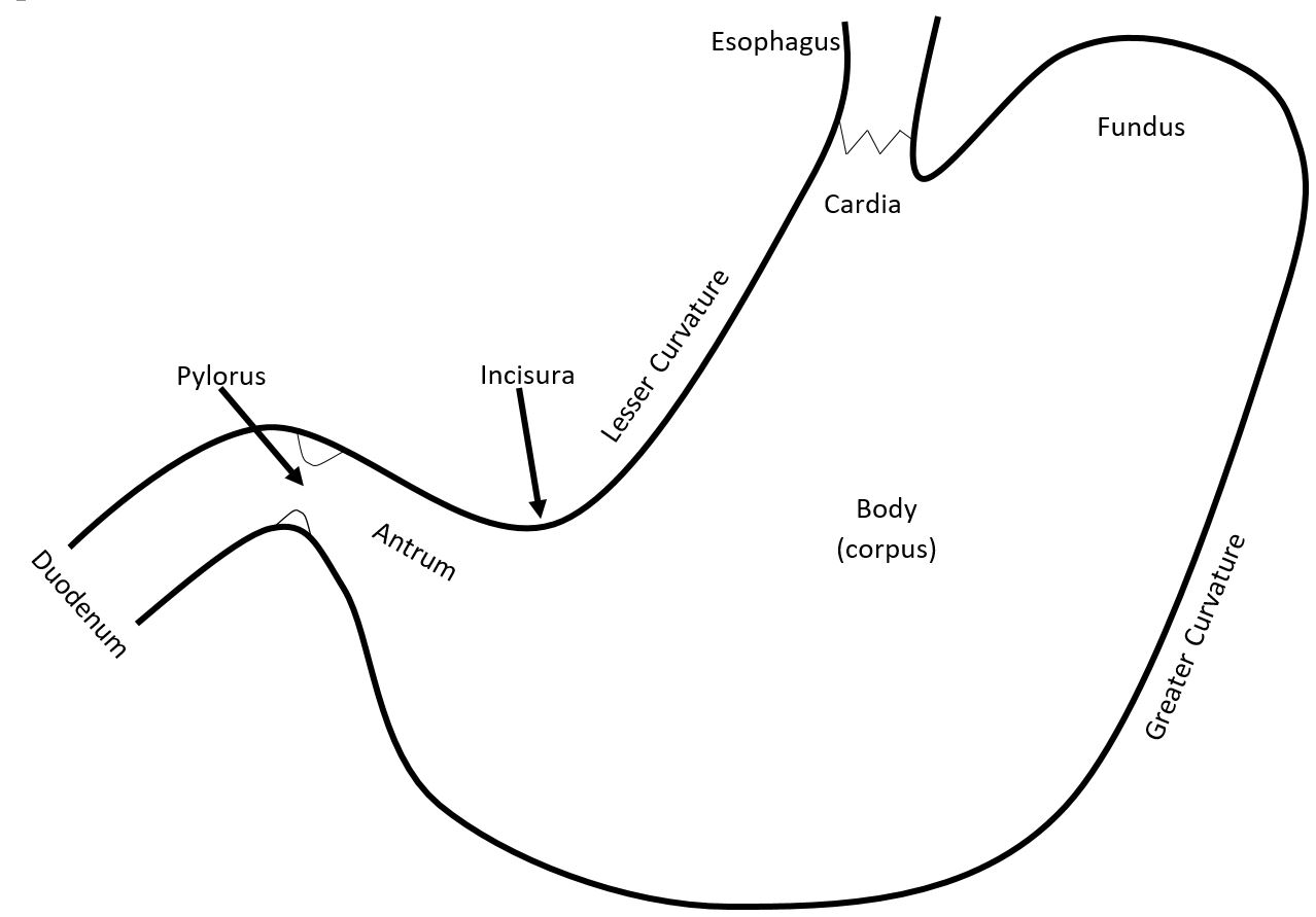
- Cardia
- Narrow conical portion distal to gastroesophageal junction
- Many authors claim that cardiac mucosa is reflux associated epithelia and not normally present
- Of note in the AJCC Cancer Staging Manual, 8th Edition (2017), carcinomas of the esophagus and the esophagogastric junction (tumor with epicenters ≤ 2 cm into cardia) are staged identically
- Fundus
- Dome shaped proximal stomach
- Body / corpus
- Remainder of stomach to incisura angularis
- Incisura angularis
- Where stomach narrows before it joins duodenum
- Antrum
- Incisura angularis to pyloric sphincter (3 - 4 cm)
- Pylorus
- Muscular ring that controls flow of food content into proximal duodenum
- Lesser curvature
- Medial curvature of stomach on the right
- Greater curvature
- Lateral curvature of stomach
Physiology
- General functions: digestion, motility, microbial defense (Statpearls: Physiology, Stomach [Accessed 28 May 2020])
- Digestive system organ that receives contents from the esophagus via the gastroesophageal sphincter and empties its contents into the duodenum via the pyloric sphincter
- Partially digests food boluses received from the esophagus
- Mechanical digestion: back and forth churning by inner oblique layer of muscularis propria
- Chemical digestion: acidic milieu breaks down proteins and kills food derived microbes
- Parietal cells secrete hydrochloric acid (HCl), which maintains acidic pH of stomach and denatures proteins
- Chief cells secrete pepsinogen, which breaks down proteins when activated to pepsin by the acidic environment
- Microenvironment of the stomach is largely regulated by enteroendocrine cell hormone secretion and vagus nerve innervation
- Empties chyme (partially digested food) into the duodenum
- Gastric emptying largely facilitated by the inner circular and outer longitudinal layers of muscularis propria
- Smooth muscle contractions (peristalsis) are controlled by the interstitial cells of Cajal (J Physiol 2006;571:1)
- Majority of nutrient absorption will occur in small bowel
- Gastric emptying largely facilitated by the inner circular and outer longitudinal layers of muscularis propria
- Partially digests food boluses received from the esophagus
Diagrams / tables
Contributed by Kelsey E. McHugh, M.D.
Anatomic regions
Layers of gastric wall
Gastric mucosa
Clinical images
Contributed by Kelsey E. McHugh, M.D.
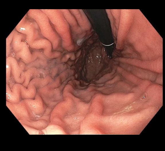
Gastric body with
unremarkable
rugal folds
Gross description
- A malleable gastric wall with pink, smooth, glistening serosa and pink-tan velvety mucosa arranged in coarse rugal folds which are more prominent in the body and fundus, whereas antral mucosa is more flat (Am J Surg Pathol 1986;10:48)
Gross images
Contributed by Leon Metlay, M.D. and Kelsey E. McHugh, M.D.
Anatomy:
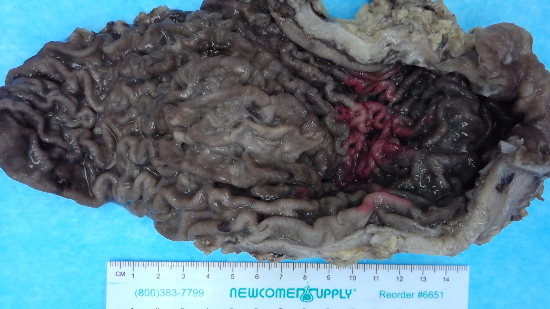
Formalin fixed gastric body
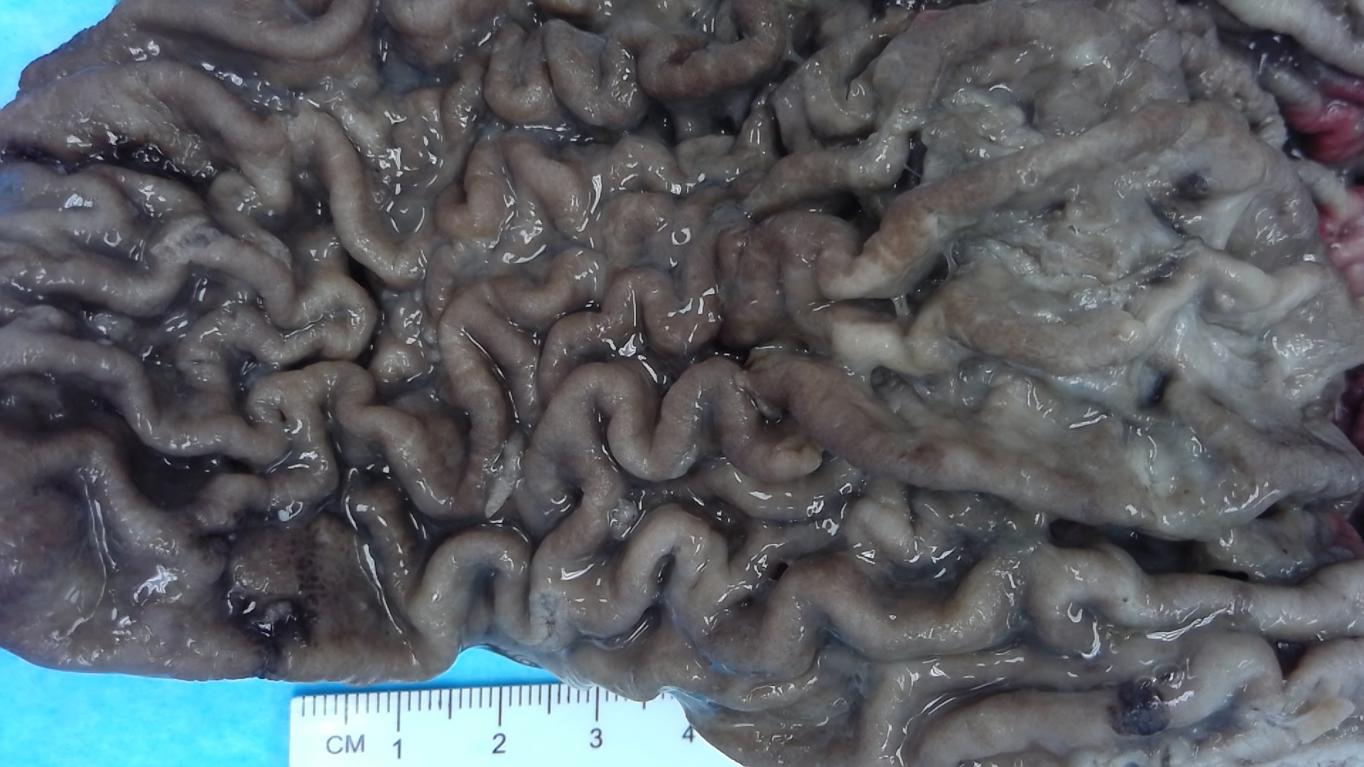
Formalin fixed gastric rugal folds
Images hosted on other servers:
Anatomy:

Normal appearance of the stomach

Normal appearance of the gastric antrum


Total gastrectomy anatomic regions
Microscopic (histologic) description
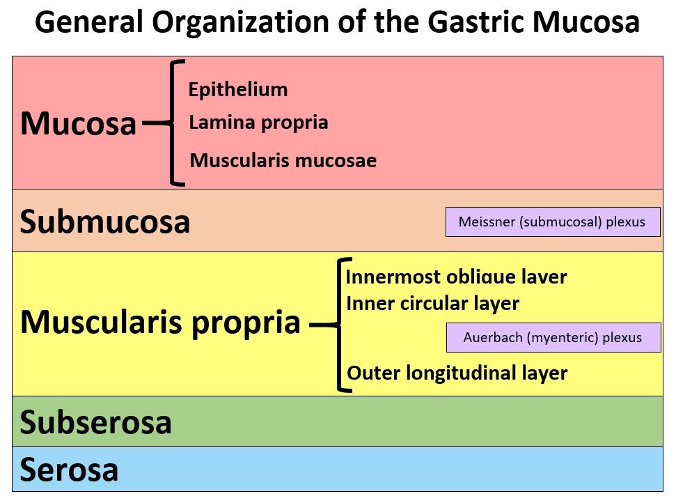
- Layers:
- Mucosa (with muscularis mucosae)
- Submucosa (with Meissner plexus)
- Muscularis propria (outer longitudinal layer, Auerbach / myenteric plexus, inner circular layer, innermost oblique layer)
- Subserosa
- Serosa (Am J Surg Pathol 1986;10:48)
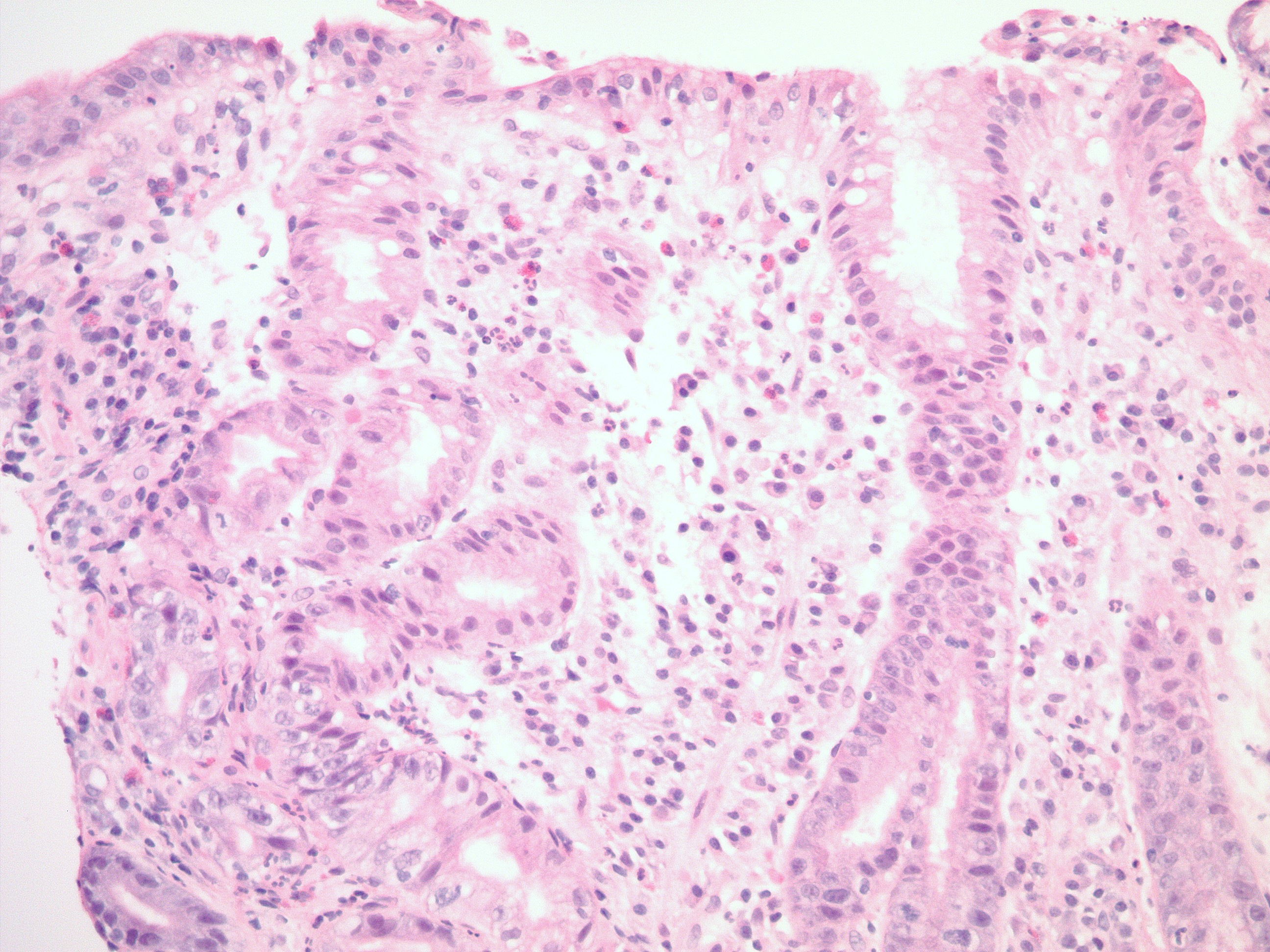
- Anatomic regions:
- Cardia: variable length extending from 1 - 15 mm (average 5 mm) (Am J Surg Pathol 2002;26:1207)
- Loosely packed mucous secreting glands (Am J Surg Pathol 2000;24:402)
- Ratio of pit to gland volume, 50:50
- Cystic dilatation of glands and mild chronic inflammation common
- May be expanded in individuals with acid reflux (Am J Surg Pathol 2000;24:344)
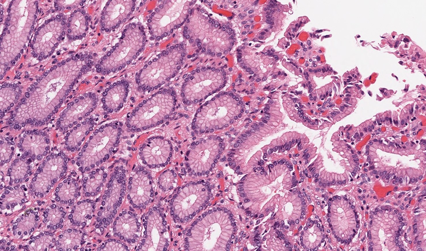
- Body (corpus) and fundus: known as oxyntic mucosa
- Tightly packed glands acid and enzyme secreting glands
- Ratio of pit to gland volume, 25:75
- Parietal cells and chief cells are the glandular constituents
- Can have interspersed mucin cells, especially in glandular isthmus / neck
- Corpus antrum boundary moves proximally with age due to reduction of oxyntic mucosal volume
- Antrum / pylorus: distal 3 - 4 cm
- Loosely packed mucous secreting glands
- Ratio of pit to gland volume, 50:50
- Usually no cystic dilatation of glands
- G cells are an endocrine cell unique to this anatomic region
- Proximal extent along the lesser curvature exceeds that along the greater curvature (Gastroenterology 1972;63:584)
- Cardia: variable length extending from 1 - 15 mm (average 5 mm) (Am J Surg Pathol 2002;26:1207)
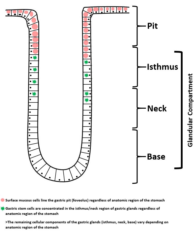
- Components of gastric mucosa:
- Gastric pits: surface invaginations that function as conduits of secretions; entirely lined by surface mucous (foveolar) cells regardless of anatomic region (Am J Surg Pathol 1986;10:48)
- Gastric glands: synthesize acids, enzymes and mucins; constituents and their products vary depending on anatomic region of the stomach
- Glands are organized into isthmus, neck and base (Am J Surg Pathol 1986;10:48)
- Gastric stem cells are housed in the isthmus and neck portion of glandular mucosa (Wiley Interdiscip Rev Dev Biol 2017;6:e261)
- Gastric cell types
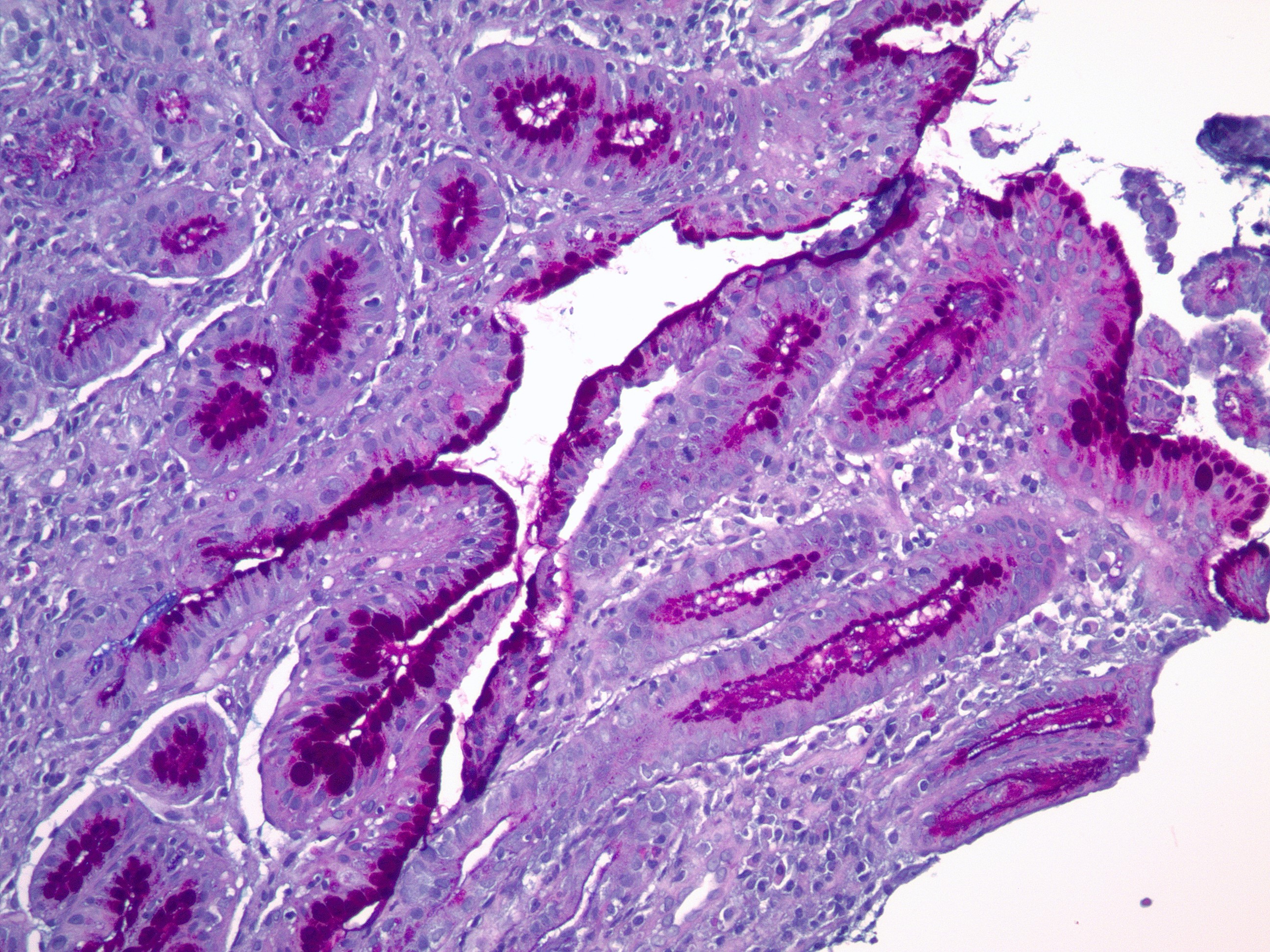
- Mucous cells
- Secrete bicarbonate rich mucus that coats and lubricates the gastric surface
- Serves protective function against autodigestion
- Surface (foveolar) epithelium contains cytoplasmic neutral mucins
- Lightly eosinophilic apical mucin cap
- Mucous glands contain cytoplasmic neutral and acidic mucin (Arch Pathol 1968;85:580)
- Lightly eosinophilic to clear bubbly / vacuolated cytoplasm
- Lack an apical mucin cap
- All mucous cells have round, basally oriented nuclei
- Secrete bicarbonate rich mucus that coats and lubricates the gastric surface
- Parietal cells
- Produce hydrochloric acid via H+ / K+ -ATPase pump (Varela: Histology, Parietal Cells, 2019)
- Hydrochloric acid maintains gastric acidity (pH 1.5 to 3.5), which:
- Activates pepsinogen to active pepsin enzyme
- Kills food derived bacteria
- Facilitates food digestion
- Promotes absorption of minerals (e.g. phosphate, calcium, iron)
- Hydrochloric acid maintains gastric acidity (pH 1.5 to 3.5), which:
- Produce intrinsic factor, which is critical for vitamin B12 absorption in small bowel (Varela: Histology, Parietal Cells, 2019)
- Secretion highly regulated by extrinsic and intrinsic neuroendocrine system (Physiol Rev 2020;100:573)
- Stimulators of gastric acid secretion include:
- Vagus nerve (via acetylcholine neurotransmitter)
- Gastrin hormone
- Hypergastrinemia can be induced by glucocorticoids
- Histamine
- Grehlin
- Inhibitors of gastric acid secretion include:
- Somatostatin
- Pharmacologic agents (e.g. proton pump inhibitors)
- Stimulators of gastric acid secretion include:
- Relatively large, triangular cells with eosinophilic cytoplasm due to abundant mitochondria
- Centrally placed nuclei with evenly distributed chromatin
- Predominantly occupy the superficial half of body / fundic glands
- Produce hydrochloric acid via H+ / K+ -ATPase pump (Varela: Histology, Parietal Cells, 2019)
- Chief cells
- Produce pepsinogen I and II propeptides (Gastroenterology 1992;102:699)
- Activated to pepsin enzyme via high acidity (low pH) environment
- Cuboidal cells with basophilic to amphophilic cytoplasm due to abundant rough endoplasmic reticulum
- Predominantly occupy the basal half of corpus glands
- Produce pepsinogen I and II propeptides (Gastroenterology 1992;102:699)
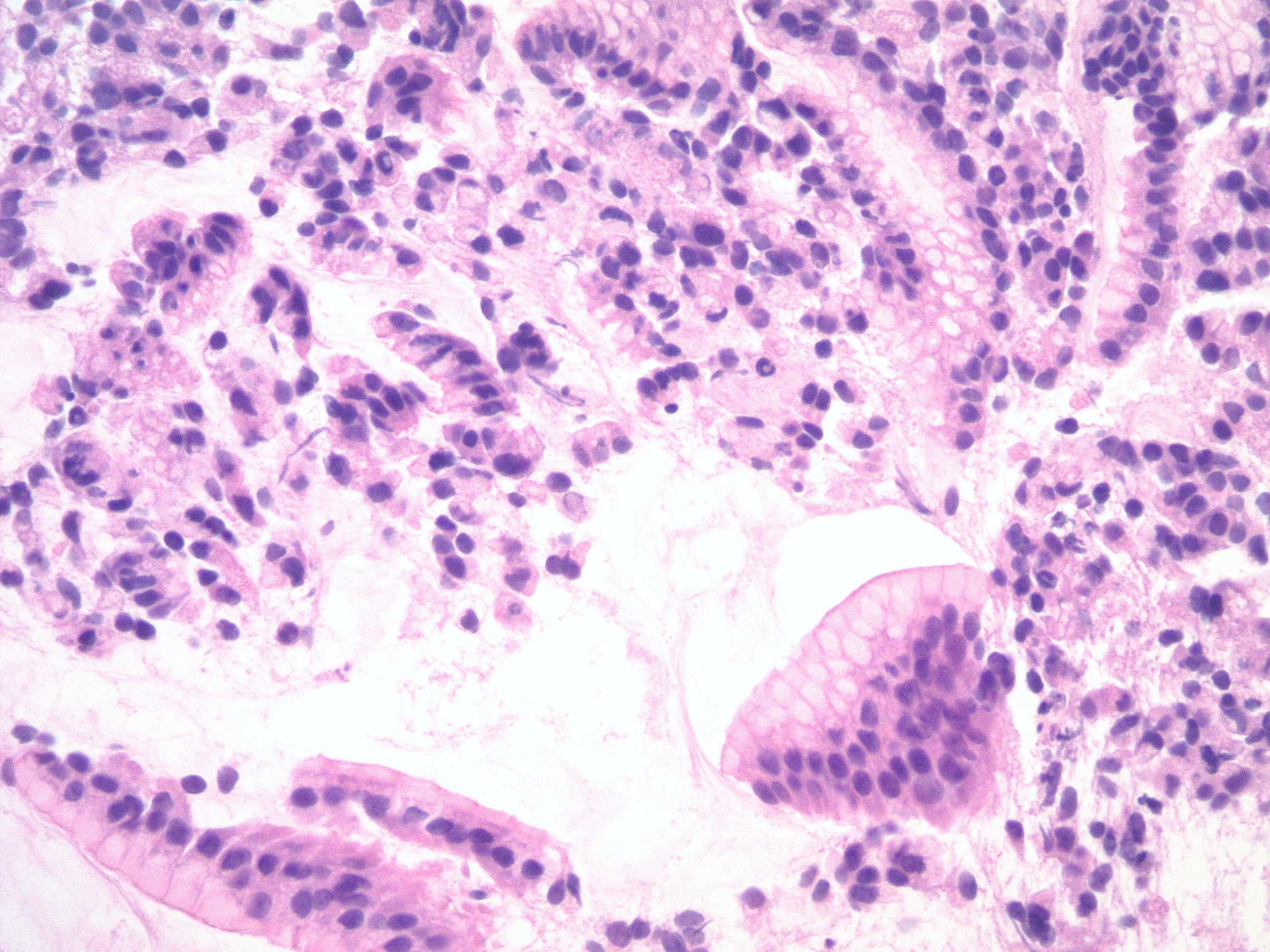
- Endocrine cells
- Generally inconspicuous, round cells with clear cytoplasm
- Typically fewer than 20 endocrine cells per gland
- Cell types include: (Regul Pept 2000;93:31)
- G cells: produce gastrin
- Limited to the gastric antrum
- D cells: produce somatostatin
- Distributed throughout whole stomach
- Enterochromaffin (EC) cells: produce serotonin
- Distributed throughout whole stomach
- Enterochromaffin-like (ECL) cells: produce histamine (Int J Mol Sci 2019;20:E2444)
- Limited to the gastric body and fundus
- Secrete histamine in response to gastrin produced by G cells
- Represent 30% of all endocrine cells
- Long term gastrin stimulation causes enterochromaffin-like cell hyperplasia (e.g. in atrophic gastritis)
- X cells: produce grehlin (Int J Pept 2010;2010:945056)
- Limited to the gastric body and fundus
- G cells: produce gastrin
- Concentrated within mucous neck region in antrum / pylorus
- 50% G cells, 30% enterochromaffin cells, 15% D cells, 5% other
- Scattered throughout oxyntic glands in body and fundus
- Majority enterochromaffin-like cells; minority enterochromaffin cells, X cells, D cells
- Mucous cells
- Gastric mucosa - general:
- There are no visibly (grossly or microscopically) distinct boundaries between mucosal zones / anatomic regions (Am J Surg Pathol 1986;10:48)
- Gastric mucosa is very metabolically active
- Gastric surface epithelium is normally replaced every 4 - 8 days (Gastroenterology 1977;72:962)
- Gastric parietal and chief cells turn over more slowly: every 1 - 3 years (J Natl Cancer Inst 1969;42:9)
- Undifferentiated stem cells are concentrated in the isthmus / neck of gastric glands throughout the entire stomach
- Lamina propria provides structural support (Am J Surg Pathol 1986;10:48)
- Structural components: reticulin, collagen and elastin fibers; capillaries and arterioles; nerve fibers; few smooth muscle fibers
- Cell types: fibroblasts, histiocytes, plasma cells, lymphocytes; occasional mast cells and eosinophils
- Lymphoid tissue is sparse in lamina propria (Hum Pathol 1993;24:577)
- Superficial lamina propria with small numbers of lymphocytes (B cells > T cells) and plasma cells
- Can see small lymphoid aggregates (no germinal centers) in gastric antrum immediately superficial to muscularis mucosae
- Intraepithelial lymphocytes absent or sparse
- Lymphatic channels are present within gastric mucosa, generally immediately superficial to muscularis mucosae
- Gastric mucosa protects itself against autodigestion / high acidity (Physiol Rev 1993;73:823)
- Mucus secretion: mucus is relatively impermeable to H+; also fluid with acid or pepsin exits gastric glands as jets and penetrates surface mucus layer without contacting surface epithelial cells (Am J Physiol Gastrointest Liver Physiol 2017;313:G599)
- Bicarbonate secretion creates pH neutral microenvironment adjacent to cell surface
- Intercellular tight junctions prevent back diffusion of H+ and disruptions are quickly repaired
- Rich blood flow supplies bicarbonate and nutrients and removes acid
- Muscularis mucosae limits injury; if intact, mucosal repair occurs in hours / days versus weeks if not intact
Microscopic (histologic) images
Contributed by Kelsey E. McHugh, M.D.
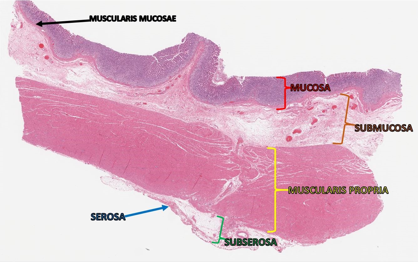
Gastric wall layers
Oxyntic mucosa
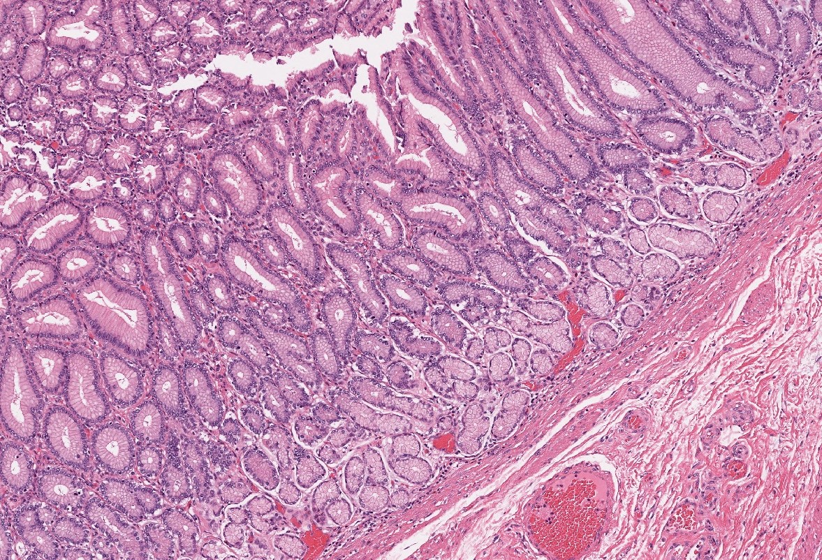
Antral mucosa
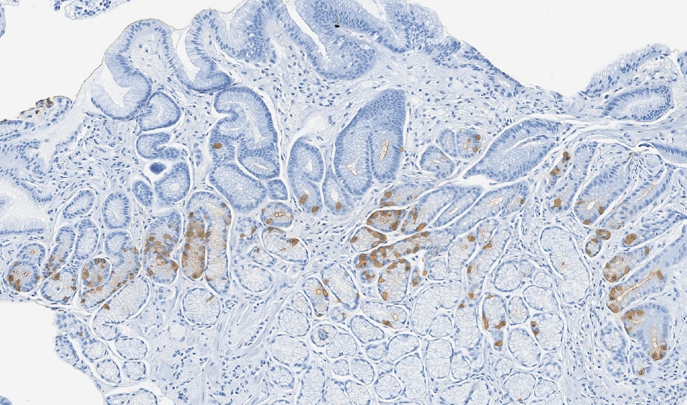
Gastrin immunostain
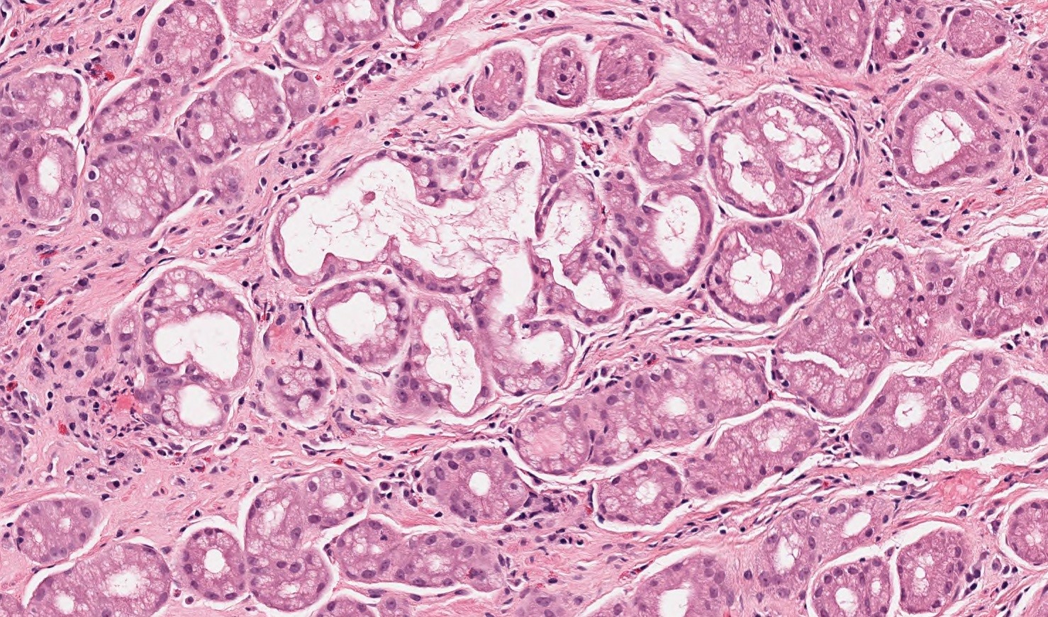
Cardia mucosa
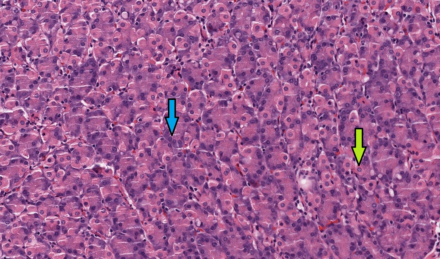
Parietal and chief cells
Gastric pits
Pseudo-signet ring cell artifact
Distended foveolar cells
PAS / AB in distended foveolar cells
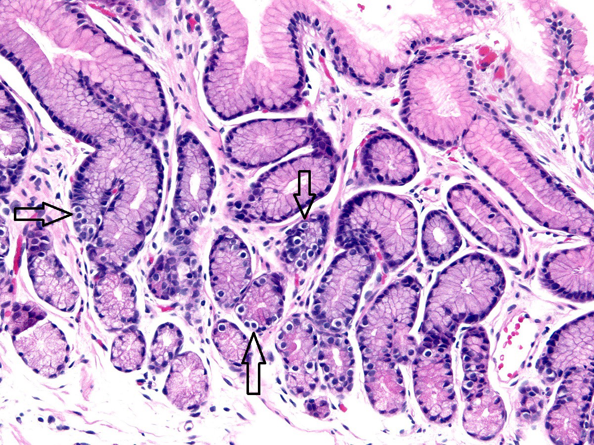
Neuroendocrine cells in gastric antrum
Virtual slides
Images hosted on other servers:

Unremarkable gastric body
Cytology description
- Mucous cells: tightly cohesive columnar cells with basal round to ovoid nuclei in orderly, honeycombed sheets
- Usually the predominant cell type
- Chief cells: small cuboidal cells with round, smooth nuclei, fine nuclear chromatin and basophilic cytoplasmic granules in honeycombed sheets
- Comparable in appearance to pancreatic or salivary acinar cells
- Parietal cells: pyramidal to flask shaped cells with round nuclei, coarse nuclear chromatin and abundant intensely eosinophilic cytoplasm
- Larger than chief cells
Cytology images
Contributed by Kelsey E. McHugh, M.D.
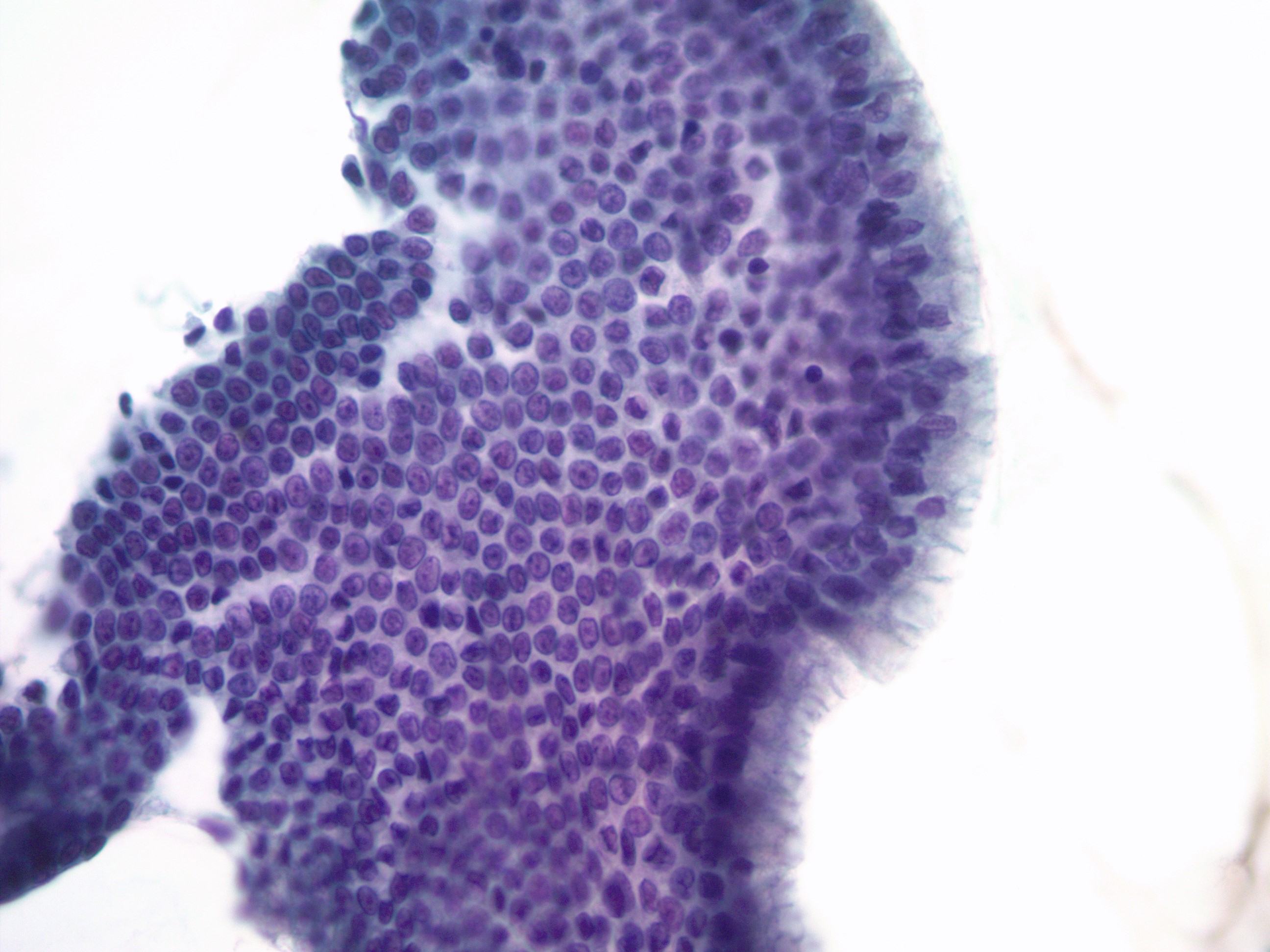
Surface foveolar epithelium
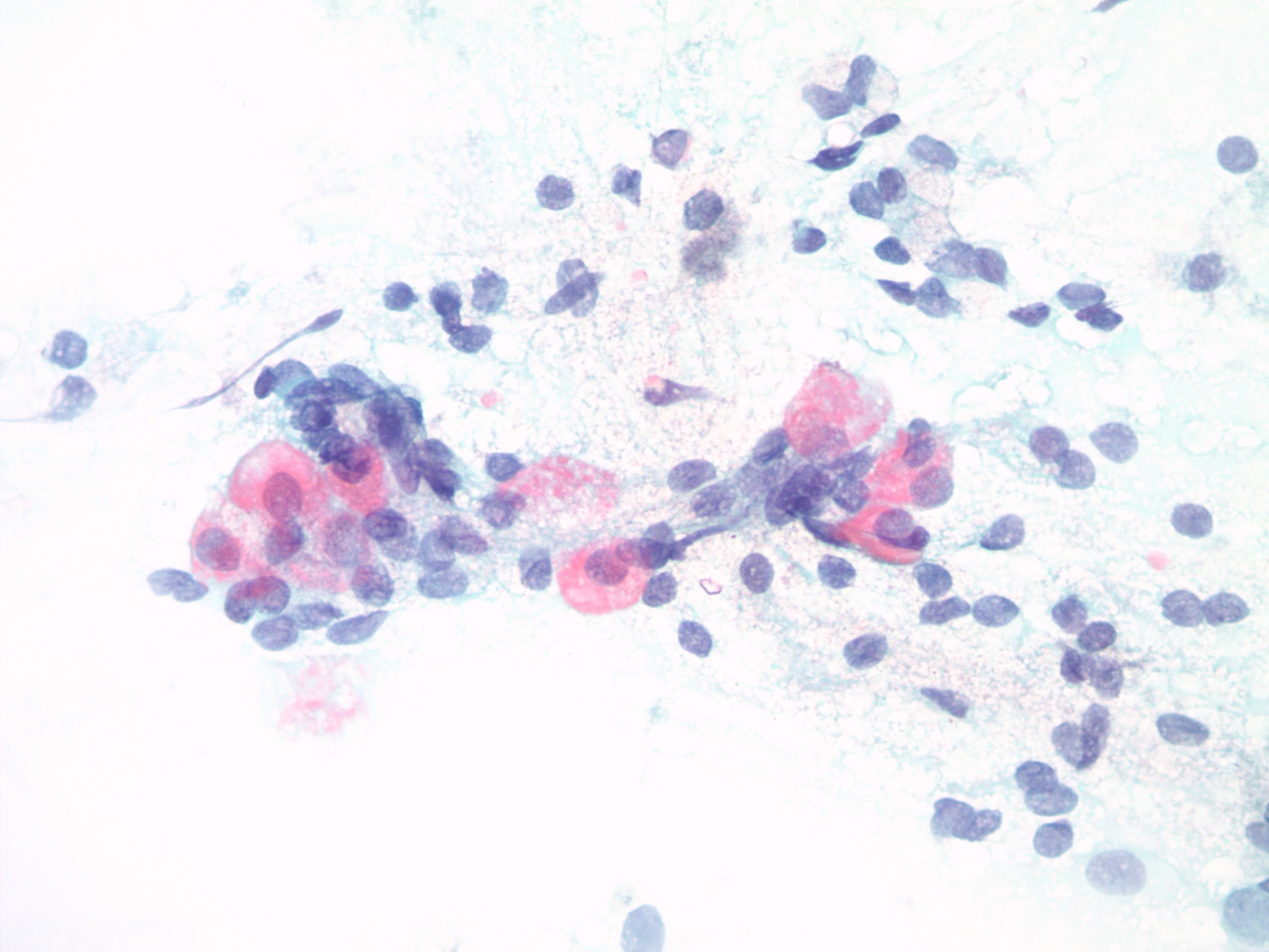
Parietal cells
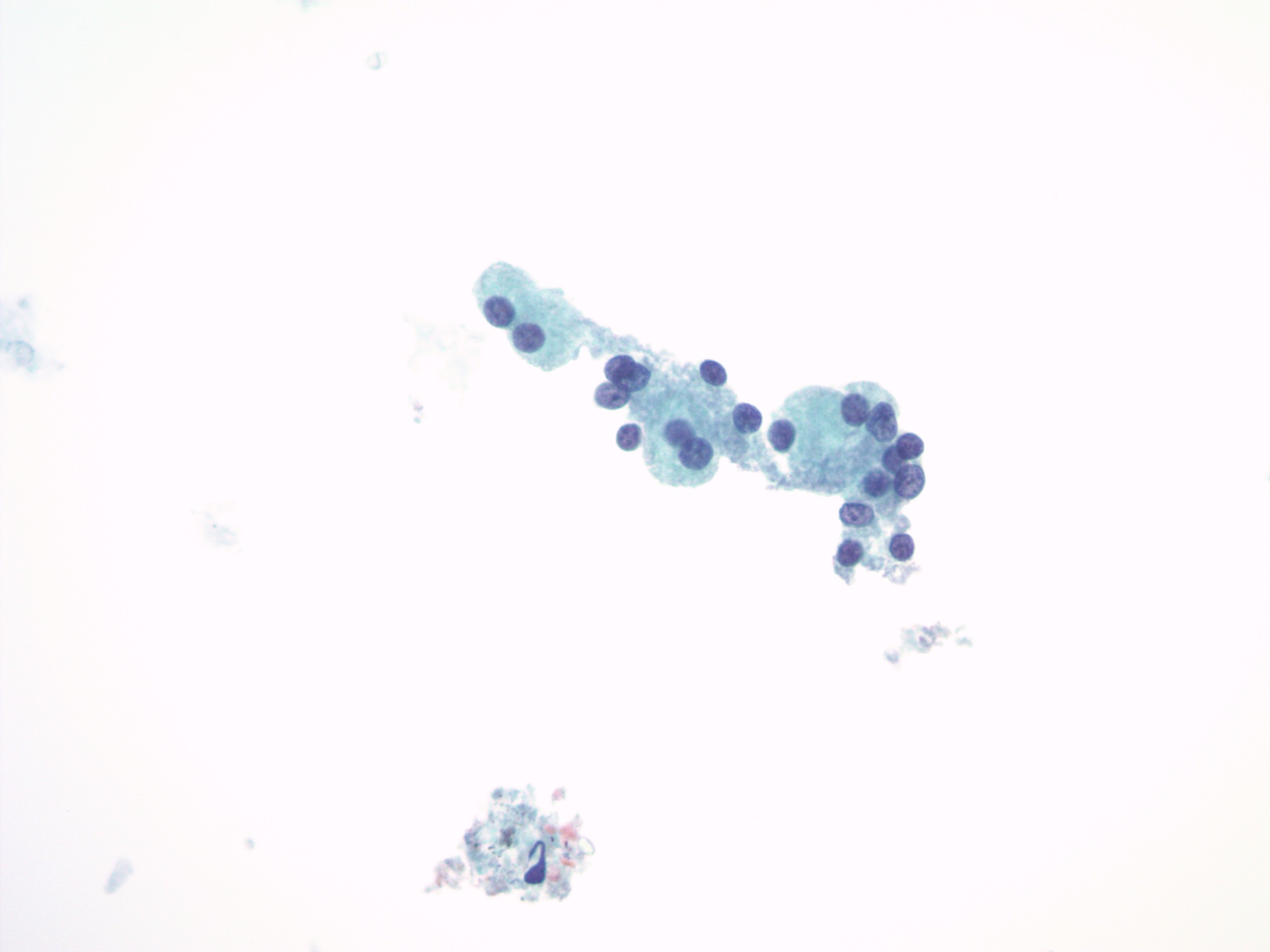
Chief cells
Positive stains
- Mucous cells: PAS (Indian J Med Paediatr Oncol 2013;34:229)
- Endocrine cells / enterochromaffin-like cells: chromogranin, synaptophysin, CD56, INSM1 (Am J Clin Pathol 2020;153:811)
- G cells: gastrin (J Cancer Res Clin Oncol 2006;132:85)
Sample pathology report
- Stomach, cardia, biopsy:
- Gastric cardia mucosa with no significant diagnostic alteration
- No evidence of Helicobacter pylori microorganisms
- Stomach, body / fundus, biopsy:
- Gastric oxyntic mucosa with no significant diagnostic alteration
- No evidence of Helicobacter pylori microorganisms
- Stomach, antrum, biopsy:
- Gastric antral mucosa with no significant diagnostic alteration
- No evidence of Helicobacter pylori microorganisms
Additional references
Practice question #1
- Which gastric cell type, pictured above, produces intrinsic factor, an integral glycoprotein in the luminal absorption of vitamin B12?
- Chief cells
- Endocrine cells
- Enterochromaffin-like cells
- Mucous cells
- Parietal cells
Practice answer #1
Practice question #2
- The Meissner plexus is located within which layer of the stomach?
- Mucosa
- Muscularis propria
- Serosa
- Submucosa
- Subserosa
Practice answer #2
Practice question #3
- Which of the following ratios most accurately described the gastric pit to gastric gland ratio within the corpus (body / fundus) of the stomach?
- 10:90
- 25:75
- 50:50
- 75:25
- 90:10
Practice answer #3
