
Breast
Other benign tumors
Hemangioma
Authors: Indu Agarwal, M.D., Luis Blanco, Jr., M.D.
Editorial Board Member: Julie M. Jorns, M.D.
Deputy Editor-in-Chief: Gary Tozbikian, M.D.


Last author update: 9 August 2022
Last staff update: 9 August 2022
Copyright: 2003-2024, PathologyOutlines.com, Inc.
PubMed Search: Hemangioma breast


Table of Contents
Definition / general | Essential features | Terminology | ICD coding | Epidemiology | Sites | Pathophysiology | Etiology | Diagrams / tables | Clinical features | Diagnosis | Radiology description | Radiology images | Prognostic factors | Case reports | Treatment | Clinical images | Gross description | Gross images | Microscopic (histologic) description | Microscopic (histologic) images | Positive stains | Negative stains | Videos | Sample pathology report | Differential diagnosis | Additional references | Board review style question #1 | Board review style answer #1 | Board review style question #2 | Board review style answer #2Cite this page: Agarwal I, Blanco L. Hemangioma. PathologyOutlines.com website. https://www.pathologyoutlines.com/topic/breasthemangioma.html. Accessed May 14th, 2024.
Definition / general
- Hemangioma:
- Benign vascular lesions, microscopically well circumscribed and lacking endothelial cell atypia (Semin Diagn Pathol 2017;34:410)
- Origination from large nonneoplastic "feeding" vessels, may be seen at the periphery of the lesion
- The following categories are recognized:
- Capillary (composed of compact, lobular, collections of small blood vessels)
- Cavernous (dilated vessels filled with erythrocytes)
- Venous (featuring vascular structures containing muscular walls of varying thickness) (Am J Surg Pathol 1985;9:659)
- Perilobular (≤ 2 mm, capillary hemangiomas, intra or interlobular location)
- Angiomatosis:
- Often clinically present as large mass (9 - 22 cm)
- Histologically benign vascular proliferation affecting a large segment of the breast
- Lacks circumscription of hemangioma and can involve the subcutaneous tissue and skin (Am J Surg Pathol 1985;9:652, Cancer 1988;62:2392)
Essential features
- Proliferation of well differentiated vessels of varying sizes
- Angiomatosis shows diffuse growth of blood vessels of varying sizes that surrounds ducts / lobules but without invasion of the intralobular stroma
- No complex anastomotic channels, cytologic atypia, cellular multilayering, solid areas, mitotic activity, necrosis or hemorrhage (blood lakes), features differentiating from angiosarcoma
Terminology
- Acceptable: angioma
ICD coding
- ICD-O: 9120/0 - hemangioma, NOS
- ICD-11: 2F30.Y & XH5AW4 - other specified benign neoplasm of breast and hemangioma, NOS
Epidemiology
- Small incidental hemangiomas of the breast are not uncommon (Eur J Surg 1992;158:503, J Clin Ultrasound 2012;40:512)
- Angiomatosis of the breast is very rare, most in younger women (< 40) years old
- In general, benign vascular tumors of breast are uncommon
- Reported in all ages, range 18 months to 82 years (J Clin Ultrasound 2012;40:512)
- Most common type: perilobular hemangioma, typically incidental and present in 1.2 - 11% of breast specimens (Arch Pathol Lab Med 1983;107:308)
Sites
- Mammary parenchyma, subcutaneous tissue or dermis
Pathophysiology
- Unknown
Etiology
- Nonneoplastic vascular malformations
Diagrams / tables
Images hosted on other servers:

Normal arterial and venous anatomy of the breast
Clinical features
- All age groups, mostly nonpalpable, found on imaging
- Occasionally a palpable lesion (Br J Radiol 2006;79:e177)
Diagnosis
- Biopsy or excision
Radiology description
- Mostly nonpalpable, found on imaging (MRI or mammography)
- No pathognomonic features, may show mammographic density or mass on mammogram or ultrasound, with oval or lobular shape and well circumscribed or microlobulated margins (AJR Am J Roentgenol 2008;191:W17)
Radiology images
Contributed by Indu Agarwal, M.D.
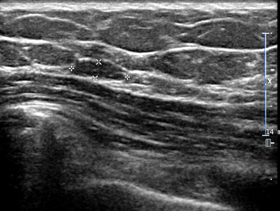
Cavernous hemangioma, ultrasound
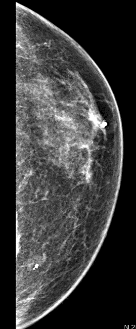
Cavernous hemangioma, mammography
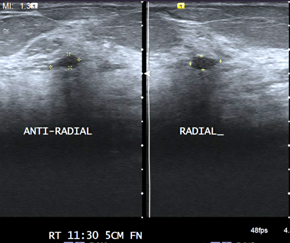
Capillary hemangioma, ultrasound
Prognostic factors
- Can recur; no metastasis reported
Case reports
- 18 year old woman with breast angiomatosis (J Clin Diagn Res 2012;6:709)
- 33 year old woman with breast hemangioma (J Clin Imaging Sci 2012;2:53)
- 43 year old woman, gravida 4, para 2, with cavernous breast hemangioma mimicking an invasive lesion on contrast enhanced MRI (Case Rep Surg 2019;2019:2327892)
- 60 year old woman with breast hemangioma mimicking carcinoma (Breast 2002;11:357)
- 64 year old man with hemangioma after thorn injury (BJR Case Rep 2021;7:20200187)
Treatment
- Excision has been done in the past; excision may not be mandatory if radiological / pathologic correlation is available (Histopathology 2017;71:795)
- Complete surgical excision necessary for angiomatosis (Radiol Case Rep 2017;12:219)
Clinical images
Contributed by Mark R. Wick, M.D.
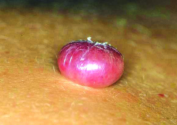
Capillary hemangioma, breast skin
Images hosted on other servers:

Diffuse dermal angiomatosis
Gross description
- Larger lesions: circumscribed red or dark-brown spongy lesion
- Small lesions may not be appreciated grossly
- Angiomatosis presents as a large, red, hemorrhagic, cystic, spongy mass lesion
Gross images
AFIP images
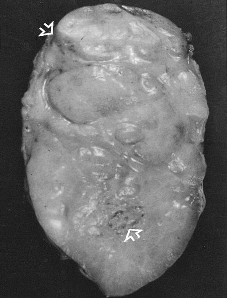
Angiomatosis composed of vessels / breast parenchyma
Images hosted on other servers:

Soft tissue mass with smooth external surface

Cut surface shows congestion and slit-like areas
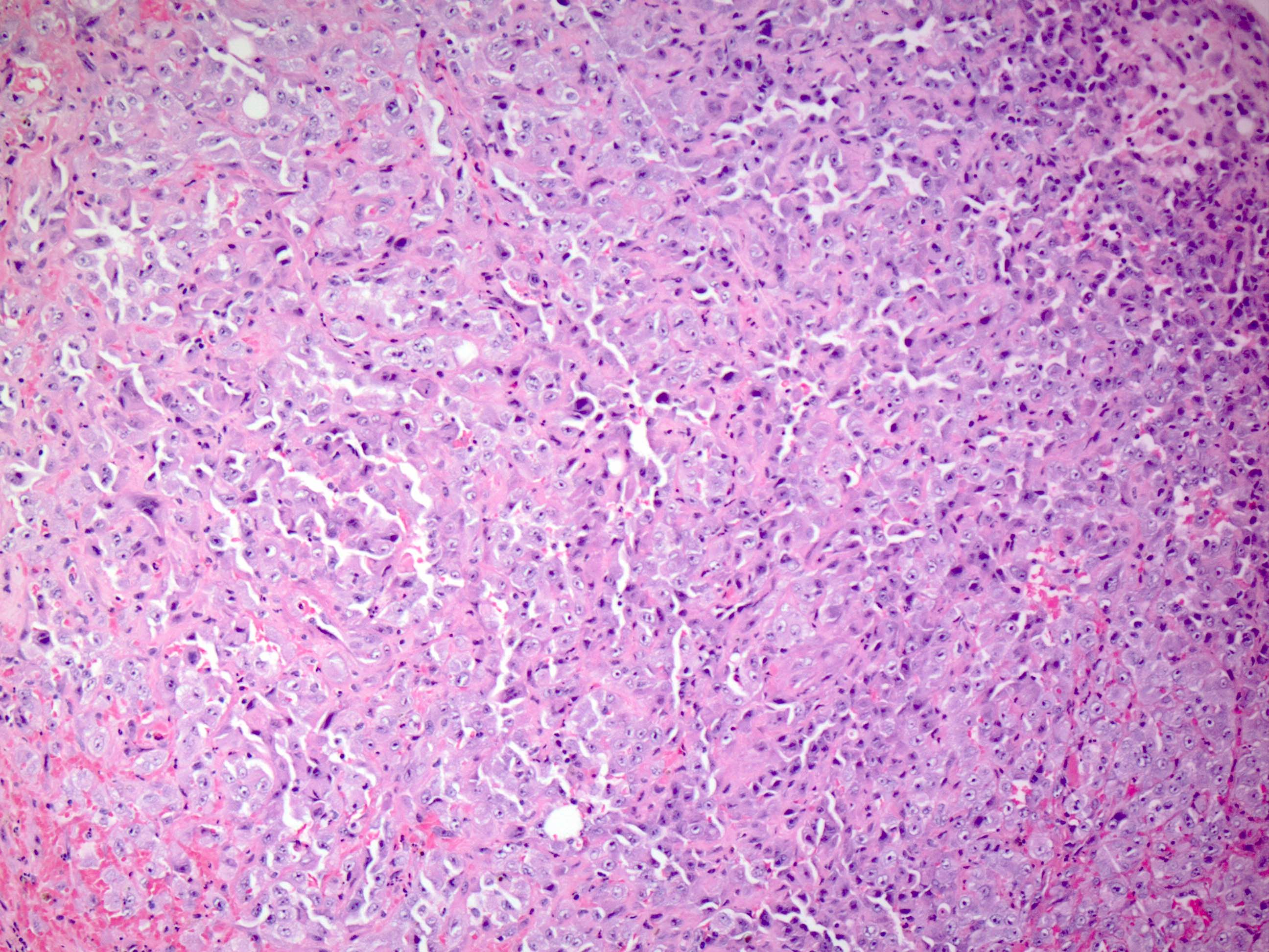
Microscopic (histologic) description
- Hemangioma:
- Proliferation of well differentiated blood vessels of varying sizes, generally small (< 2 cm), usually nonanastomosing
- Largely well circumscribed
- Cytologic atypia, hemorrhage, mitotic activity and necrosis are absent
- Within interlobular stroma, except in perilobular hemangioma which involves intralobular stroma
- Perilobular hemangioma: usually incidental microscopic lesions, ≤ 2 mm, conglomerate of small, thin walled capillary vessels in and around a lobule (Am J Surg Pathol 1985;9:491)
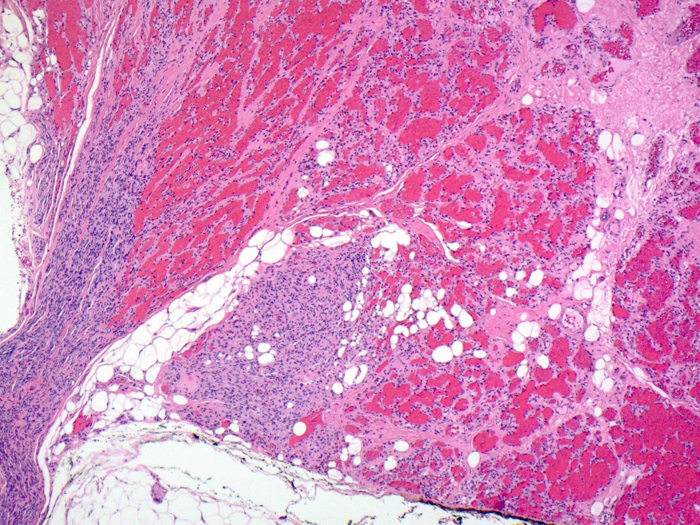
- Angiomatosis:
- Proliferation of vascular channels with wide variation in caliber, diffusely growing throughout the breast with some anastomosis
- Unencapsulated with infiltrative borders
- No invasion of intralobular stroma
- May extend to overlying skin (Clin Breast Cancer 2016;16:e7)
- Absence of destructive invasion, solid areas, hemorrhage and necrosis, differentiating it from angiosarcoma
- Low mitotic rate, Ki67 proliferation index < 2%
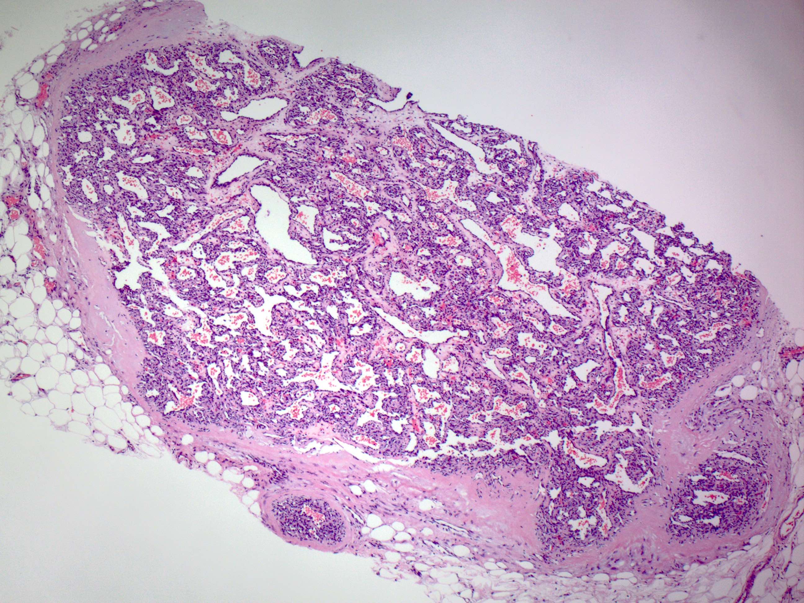
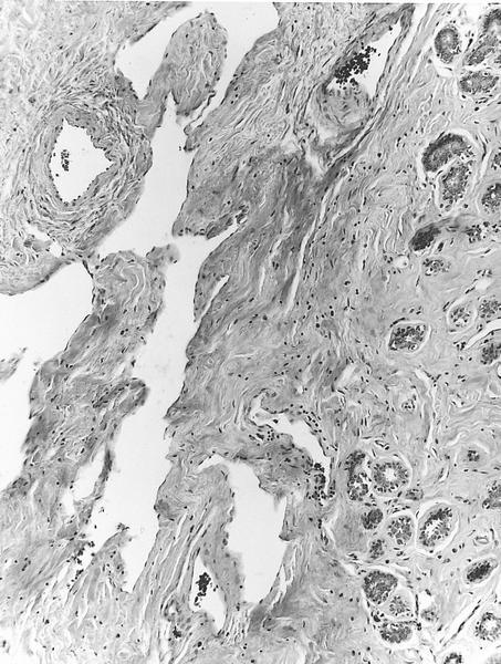
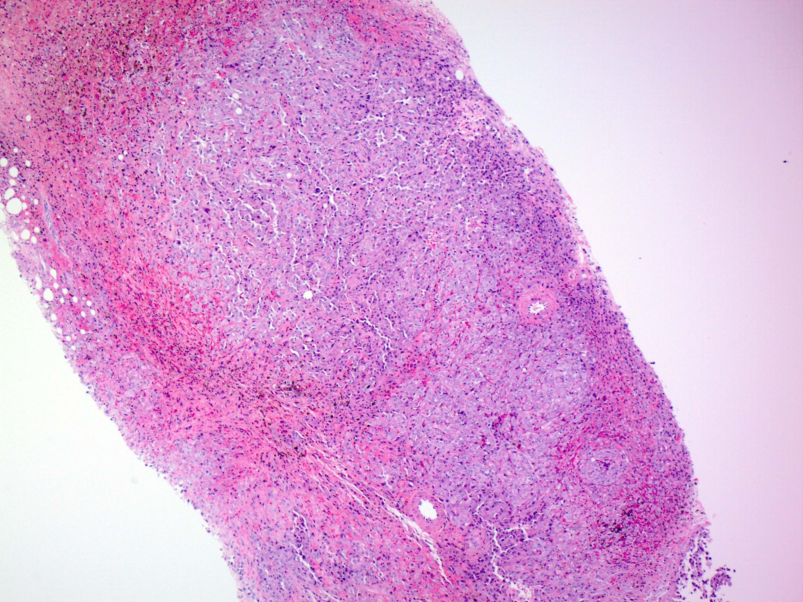
Microscopic (histologic) images
Contributed by Indu Agarwal, M.D. and AFIP images
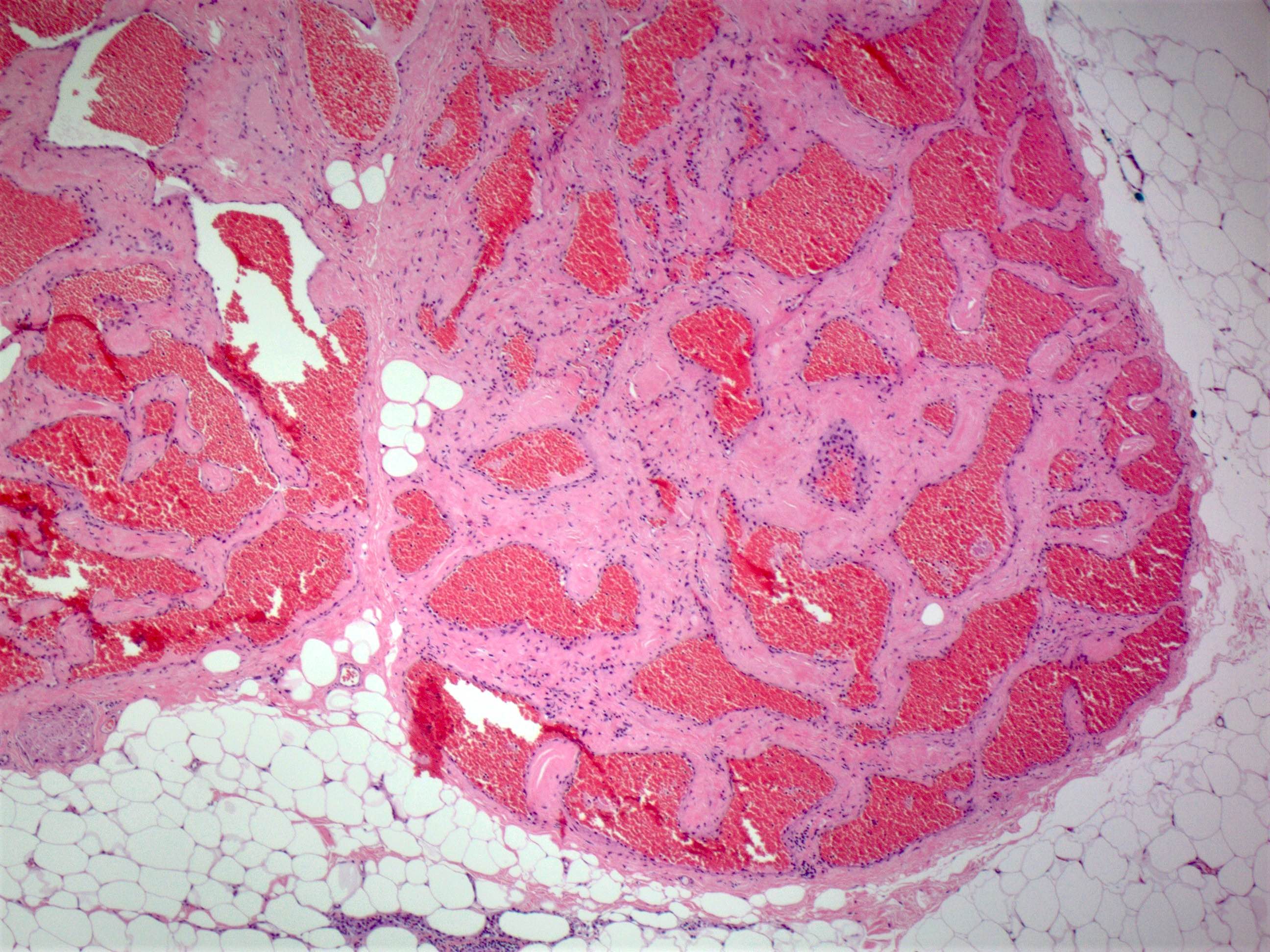
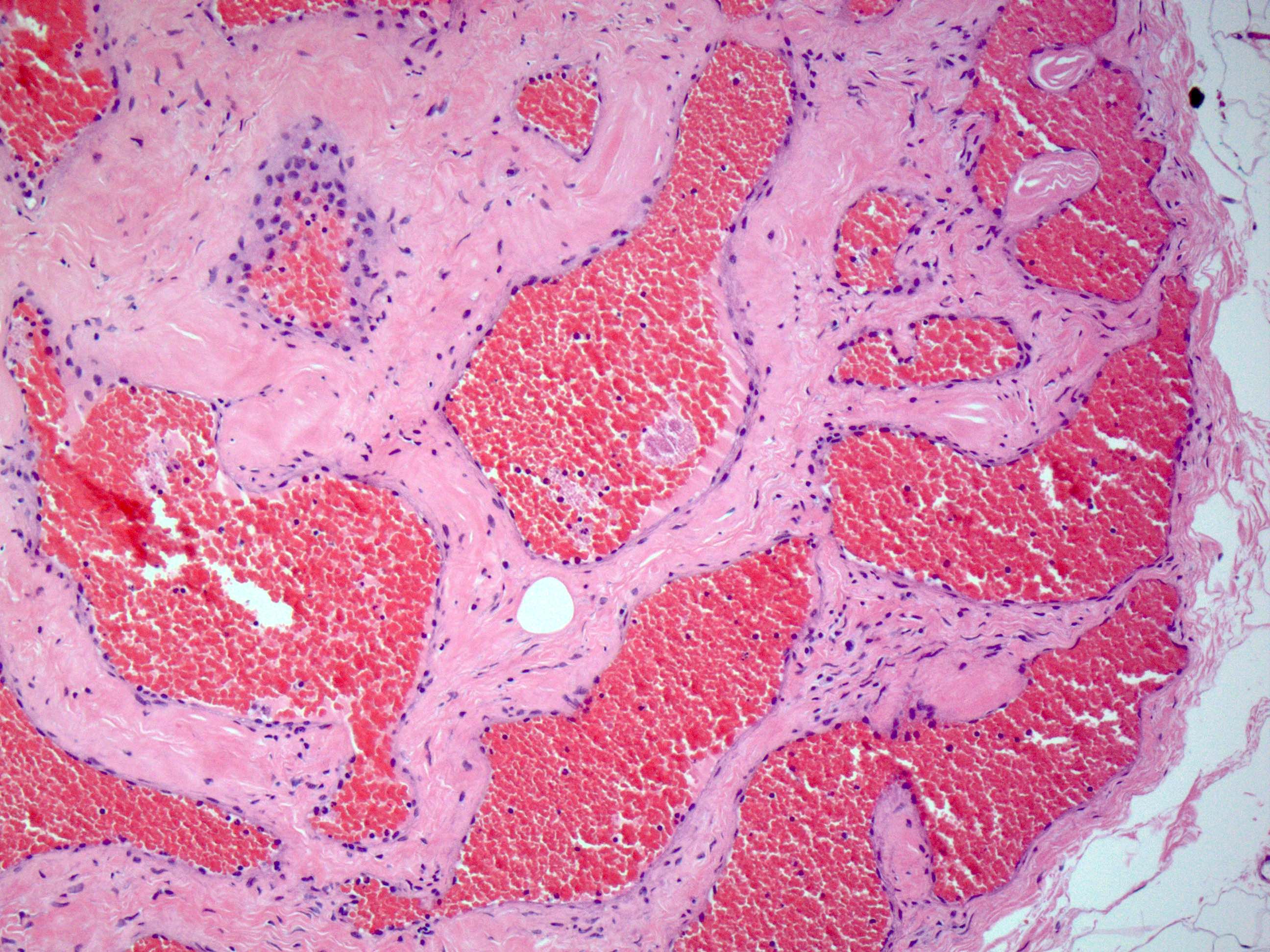
Cavernous hemangioma of breast
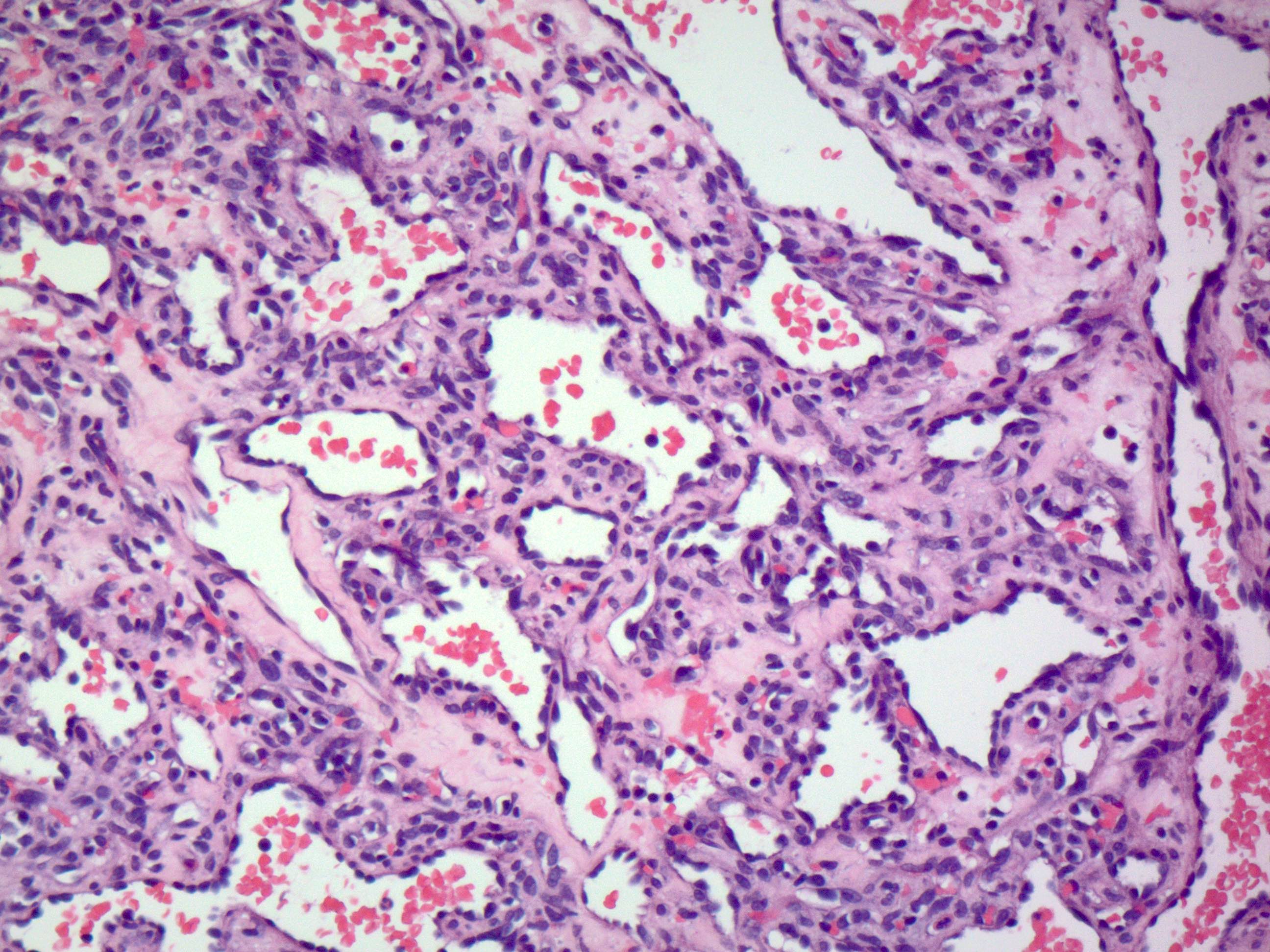
Capillary hemangioma
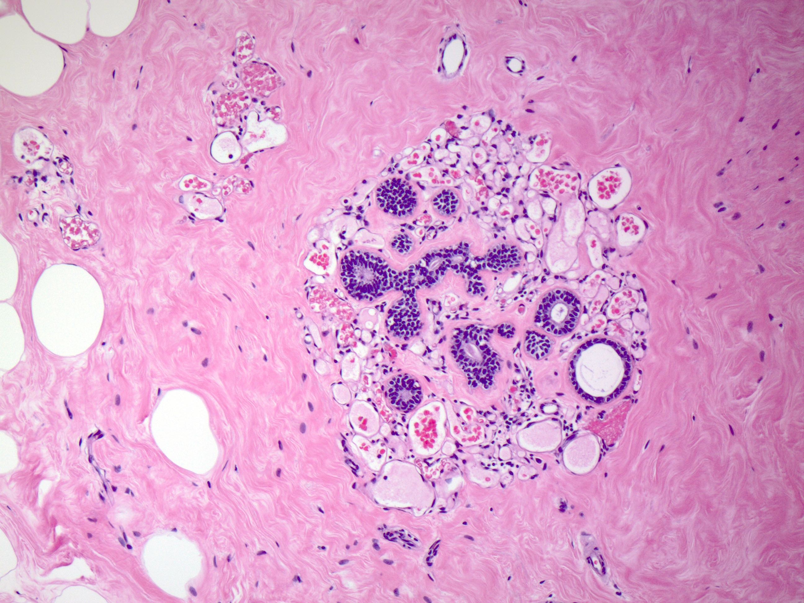
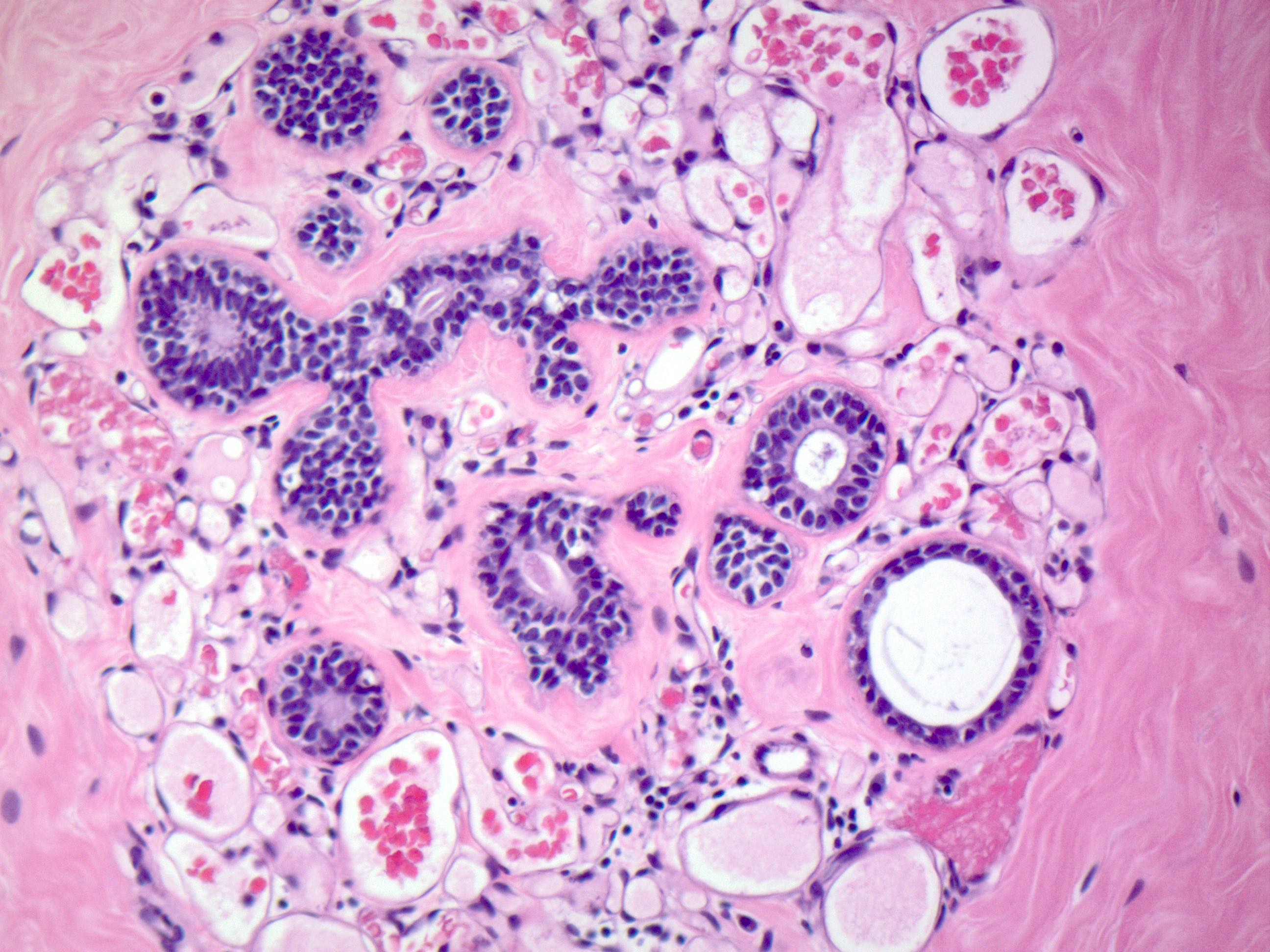
Perilobular hemangioma
Complex hemangioma
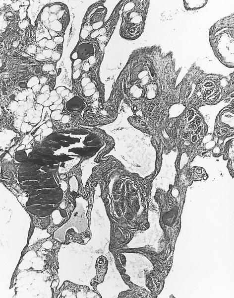
Angiomatosis
Negative stains
Videos
Histopathology breast, soft tissue - hemangioma
Sample pathology report
- Left breast, MRI guided core needle biopsy:
- Breast tissue with perilobular hemangioma
Differential diagnosis
- Angiolipoma:
- Has intermixed adipose tissue, clusters of capillary sized vessels, some with microthrombi
- Angiosarcoma:
- Has solid areas, mitotic activity, infiltrative pattern of growth and complex anastomosing vascular channels
- Atypical vascular lesion:
- Often past history of radiation
- Anastomosing thin walled vessels, unlike well formed vessels of hemangioma
Additional references
Board review style question #1
A 58 year old woman had a 4.0 cm breast mass with overlying erythematous skin patch. A stereotactic biopsy showed the histology above. What is the best treatment recommendation for this patient?
- Excision with wide margins
- Lumpectomy with hormone therapy
- Lumpectomy with radiation
- No further treatment needed
Board review style answer #1
A. Excision with wide margins. This is an angiosarcoma with a predominantly solid appearance and the cells demonstrate nuclear atypia, unlike a hemangioma, which lacks these features. Wide excision, preferably mastectomy, should be performed.
Comment here
Reference: Hemangioma
Comment here
Reference: Hemangioma
Board review style question #2
A 35 year old woman presents with a 10 cm palpable breast mass. Core needle biopsy shows the lesion occupying the entire biopsy tissue and consisting of large, thin walled vessels lined by bland endothelial cells. The lesion surrounds but does not invade the normal breast lobular units. No cytologic atypia, necrosis, solid areas or mitotic activity is seen. The Ki67 proliferation index is 1%. What is the most likely diagnosis?
- Angiomatosis
- Angiosarcoma
- Cavernous hemangioma
- Lymphangioma
Board review style answer #2
A. Angiomatosis. This is a case of angiomatosis of breast. The lesion is large and consists of large, thin walled vessels, which lack atypia, mitosis and other malignant cytologic features, and do not invade the normal breast lobular stroma.
Comment here
Reference: Hemangioma
Comment here
Reference: Hemangioma
