
Pancreas
Exocrine carcinomas
Undifferentiated carcinoma with osteoclast-like giant cells
Author: Claudio Luchini, M.D., Ph.D.
Editorial Board Members: Naziheh Assarzadegan, M.D., Wei Chen, M.D., Ph.D.
Editor-in-Chief: Debra L. Zynger, M.D.

Last author update: 31 October 2022
Last staff update: 31 October 2023
Copyright: 2002-2024, PathologyOutlines.com, Inc.
PubMed Search: Undifferentiated carcinoma with osteoclast-like giant cells pancreas
Table of Contents
Definition / general | Essential features | Terminology | ICD coding | Epidemiology | Sites | Pathophysiology | Clinical features | Diagnosis | Radiology description | Radiology images | Prognostic factors | Case reports | Treatment | Gross description | Gross images | Frozen section description | Frozen section images | Microscopic (histologic) description | Microscopic (histologic) images | Cytology description | Cytology images | Positive stains | Negative stains | Electron microscopy description | Electron microscopy images | Molecular / cytogenetics description | Sample pathology report | Differential diagnosis | Additional references | Board review style question #1 | Board review style answer #1 | Board review style question #2 | Board review style answer #2Cite this page: Luchini C. Undifferentiated carcinoma with osteoclast-like giant cells. PathologyOutlines.com website. https://www.pathologyoutlines.com/topic/pancreasosteoclastic.html. Accessed April 26th, 2024.
Definition / general
- Undifferentiated carcinoma of the pancreas with no glandular differentiation and with prominent infiltration by histiocytes and osteoclast-like giant cells
- May be found in association with a differentiated component of pancreatic ductal adenocarcinoma
Essential features
- Undifferentiated carcinoma with atypical neoplastic cells and lacking gland formation
- Presence of multinucleated giant cells resembling osteoclasts
Terminology
- Undifferentiated carcinoma with osteoclast-like giant cells (UCOGC, UCOCGC)
- Old terminology, not in use: giant cell tumor of the pancreas, osteoclastoma
ICD coding
Epidemiology
- M:F = 0.9:1 (Am J Surg Pathol 2016;40:1203, J Pathol 2017;243:148)
- Commonly presents in the sixth to seventh decades (Am J Surg Pathol 2016;40:1203, J Pathol 2017;243:148)
- 1.4% of all pancreatic cancers (Am J Surg Pathol 2016;40:1203)
Sites
- Head of the pancreas (60 - 75% of cases), body / tail (25 - 40%) (Am J Surg Pathol 2016;40:1203, J Pathol 2017;243:148)
Pathophysiology
- Unknown; a role may be played by hemorrhagic / chronic inflammatory foci with recruitment of tumor supporting macrophages within the pancreas or within a pancreatic ductal adenocarcinoma (Hum Pathol 2018;81:157)
Clinical features
- Presenting symptoms include jaundice (tumors in the pancreatic head), abdominal or back pain, weight loss and nausea (Am J Surg Pathol 2016;40:1203, J Pathol 2017;243:148)
Diagnosis
- CT scan and MRI are the preferred imaging modalities
- Diagnosis is by biopsy or surgical resection (more difficult with cytology but may be possible) (Cancer Cytopathol 2017;125:563)
Radiology description
- Usually mass lesions with delayed enhancement (Am J Surg Pathol 2016;40:1203)
Radiology images
Images hosted on other servers:

Ultrasound


CT
Prognostic factors
- Presence of an associated conventional ductal adenocarcinoma component is associated with a poorer prognosis and is one of the most important prognostic moderators (Am J Surg Pathol 2016;40:1203, J Pathol 2017;243:148)
- PDL1 expression by tumor cells is associated with poor prognosis (Hum Pathol 2018;81:157)
Case reports
- 54 year old man with a huge (28 cm) mass for which radical resection was possible (Int Cancer Conf J 2017;6:193)
- 54 year old woman with a large cystic mass in the pancreas (Int J Clin Exp Pathol 2015;8:11785)
- 61 year old woman with long term survival after surgical resection and chemotherapy (Case Rep Gastroenterol 2016;10:472)
- 62 year old woman with metastatic disease showing durable response to immunotherapy (tumor with high tumor mutational burden) (J Natl Compr Canc Netw 2021;19:247)
- 65 year old man with weight loss (Medicine (Baltimore) 2018;97:e13516)
- 68 year old woman with a prominent invasion of pancreatic ductal tree (JOP 2011;12:170)
Treatment
- Surgical resection if possible, gemcitabine based chemotherapy (Case Rep Gastroenterol 2016;10:472)
- Immunotherapy is a novel opportunity to be explored in this type of cancer (BMC Gastroenterol 2020;20:220, J Natl Compr Canc Netw 2021;19:247)
Gross description
- Larger than conventional ductal adenocarcinoma (J Pathol 2017;243:148)
- Brownish (not whitish) and with pushing borders
- Frequent cyst formation
- Hemorrhage or necrosis is typically present
Gross images
Contributed by Claudio Luchini, M.D., Ph.D.
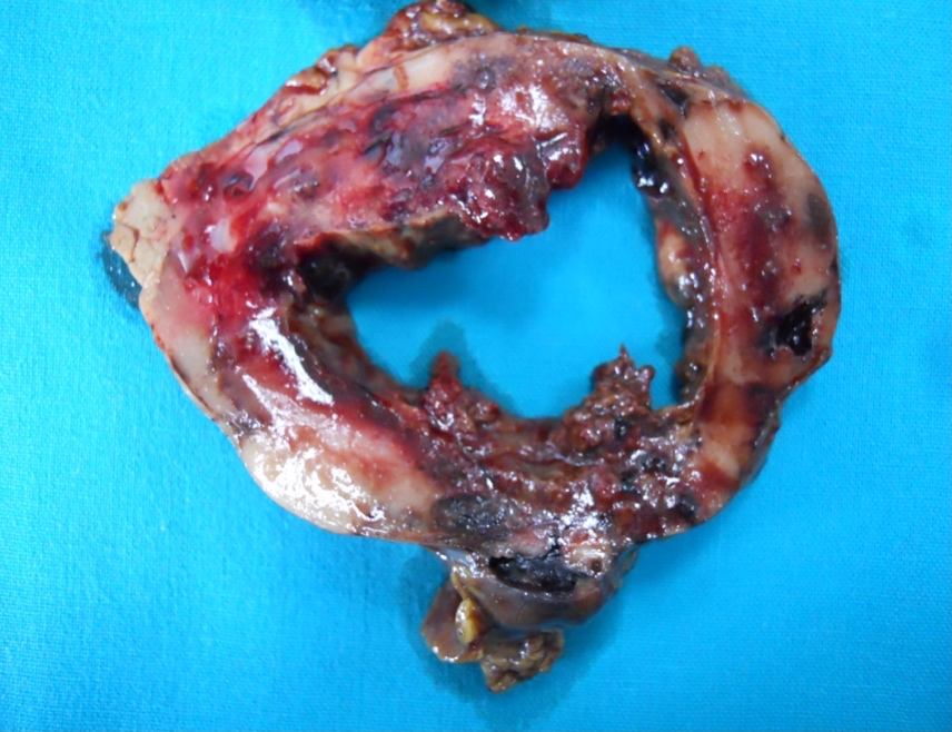
Cystic tumor
Images hosted on other servers:

Hemorrhagic mass

Macrophotograph revealing cystic mass

Cystic mass with gastric invasion
Frozen section description
- Hypercellular neoplasm with atypical cells
- Multinucleated giant cells, although they cannot always be documented in small biopsy sent for frozen section (may be focal component)
Frozen section images
Contributed by Claudio Luchini, M.D., Ph.D.
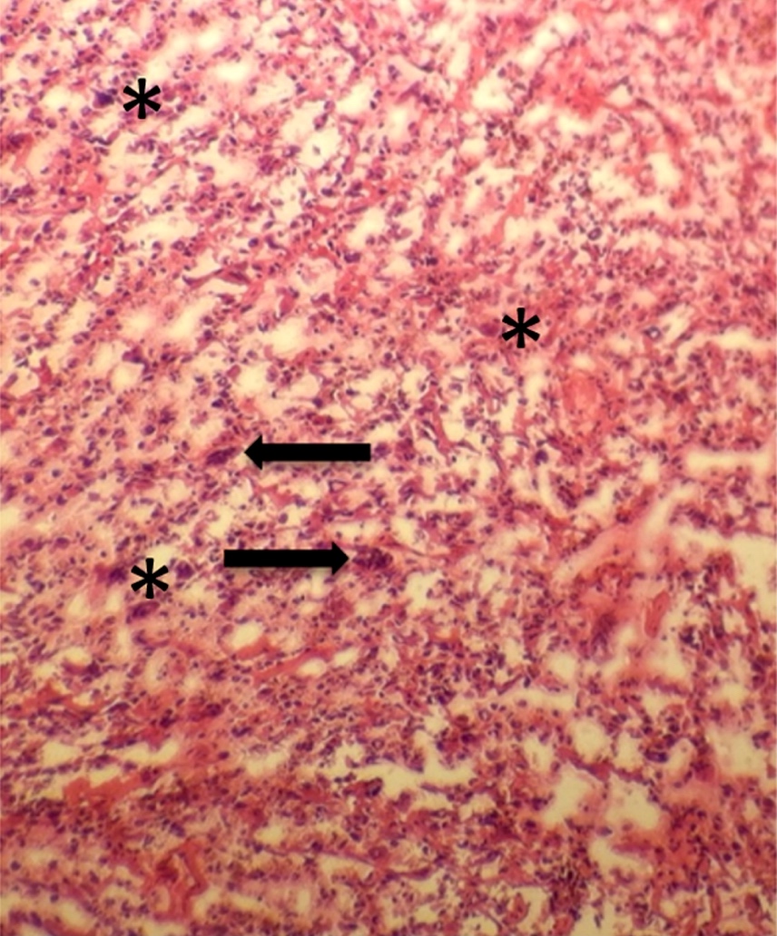
Osteoclast-like giant cells and neoplastic cells
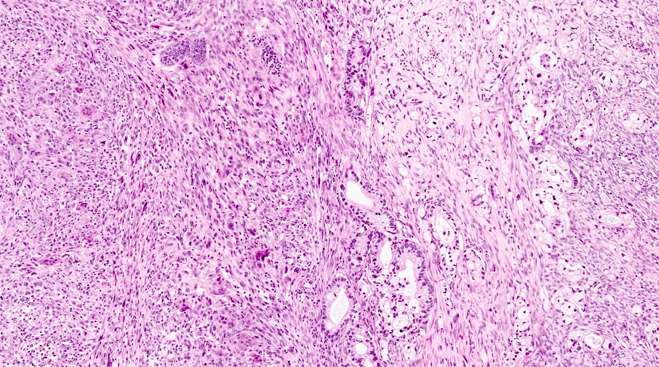
Microscopic (histologic) description
- Composed of 3 cell types:
- Mononuclear neoplastic cells, sometimes very atypical
- Mononuclear histiocytes (nonneoplastic)
- Multinucleated giant cells (osteoclast-like giant cells) (nonneoplastic)
- Foci of hemorrhage and necrosis are very common
- Typical involvement of the pancreatic ductal tree, sometimes with a pseudopolypoid growth pattern (J Pathol 2017;243:148)
- May arise in association with mucinous cystic neoplasm of the pancreas and intraductal papillary mucinous neoplasm of the pancreas (Am J Surg Pathol 2016;40:1203, World J Surg Oncol 2011;9:100, Am J Surg Pathol 2015;39:179)
- Osteoid material may be found (J Pathol 2017;243:148)
Microscopic (histologic) images
Contributed by Claudio Luchini, M.D., Ph.D.
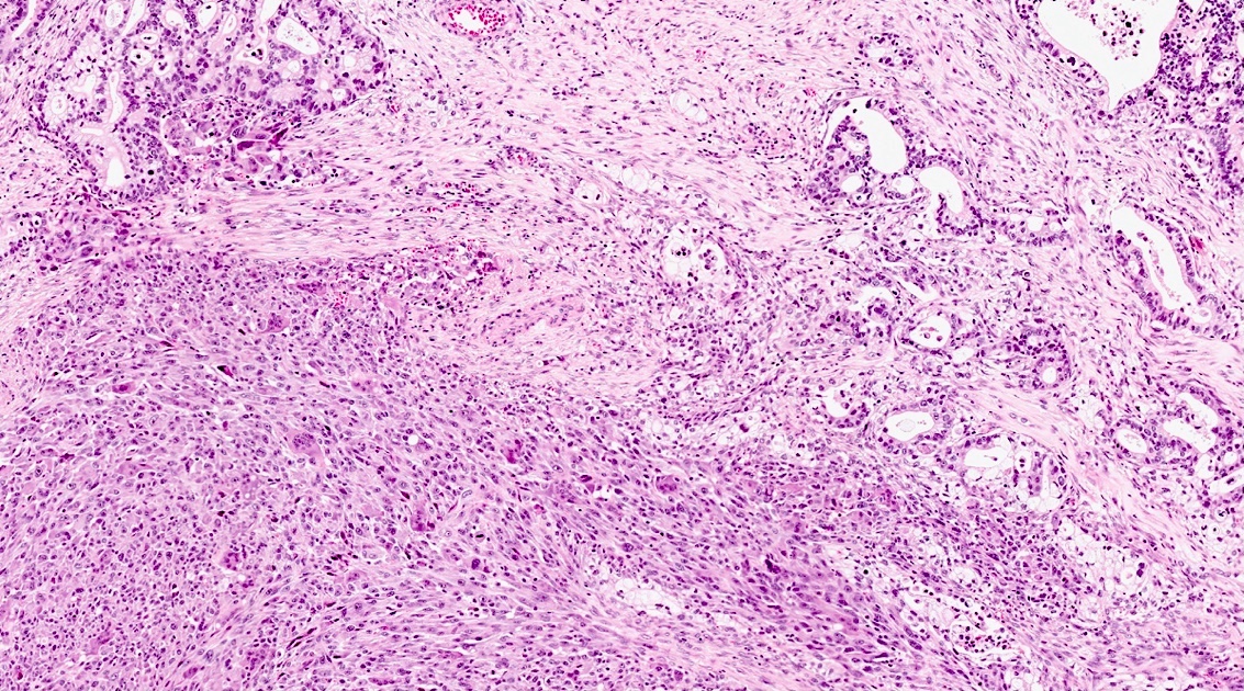
Associated conventional adenocarcinoma
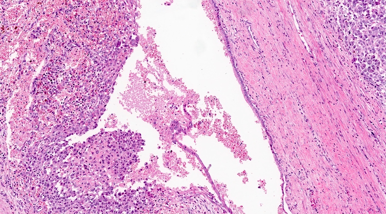
Ductal tree involvement
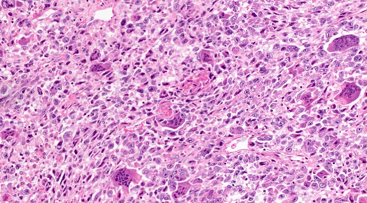
Microhemorrhagic foci
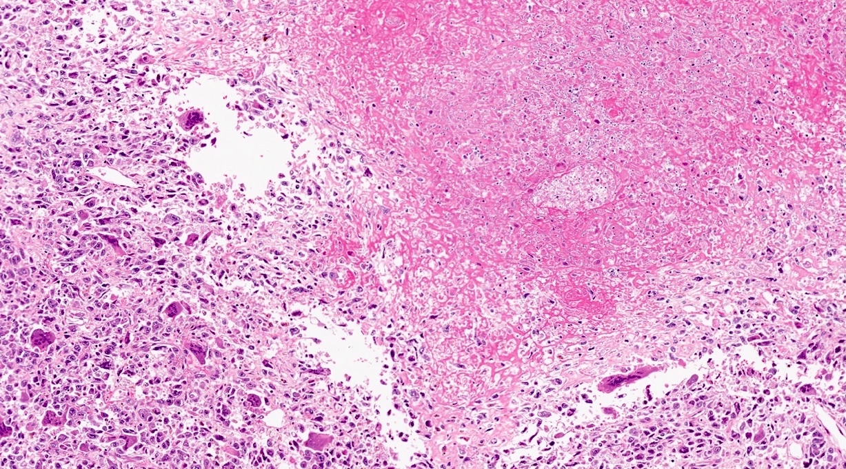
Extensive necrotic hemorrhage
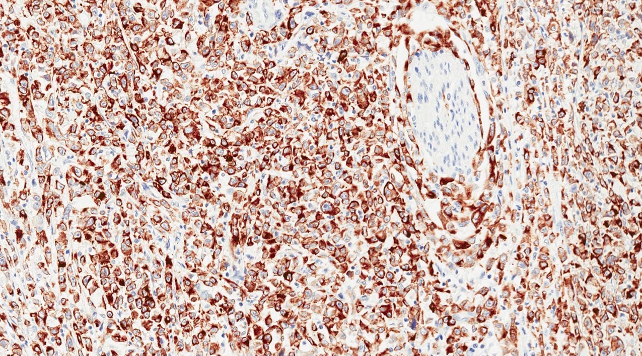
CK8/18+
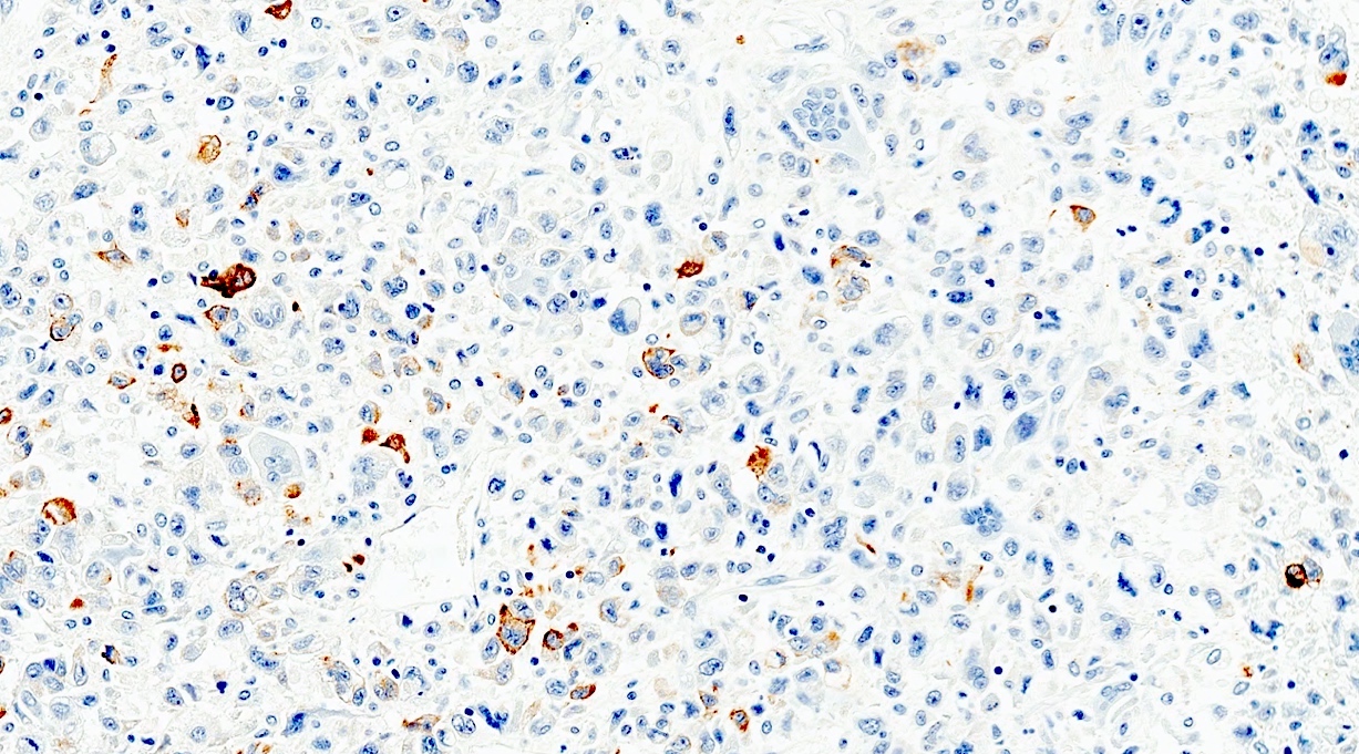
Focal CK8/18+

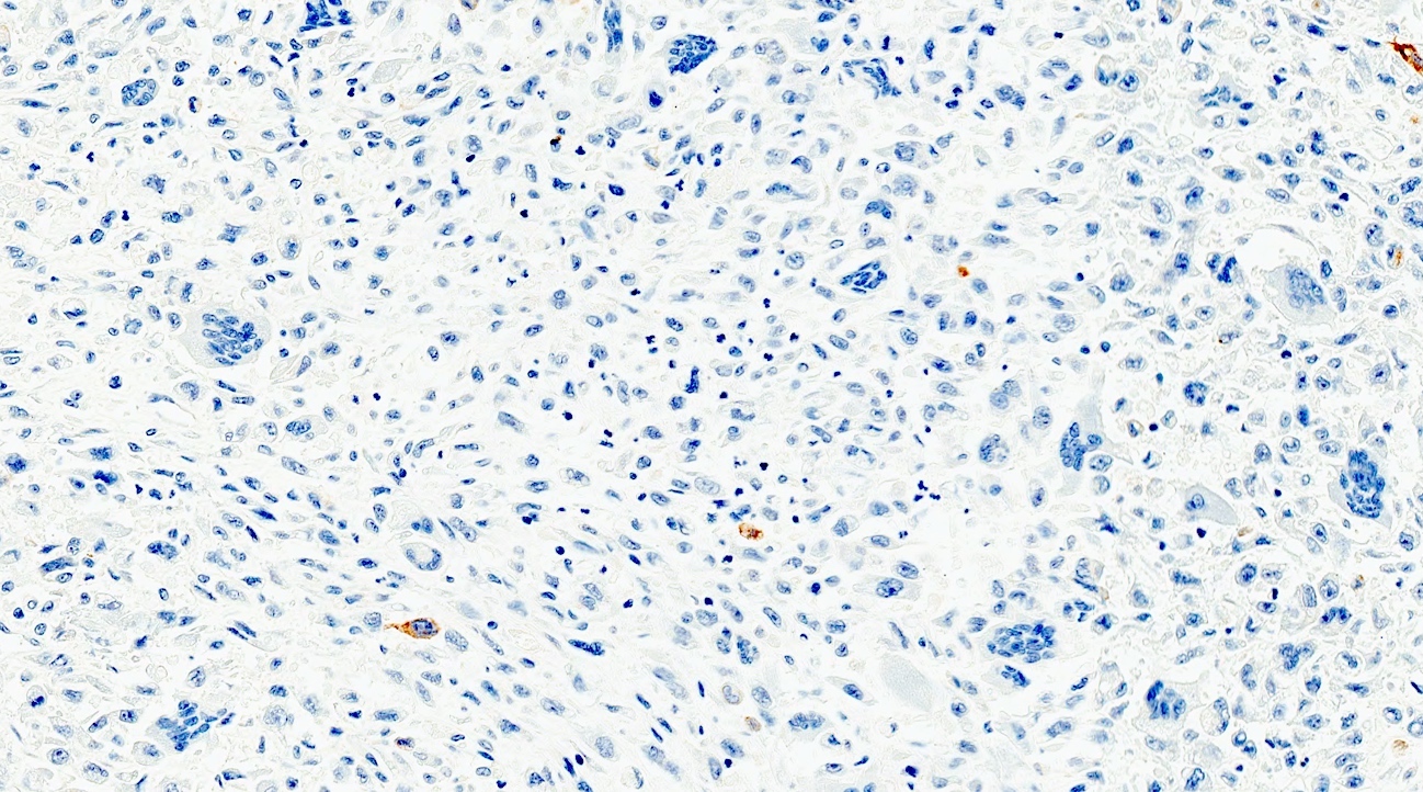
CK8/18-
Cytology description
- 3 cell types (osteoclast-like giant cells, neoplastic cells and histiocytes) are present in most cases (~80%)
- Cytology has a diagnostic value for this tumor in ~80% of cases (Cancer Cytopathol 2017;125:563)
Cytology images
Images hosted on other servers:

Romanowsky stain

Papanicolaou stain
Positive stains
- Neoplastic cells:
- Cytokeratin AE1 / AE3, CK7, CK8/18 are usually positive but may be totally negative
- p53 in > 50% of cases
- Ki67 high proliferation index
- Histiocytes:
- Multinucleated giant cells:
Negative stains
Electron microscopy description
- Neoplastic cells: abundant mitochondria, no microvilli or desmosomes (J Korean Med Sci 2005;20:516)
- Multinucleated osteoclast-like giant cells: multiple nuclei with rim of chromatin; abundant mitochondria; free ribosomes; dilated, empty, rough endoplasmic reticulum cisternae (J Korean Med Sci 2005;20:516)
Electron microscopy images
Images hosted on other servers:

Neoplastic cell

Giant cell
Molecular / cytogenetics description
- Overlaps with the genetic profile of conventional ductal adenocarcinoma, with the most common mutated genes being KRAS, TP53, CDKN2A and SMAD4 (J Pathol 2017;243:148)
- GLI3, a gene belonging to the hedgehog signaling pathway, may be found mutated in this tumor
- The same mutation (290 M > L) affecting the gene SERPINA3 has been documented in 25% of cases in a whole exome sequencing analysis (J Pathol 2017;243:148)
Sample pathology report
- Pancreas, pancreatectomy:
- Pancreatic undifferentiated carcinoma with osteoclast-like giant cells (see synoptic report)
Differential diagnosis
- Undifferentiated / anaplastic carcinoma of the pancreas:
- Absence of osteoclast-like giant cells
- Paraduodenal (groove) pancreatitis:
- Multinucleated giant cells may be abundant but neoplastic cells are lacking (Cytopathology 2015;26:122)
- Melanoma:
- Contains melanin pigment instead of or in addition to hemosiderin
- S100+
Additional references
Board review style question #1
This is an immunohistochemical slide (CK8/18) from a pancreatic tumor in an elderly woman with jaundice. S100 was negative throughout and the multinucleated cells were CD68 positive. Which of the following is the most likely diagnosis?
- Anaplastic lymphoma
- Melanoma
- Pancreatic undifferentiated carcinoma with osteoclast-like giant cells
- Pancreatitis
- Primitive mesenchymal tumor of the pancreas
Board review style answer #1
C. This is a pancreatic undifferentiated carcinoma with osteoclast-like giant cells. These tumors may be composed of neoplastic cells that are totally negative for cytokeratin. This finding, which is not uncommon, cannot be used to exclude this tumor entity.
Comment Here
Reference: Undifferentiated carcinoma with osteoclast-like giant cells
Comment Here
Reference: Undifferentiated carcinoma with osteoclast-like giant cells
Board review style question #2
Which cell types usually form an undifferentiated carcinoma with osteoclast-like giant cells?
- Osteoclast-like giant cells, which are tumor cells
- Tumor cells and histiocytes
- Tumor cells arranged in glands and osteoclast-like giant cells
- Tumor cells, histiocytes and osteoclast-like giant cells
- Tumor cells, lymphocytes and osteoclast-like giant cells
Board review style answer #2
D. Undifferentiated carcinoma of the pancreas with osteoclast-like giant cells typically contains 3 types of cells: tumor cells, histiocytes and osteoclast-like giant cells.
Comment Here
Reference: Undifferentiated carcinoma with osteoclast-like giant cells
Comment Here
Reference: Undifferentiated carcinoma with osteoclast-like giant cells
