
Colon
Other nonneoplastic
Amyloidosis
Author: Raul S. Gonzalez, M.D.

Last author update: 14 April 2021
Last staff update: 22 April 2022
Copyright: 2003-2024, PathologyOutlines.com, Inc.
PubMed search: Colon[TIAB] amyloidosis[TI]


Table of Contents
Definition / general | Essential features | Terminology | Epidemiology | Pathophysiology | Etiology | Clinical features | Diagnosis | Radiology description | Case reports | Treatment | Gross description | Gross images | Microscopic (histologic) description | Microscopic (histologic) images | Positive stains | Sample pathology report | Differential diagnosis | Additional references | Board review style question #1 | Board review style answer #1Cite this page: Gonzalez RS. Amyloidosis. PathologyOutlines.com website. https://www.pathologyoutlines.com/topic/colonamyloidosis.html. Accessed April 27th, 2024.
Definition / general
- Extracellular deposition of amyloid protein, often around blood vessels
Essential features
- Amyloid deposition in the colon, confirmable with Congo red
- Usually around blood vessels, which can lead to vascular injury
Terminology
- Localized (limited to the colon) or diffuse (present in numerous organs)
Epidemiology
- Can be primary, secondary, hereditary or endocrine related
Pathophysiology
- Overproduction of amyloid protein (AL, AA, ATTR, etc.) due to various causes
- Senile amyloid is often present in GI tract of elderly patients (Pathol Res Pract 1994;190:641)
Etiology
- Can have multiple causes, including malignancy, chronic inflammation, dialysis and endocrine abnormalities; sometimes associated with hemodialysis (Gastroenterology 1989;96:230, Clin Nephrol 2000;53:394, Mod Pathol 1995;8:577)
Clinical features
- Gastrointestinal involvement is seen in most patients with systemic amyloidosis
- May be asymptomatic or cause bleeding, obstruction, perforation or abnormal motility
- Amyloid tumor may clinically resemble carcinoma (AJR Am J Roentgenol 2002;179:536)
- Uncommonly, amyloid is localized to colon and does not require systemic treatment (Amyloid 2003;10:36)
Diagnosis
- Can diagnose with rectal biopsy that includes submucosa (85% sensitivity), though amyloid deposition may be initially discovered in a resection specimen
Radiology description
- Can cause various abnormalities on barium enema (Gastrointest Radiol 1991;16:133)
Case reports
- 65 year old man with amyloid tumor with synchronous adenocarcinoma (J Clin Pathol 1995;48:592)
Treatment
- If systemic, depends on type of amyloid but generally targeted at the cause (myeloma, kidney failure, etc.)
Gross description
- Mucosa may be normal or finely granular
Gross images
Images hosted on other servers:

Amyloid tumor (above)
and adenocarcinoma
arising from villous
adenoma (below)
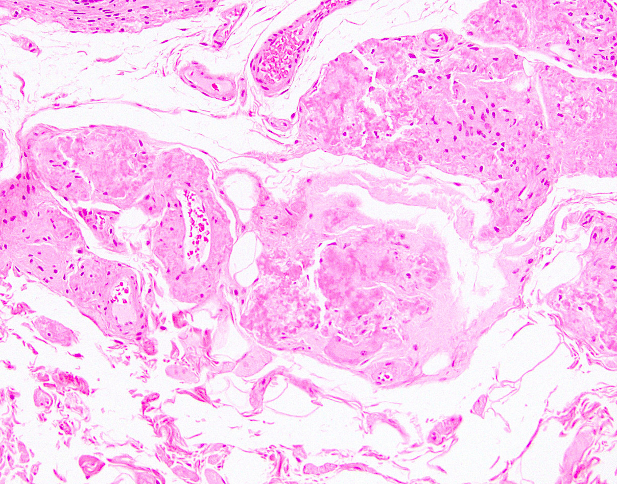
Microscopic (histologic) description
- Amyloid present in blood vessel walls and muscularis propria; may be subepithelial; may cause ischemic changes or frank hemorrhage
Microscopic (histologic) images
Contributed by Raul S. Gonzalez, M.D.
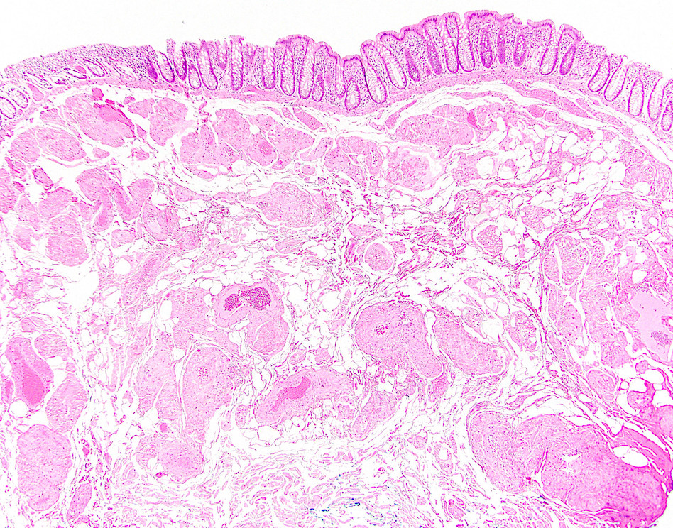
Colonic amyloid
Images hosted on other servers:

Submucosal vessel involvement

With Congo red stain

Congo red stain
highlights vessel
wall and free
submucosal amyloid

Congo red stain

Subepithelial
deposits resembling
collagenous colitis
Positive stains
- Congo red (stains deep pink and demonstrates apple green birefringence, as in other body sites)
Sample pathology report
- Colon, splenic flexure, biopsy:
- Amyloidosis (see comment)
- Comment: The biopsy shows amorphous eosinophilic material present around submucosal blood vessels. On Congo red stain, the material demonstrates apple green birefringence.
Differential diagnosis
- Collagenous colitis:
- Surface epithelial damage, epithelial lymphocytes
- Elastofibromatous change:
- Lacks apple green birefringence on Congo red, elastin stain positive
- Lifting agent granuloma:
- Can contain inflammatory component
- Negative on Congo red
- Pulse granuloma:
- Contains pulse material and inflammatory component
- Negative on Congo red
Additional references
Board review style question #1
Which of the following stains would be positive in the amorphous perivascular material seen in this colon polyp?
- AFB
- Congo red
- GMS
- von Kossa
Board review style answer #1
B. The material is amyloid. It would stain a salmon pink color with Congo red and demonstrate apple green birefringence.
Comment Here
Reference: Amyloidosis
Comment Here
Reference: Amyloidosis
